
Abstract
The RNS® System is the first commercially available device to provide closed-loop responsive brain stimulation. The system includes a cranially implanted neurostimulator that continually monitors the electrocorticogram through one or two depth and/or subdural cortical strip leads that are placed at the seizure focus. When abnormal electrographic activity is detected, the neurostimulator delivers brief pulses of electrical stimulation to the seizure focus through the implanted leads. In November 2013, the US FDA approved the RNS System as an adjunctive therapy for patients with drug resistant, partial onset seizures who have undergone diagnostic testing that localized no more than 2 epileptogenic foci. Safety and effectiveness of the RNS System for the indicated patient population was demonstrated in a multicenter, randomized, sham-stimulation controlled 2-year pivotal study. An ongoing, prospective, long-term treatment study is currently gathering an additional 7 years of prospective safety and effectiveness data of the RNS System.
Epilepsy is one of the most common serious neurological disorders, affecting approximately 1% of the population Citation[1]. Partial-onset epilepsy, also known as focal-onset epilepsy, is the most common type of epilepsy and the most challenging type to control Citation[1,2]. The primary treatment option for patients with epilepsy is pharmacotherapy. Antiepileptic drugs (AEDs) are effective in controlling seizures in approximately 60% of persons with partial-onset epilepsy Citation[2]. However, there remain a large proportion of patients who continue to have seizures despite medication Citation[2]. While there are now more than two dozen US FDA approved AEDs for the treatment of partial-onset epilepsy, once a patient has failed two or more trials of AEDs, the likelihood of becoming seizure-free with any subsequent trial of AED is less than 5% Citation[2–4]. Thus, guidelines recommend that patients and physicians consider non-pharmacotherapy options, such as resective surgery after patients have failed two or more trials of AEDs Citation[3,5,6]. Resective surgery is an option for some but not all patients with drug-resistant partial-onset epilepsy. Ideal surgical candidates have a single well-localized seizure focus in an area of the brain that may be safely removed with minimal neurological deficit Citation[6–8]. Not all patients who undergo resective procedures achieve seizure freedom. Approximately 30–40% of patients who undergo temporal lobectomies, which is the most common and most successful type of resective surgery, continue to have disabling seizures at 1 year after surgery Citation[6,9,10], and 30–60% of patients continue to have seizures after other types of resective surgeries Citation[6,8].
Neurostimulation is an option for those patients who have drug-resistant epilepsy and have either failed or are not candidates for resective surgery. There are currently two FDA-approved neurostimulation therapies for treatment of partial-onset epilepsy: vagus nerve stimulation (VNS) and responsive cortical stimulation. A third neurostimulation modality, open-loop deep brain stimulation (DBS) of the anterior thalamic nucleus is not approved in the US, but is approved in other countries as an adjunctive treatment for partial-onset seizures in adults with medically refractory epilepsy Citation[11].
VNS using the VNS Therapy® System (Cyberonics, Houston, TX, USA) was approved by the FDA in 1997 as an adjunctive therapy in reducing the frequency of seizures in patients with partial-onset seizures that are refractory to medication. The VNS Therapy System provides scheduled open-loop stimulation to the left vagus nerve using a pulse generator that is implanted subcutaneously in the subclavicular fossa and electrodes wrapped around the vagus nerve Citation[12]. In the randomized controlled trials, the average reduction in seizures in the treatment group was 24–28% Citation[13,14].
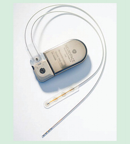
Responsive cortical stimulation using the RNS® System (NeuroPace, Inc., Mountain View, CA) has recently been approved by the FDA for use as an adjunctive therapy in patients with frequent and disabling drug-resistant partial-onset seizures localized to one or two epileptogenic foci. The RNS System is the first commercially available closed-loop neurostimulation system designed to treat partial-onset seizures. A cranially implanted programmable neurostimulator senses and records brain activity through depth and/or subdural cortical strip leads that are placed at that patient’s seizure focus . The neurostimulator detects electrographic patterns identified by the physician as abnormal and then provides brief pulses of electrical stimulation through the leads to interrupt those patterns. Here, we present the RNS System technology and review the clinical profile of the RNS System in the context of other treatment options.
Figure 1. The RNS® neurostimulator and NeuroPace® leads.
The RNS System
Indication
The RNS System is FDA approved for use as an adjunctive therapy in reducing the frequency of seizures in individuals 18 years of age or older with partial-onset seizures who have undergone diagnostic testing that localized no more than two epileptogenic foci, are refractory to two or more antiepileptic medications and currently have frequent and disabling seizures (motor partial seizures, complex partial seizures and/ or secondarily generalized seizures). The RNS System has demonstrated safety and effectiveness in patients who average three or more disabling seizures per month over the three most recent months (with no month with fewer than two seizures) and has not been evaluated in patients with less frequent seizures.
System overview
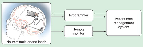
The RNS System is the first device to provide closed-loop responsive brain stimulation. Implantable components of the system include a neurostimulator and depth and/or subdural cortical leads. Non-implanted components of the RNS System include a programmer, a patient remote monitor and an internet-based interactive database (the Patient Data Management System [PDMS]) .
Figure 2. The RNS System. The implantable components of the RNS System include a neurostimulator and depth and cortical strip leads. The programmer is a laptop computer with proprietary software and custom telemetry components that the physician uses to communicate with the neurostimulator. The programmer can configure neurostimulator settings and retrieve data stored on the neurostimulator. The remote monitor is a home-use monitoring device used by the patient to retrieve data stored on the neurostimulator. The PDMS is a centralized database, which contains data uploaded from the programmer and remote monitor. Neurostimulator data and detection settings can also be transferred from PDMS to the programmer.
The RNS neurostimulator
The RNS neurostimulator is a programmable responsive neurostimulator that is surgically implanted in the cranium. The neurostimulator contains electronic circuitry and a battery that are hermetically sealed within a flat-curved titanium enclosure. The neurostimulator can be connected to one or two leads that are surgically placed at the seizure focus.
The neurostimulator continuously senses electrocorticographic (ECoG) activity through the leads. In response to the detection of patterns previously identified by the physician as abnormal (such as epileptiform activity), the neurostimulator delivers short trains of current-controlled, charge-balanced pulses through the leads. The physician may adjust detection and stimulation settings noninvasively using a programmer that communicates with the neurostimulator via a short-range wireless radiofrequency link.
The neurostimulator stores information regarding the time and date of detections and stimulations. The physician can also program the neurostimulator to store segments of the ECoG activity immediately before and after prespecified events such as a detection of abnormal electrographic activity, responsive stimulation, a magnet swipe (used by the patient to indicate a clinical event) or according to time of day.
Leads
The leads are insulated wires that provide an interface through which electrical activity of the brain can be sensed by the neurostimulator and through which responsive electrical stimulation can be delivered. Each lead has four platinum/iridium electrodes at the distal end, which are implanted at the seizure onset region, and four contacts at the proximal end, which connect to the neurostimulator. There are two types of leads: NeuroPace® Depth Leads, which are implanted into the brain, and NeuroPace Cortical Strip Leads, which are placed on the surface of the brain.
NeuroPace programmer
The programmer is a laptop computer with proprietary software and custom telemetry components to communicate with the neurostimulator. The physician uses the programmer to program the neurostimulator detection and stimulation settings and to retrieve data stored on the neurostimulator such as battery measurements, lead impedances, programmed settings, data regarding the occurrences of detection and stimulation, and ECoG segments. Using a secure internet connection, the programmer can transmit these data to the PDMS for storage and subsequent physician review.
NeuroPace remote monitor
The remote monitor is a home-use monitoring device that can communicate with the neurostimulator using a short-range wireless radiofrequency link. The patient is provided with a remote monitor, which can be used by the patient or caretaker to upload neurostimulator data to the PDMS via a secure internet connection. This allows the physician to remotely monitor the neurostimulator function between office visits. The remote monitor cannot be used by the patient to program the neurostimulator.
NeuroPace PDMS
The PDMS is a centralized database that contains neurostimulator data uploaded from the programmer and remote monitor. Using a secure web browser, the physician may access and review the patient’s neurostimulator data from the PDMS at any time. Neurostimulator data and detection settings can also be transferred from the PDMS to the programmer so that the physician can review ECoG data and detection on the programmer as well as on the PDMS. Data transferred to and from the PDMS are encrypted to ensure security and integrity of the data.
Neurostimulator functions
The neurostimulator performs three main functions: sensing, detection and stimulation. Each of these functions can be configured and adjusted by the physician during office visits.
Sensing & storage
The neurostimulator continuously senses and monitors electrographic activity through the leads. Sensing is performed using a bipolar montage between any two electrode contacts. The typical configuration is to sense between neighboring electrode contacts. For example, the first sensing channel would be programmed to sense the voltage difference between the first and second electrode contacts on the first lead. Up to four bipolar channels can be sensed simultaneously. The sensed signals are digitized at 250 Hz for recording and detection analysis.
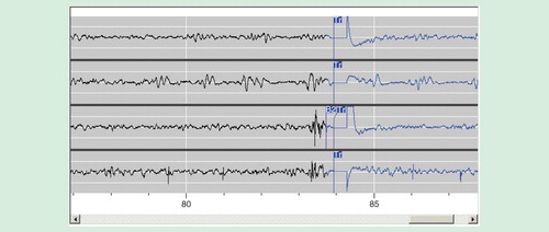
The neurostimulator can be programmed by the physician to store segments of the ECoG when specific events occur. The types of events that can trigger ECoG storage include detection, responsive stimulation, time of day, long episode (indicating that the detected event has continued beyond a preset duration), magnet swipe (used by the patient to indicate a clinical event), saturation (indicating high-amplitude activity) and noise (indicating 60 Hz signal was detected). When such an event occurs, the ECoG activity immediately before and after the event is stored on the neurostimulator. An example of a typical epileptiform discharge stored by the neurostimulator is shown in . Due to the storage capacity of the neurostimulator, approximately four 90-s four-channel ECoG segments can be stored on the neurostimulator at any time. Once this limit is exceeded, the oldest stored ECoGs are replaced by the newly stored ECoGs.
Figure 3. Example of a four-channel ECoG segment stored by the neurostimulator as displayed on the PDMS. In this example, the neurostimulator has been programmed to detect paroxysmal high-amplitude sharply contoured discharges on channel 3, which is typical of this patient’s interictal epileptiform activity. ‘B2’ denotes detection of epileptiform activity on the third channel. ‘Tr’ indicates delivery of responsive stimulation. Following the initial detection, the electrocoticogram is displayed in blue. When responsive stimulation is delivered, and immediately afterward, there is an artifact in the recording.
Stored ECoG segments can be uploaded from the neurostimulator to the programmer or remote monitor and then to PDMS. After the ECoG segments are uploaded, the neurostimulator ECoG memory is cleared to store more ECoGs. The physician can view previously stored ECoG segments to assess detection and the effects of stimulation. The physician may decide the detection is adequate or may decide to reprogram the neurostimulator so that the detection occurs earlier or later in the discharge.
The neurostimulator can also be programmed to display and store ECoG segments in real time. Real-time ECoGs are useful during the implantation procedure for evaluating the ECoG signals and adjusting amplifier gain settings. Real-time ECoGs can also be displayed and stored as the physician tests new stimulation settings and are useful for assessing acute effects of stimulation.
Detection
The detection algorithms implemented in the neurostimulator are designed to be computationally efficient and are optimized to perform real-time ECoG pattern detection (such as epileptiform activity) within the power and processing constraints of currently available implantable technology. Three detection tools (line length, area and bandpass) are provided. The detection tools are highly configurable and can be adjusted by the physician to optimize detection for each individual patient. Up to two independent detectors can be programmed for each of two sensing channels.
The line length detection tool Citation[15] is used to identify changes in both amplitude and frequency. Average line length, which is a measurement of the signal waveform length within a given time period, is calculated as the sum of the sample-to-sample differences within a window divided by the number of samples in the window. The line length detection tool works by comparing the average line length within a short-term sliding window to that of a long-term sliding window. Detection occurs when the short-term measurement exceeds an absolute or relative threshold as compared to the long-term measurement. A negative threshold can also be used to detect decreases in line length, which may represent an electrodecremental event or decreased frequency. Because the line length detection tool is sensitive to both amplitude and frequency changes, the line length detection tool is often used as the initial detector to detect any large change in the signal. Once an electrographic pattern is selected for detection, other detection tools may be used to provide more specific detections.
The area detection tool Citation[16,17] is used to identify changes in the overall signal energy without regard for frequency. Area is defined as the average area under the curve within a window. As with the line length detection tool, a short-term window average is compared to a long-term background window average, and detection occurs when a positive or negative threshold is exceeded.
The bandpass detection tool is similar to that described by Gotman Citation[18] and is used to detect rhythmic and spiking activity. The tool segments the electrographic signal at local minima and maxima resulting in ‘half-waves,’ the amplitude and duration of which are representative of the amplitude and frequency components of the ECoG. Half-waves that exceed a physician programmed amplitude and duration are counted; the number of these half-waves occurring within a given window length must exceed a certain threshold for detection to occur. The bandpass detection tool therefore acts like a frequency filter and can be used to detect activity within specific frequency bands (e.g., theta, alpha, beta and gamma). Moreover, because the bandpass detection tool typically provides more specific detection than the line length and area detection tools, this detection tool is often used once specific ECoG patterns for detection have been identified.
The output of the bandpass, line length, and area detection tools from the same or different EEG channels may be combined to identify specific electrographic events. In addition, the output of a tool may be assigned a persistence duration to facilitate detection of events in a sequence. For example, a seizure onset consisting of a series of spike-wave elements followed by low-voltage fast activity may be identified by a line length detection tool configured to detect a sudden increase in signal amplitude followed by a bandpass detection tool configured to detect high-frequency activity.
Responsive stimulation
The neurostimulator delivers current-controlled, charge-balanced biphasic pulses and can be programmed by the physician to deliver bursts of stimulation with pulses of between 40 and 1000 µsec, pulse frequencies between 1 and 333 Hz, current amplitudes of between 0.5 and 12 mA and burst durations between 10 and 5000 ms. The neurostimulator can be configured to deliver current between any combination of electrodes. The titanium neurostimulator canister may also be configured as part of the stimulation path.
Up to five individually configured sequential therapies of electrical stimulation may be programmed, where each therapy is composed of two independently configurable bursts. The neurostimulator will attempt to redetect the ECoG pattern after each therapy is delivered. If the pattern is still detected, the next therapy will be delivered. If the pattern is no longer detected, the remaining therapies in the sequence will not be delivered. The therapy sequence will refresh with the detection of each new episode.
Typical initial responsive therapy settings are a frequency of 200 Hz, pulse width of 160 µsec and burst duration of 100 ms. Current is initially programmed at a low amplitude (e.g., 1.0 mA) and titrated upward as tolerated to a maximum of 12 mA. In general, stimulation is delivered to the leads and electrodes from which electrographic patterns of interest are observed. For example, if electrographic activity of interest is observed on all channels, then the stimulation pathway could be configured to stimulate across all electrodes. However, if electrographic activity is observed on only two channels, then the stimulation pathway could be configured such that current is delivered through only those electrodes with electrographic activity of interest.
Clinical profile
Data from three clinical studies led to the FDA-approval of the RNS System for use as an adjunctive therapy in individuals 18 years of age or older with medically refractory partial-onset seizures localized to no more than two epileptogenic foci. These were the completed feasibility and pivotal studies, and the ongoing long-term treatment (LTT) study.
Feasibility study
The prospective, multi-center, primarily open-label feasibility study was designed to demonstrate adequate safety and provide sufficient evidence of effectiveness for the RNS System to support the commencement of a pivotal study. Sixty-five subjects were implanted with the RNS System and 59 subjects completed the 2-year study. The primary safety endpoint was met: the serious adverse event rates during the first month post-implant (6.2%) and the first 3 months post-implant (9.2%) were not worse than the pre-specified historical control (DBS for treatment of movement disorders), which were 19 and 36%, respectively Citation[19–25]. These results supported the commencement of the pivotal study.
Pivotal study
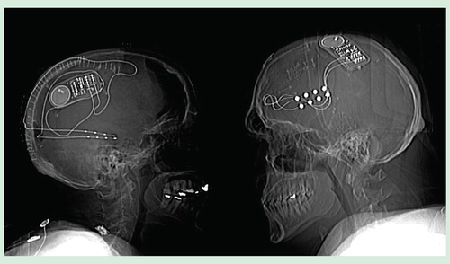
The prospective, multi-center, double-blinded, randomized, sham-stimulation-controlled pivotal study was designed to demonstrate the safety and effectiveness of the RNS System as an adjunctive treatment for adults with medically intractable partial-onset seizures arising from 1 or 2 seizure foci. During this study, 191 subjects were implanted, and 92% completed the 2-year study. X-ray images for two subjects implanted in the pivotal study are shown in . Subjects were on average 35 ± 11.6 years old and had epilepsy for 20.5 ± 11.6 years. Nearly one-third (32%) had prior therapeutic epilepsy surgery (resection, subpial transection and/or callosotomy), and over one-third (34%) had previously been treated with a vagus nerve stimulator.
Figure 4. Skull x-rays of two patients implanted with the RNS® neurostimulator and NeuroPace® leads. The patient on the left is a female, with seizures arising bilaterally from the mesial temporal lobes. The x-ray shows NeuroPace depth leads implanted longitudinally in the left and right hippocampi. The Leads are connected to the RNS neurostimulator shown implanted in the parietal bone. The patient on the right is a male, with seizures arising from the left frontal lobe. The x-ray shows NeuroPace cortical strip leads implanted over the left frontal lateral cortex. The leads are connected to the RNS neurostimulator implanted in the parietal skull.
Effectiveness of responsive stimulation was assessed by comparing the seizure reduction in the group receiving active stimulation (treatment group) versus the group receiving no stimulation (sham group) during a 12-week blinded evaluation period relative to a 12-week pre-implant baseline; throughout the trial (both pre-implant and post-implant), seizures were recorded in daily seizure diaries. The primary effectiveness endpoint was met: the reduction in seizure frequency in the treatment group (−37.9%) was significantly greater than that in the sham group (−17.3%; p = 0.012) Citation[26]. There was no difference in effectiveness in patients with mesial temporal lobe seizure onsets compared to patients with neocortical seizure onsets, in patients whose seizures arose from one compared to two foci, in patients who had been treated with VNS compared to those who had not, and in patients who had already undergone a therapeutic epilepsy surgery compared to those who had not Citation[26].
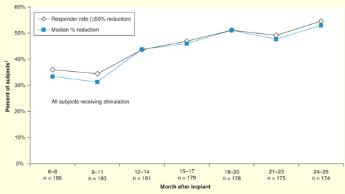
During the open-label period of the pivotal study, when all subjects had the opportunity to receive responsive stimulation, seizure reduction continued to improve. The median percent reduction in seizures was 44% at 1 year and 53% at 2 years post-implant compared to baseline . In a last observation carried forward analysis conducted at the end of the pivotal study, 54% of patients were responders (50% or greater reduction in seizures) and 9% were seizure free Citation[27].
Figure 5. Responder rate and median percent reduction in seizure frequency during the open-label period in the pivotal study.
Safety was assessed using AE data. The RNS System serious AE rate was no worse than the literature-derived serious AE rate for comparable procedures . The serious AE rate for the RNS System over the first 28 days (12.0%) was not worse than the pre-specified literature-derived comparator of 15% for implantation of intracranial electrodes for seizure localization and epilepsy surgery Citation[22,28–31]. The serious AE rate for the RNS System during the implant and first 84 days (18.3%) was not worse than the pre-specified literature-derived comparator of 36% for implantation and treatment with DBS for Parkinson's disease Citation[19,21,23–25]. Stimulation was well tolerated. During the blinded periods, there was no difference in the frequency or type of AE between the treatment and sham groups except for side effects of antiepileptic medications, which were more common in the sham group (six subjects, all mild events) than the treatment group (none) Citation[27].
Table 1. Pivotal study – primary safety endpoint.
During the open-label period, the most common serious AE was video-EEG monitoring (13 subjects; 7.0%); this was considered a serious AE because all hospitalizations were recorded as serious AEs within the trial, and video-EEG monitoring is performed as an inpatient procedure. Additionally, five subjects had lead revisions to replace damaged leads, and four subjects had lead revisions in order to improve the lead location. Three subjects had implant site infections; one had the device explanted. There were two subdural hemorrhages during the open-label period – both were attributed to seizure-related head trauma, and neither of these subjects had neurological consequences. There were six deaths in the pivotal study: four were attributed to possible, probable or definite sudden unexplained death in epilepsy, one was due to lymphoma and one was a suicide. The sudden unexplained death in epilepsy rate during the RNS System studies was not higher than expected for this population Citation[32]. The subject who died of suicide had a pre-existing history of depression including scores on the mood inventories that indicated severe depression but was considered to be clinically stable at the time of enrollment Citation[27].
Additional assessments included a quality-of-life inventory (the Quality of Life in Epilepsy Inventory [QOLIE-89]), neuropsychological evaluations (to assess visual and verbal memory, language, cognitive flexibility, learning and concentration) and mood inventories (the Beck Depression Inventory [BDI-II], Profile of Mood States [POMS] and Center for Epidemiological Studies Depression Scale [CES-D]). At 1 and 2 years after implantation, subjects reported significant improvements in quality of life overall (p < 0.001) and in nine of the primary scale scores, including memory, concentration and language Citation[27]. There was no significant deterioration in any neuropsychological measure or any of the mood inventories at the end of the blinded period compared to the baseline period, or at 1 and 2 years post-implant, indicating there were no acute, delayed or longer-term adverse effects of responsive stimulation on neuropsychological function and mood Citation[27].
LTT study
The LTT study is an ongoing, prospective, open-label study to follow subjects originally implanted in either the feasibility or pivotal studies in order to accumulate a total of 9 years of prospective data on safety and efficacy of the RNS System for the indicated patient population. At the completion of either the feasibility or pivotal studies, 97% of eligible subjects enrolled into the LTT study. Interim results of the LTT study (data as of 12 May 2011) were presented at the 22 February 2013 FDA neurological devices advisory panel Citation[33]. At that time, the median post-implant follow-up was 3.3 years (ranging from 5 weeks to 7 years) and the retention rate in the LTT study was 90.9% (209/230).
The effectiveness results were sustained over the long term: the responder rates and median percent reductions in seizures were between 50 and 60% at 4 and 5 years post-implant. Over the entire follow-up period, 27% of the subjects experienced at least one 3-month period of seizure freedom and 14.5% experienced at least one 6-month period of seizure freedom.
There were no unanticipated device-related serious AEs, and the types of AEs experienced were consistent with known risks of medical device implantation, other epilepsy therapies and with epilepsy. The most common serious AEs over the entire period of follow-up (feasibility, pivotal and LTT studies combined) were hospitalization for video-EEG monitoring (10.5%), implant site infection (6.3%) of the soft tissue, increased complex partial seizures (6.3%) and increased tonic-clonic seizures (5.5%).
Expert commentary
According to the Institute of Medicine Citation[5], at least 30% of adults with focal seizures do not have their seizures controlled with antiseizure medications, Citation[34,35], and a similar percentage experience medication-related side effects that impact quality of life, such as impaired cognition, fatigue, problems with coordination, nausea or other gastrointestinal symptoms Citation[6,34–37]. Some of these patients will consider epilepsy surgery or a VNS. However, not all patients are candidates for these treatments, and these treatments do not always provide meaningful benefit.
Responsive neurostimulation is a new approach to treating epilepsy. In the most general sense, epilepsy is a disorder in the balance of neuronal excitation and inhibition. In partial epilepsy, the disturbance may be well localized, but electrical activity spreads mono- and poly-synaptically to other brain regions to create the symptoms of the clinical seizure. The concept behind responsive stimulation is to identify the critical region and/or propagation pathways of seizures and to then provide neutralizing, disruptive or driving activity at this site or sites in order to restore normal function.
Responsive stimulation reduced the frequency of partial-onset seizures in patients with drug-resistant epilepsy who had failed, were not candidates or did not choose to pursue additional AEDs, epilepsy surgery and VNS. In the randomized controlled trials, patients receiving responsive stimulation with the RNS System had a statistically significant reduction in seizures compared to controls. The benefit of responsive stimulation therapy increased over time; the median percent reduction in seizures was 44% at 1 year post-implant and 53% at 2 years post-implant.
The clinical meaningfulness of the response to treatment with the RNS System is further supported by statistically significant improvements in overall quality of life and in individual domains of quality of life that indicate a more positive perception of cognitive function, relationships and social function, overall health and vulnerability to seizures. Improvements in health discouragement and seizure worry are strongly associated with improved quality of life in persons with intractable epilepsy Citation[38].
The patients in the RNS System studies had already failed at least two trials of AEDs; the likelihood of becoming seizure free with additional AEDs was less than 5% Citation[2–4]. However, 14.5% of patients participating in the RNS System studies experienced a seizure free period of at least 6 months or longer. This compares favorably to the 6-month seizure-free rates for recently approved AEDs in similar patient populations, which ranges from less than 2% with gabapentin to approximately 14% with levetiracetam Citation[39], and to the 4.6% seizure-free rate with VNS at last follow-up Citation[40].
The risks of the surgery to implant the RNS System compare favorably to those of other epilepsy-related procedures and to the deep brain stimulator for treatment of Parkinson’s disease. The risk for implant site infection related to the implant during the RNS System pivotal study (2.6%) was comparable to the rates reported in the literature for implantation of intracranial electrodes for epilepsy surgery evaluation (1.1–3.5%), for epilepsy resective surgery (5%) Citation[6,22,27,28,30,41], and for the rate with DBS for treatment of movement disorders (10%) Citation[42]. The rate of hemorrhage related to the implant during the RNS System pivotal study (2.1%) was also comparable to those reported in the published literature for implantation of intracranial electrodes (1.5–2.0%) and epilepsy resective surgery (5%) Citation[6,27,29,43].
Patient satisfaction with treatment with the RNS System is supported by the retention rate in the trials over many years of follow-up. Retention rate is perhaps one of the clearest reflections of the clinical meaningfulness of a therapy; patients choose to continue with a therapy only if the treatment is effective and well tolerated. During the RNS System studies, 91% of implanted subjects completed the 2-year feasibility study, 92% of implanted subjects completed the 2-year pivotal study and 97% of these subjects elected to enroll in the 7-year LTT study in order to continue receiving treatment with the RNS System. In comparison, the 2-year retention rates for recently approved AEDs ranged from 10 to 80% Citation[44–47], and the 2-year retention rate for VNS was 85% Citation[48].
For those patients who have failed at least two trials of AEDs and are not candidates for or have failed epilepsy surgery, responsive cortical stimulation with the RNS System provides an opportunity for improved seizure control with good tolerability, acceptable risk and a chance for an improved quality of life.
Five-year view
Stimulation of the brain is an emerging therapy for treatment of epilepsy and other neurological disorders. There are now two approved devices for treating epilepsy and other device therapies are being investigated for epilepsy, pain, depression, Tourette’s syndrome and other neuropsychiatric disorders. DBS of the anterior nucleus of the thalamus is currently approved for treatment of medically intractable partial-onset seizures in many countries, including Europe, and may become available in the US. Therefore, it is likely that over the next 5 years, additional brain stimulation treatments will be available for clinical use.
The RNS System is a first-of-a-kind device that provides responsive cortical stimulation to the seizure focus. The stimulation target and stimulation delivery is individualized for each patient. Experience in clinical trials with years of prospective follow-up established that responsive stimulation can reduce seizure frequency acutely and that the response is sustained and improves over years. However, at this time responsive cortical stimulation is a palliative treatment, since the majority of patients were not seizure free. As expected with any implanted medical device, there is a risk for implant site infection, although in these studies, the rate was low and all infections involved only the soft tissue. Stimulation was well tolerated. Clinical experience in the real-world setting will refine our understanding of optimal stimulation targets and stimulation parameters, as well as patient selection. Similar to the evolution of cardiac devices to treat cardiac rhythm disorders, additional clinical experience with the RNS System along with expected technological developments indicate that responsive stimulation’s early success should continue to improve.
Key issues
The RNS System is the first device to provide closed-loop responsive brain stimulation. The neurostimulator continually monitors the electrocorticogram and delivers brief pulses of electrical stimulation when abnormal activity is detected.
A multicenter, randomized, sham-stimulation-controlled 2-year pivotal study provided Class I level evidence of the safety and effectiveness of responsive stimulation using the RNS System for treating patients with drug resistant, partial-onset epilepsy.
In 2013, the US FDA approved the RNS System as an adjunctive therapy in individuals 18 years of age or older with partial-onset seizures who have undergone diagnostic testing that localized no more than two epileptogenic foci, are refractory to two or more antiepileptic medications and currently have frequent and disabling seizures (motor partial seizures, complex partial seizures and/ or secondarily generalized seizures).
An ongoing, prospective, long-term treatment study is currently gathering an additional 7 years of prospective safety and effectiveness data of the RNS System for the intended population.
Financial & competing interests disclosure
F Sun is a contractor for NeuroPace, Inc. M Morrell is an employee and officer of NeuroPace, Inc. The authors have no other relevant affiliations or financial involvement with any organization or entity with a financial interest in or financial conflict with the subject matter or materials discussed in the manuscript apart from those disclosed.
No writing assistance was utilized in the production of this manuscript.
Notes
References
- Hauser WA, Annegers JF, Rocca WA. Descriptive epidemiology of epilepsy: contributions of population-based studies from Rochester, Minnesota. Mayo Clin Proc 1996;71(6):576-86
- Kwan P, Brodie MJ. Early identification of refractory epilepsy. N Engl J Med 2000;342(5):314-19
- Kwan P, Arzimanoglou A, Berg AT, et al. Definition of drug resistant epilepsy: consensus proposal by the ad hoc Task Force of the ILAE Commission on Therapeutic Strategies. Epilepsia 2010;51(6):1069-77
- Brodie MJ, Barry SJ, Bamagous GA, et al. Patterns of treatment response in newly diagnosed epilepsy. Neurology 2012;78(20):1548-54
- IOM (Institute of Medicine). Epilepsy across the spectrum: promoting health and understanding. The National Academies Press; Washington, DC: 2012
- Engel J Jr, Wiebe S, French J, et al. Practice parameter: temporal lobe and localized neocortical resections for epilepsy: report of the Quality Standards Subcommittee of the American Academy of Neurology, in association with the American Epilepsy Society and the American Association of Neurological Surgeons. Neurology 2003;60(4):538-47
- Engel J Jr. Surgery for seizures. N Engl J Med 1996;334(10):647-52
- Englot DJ, Chang EF. Rates and predictors of seizure freedom in resective epilepsy surgery: an update. Neurosurg Rev 2014;37(3):389-405
- Englot DJ, Lee AT, Tsai C, et al. Seizure types and frequency in patients who “fail” temporal lobectomy for intractable epilepsy. Neurosurgery 2013;73(5):838-44. quiz 844
- Wiebe S, Blume WT, Girvin JP, et al. A randomized, controlled trial of surgery for temporal-lobe epilepsy. N Engl J Med 2001;345(5):311-18
- Fisher R, Salanova V, Witt T, et al. Electrical stimulation of the anterior nucleus of thalamus for treatment of refractory epilepsy. Epilepsia 2010;51(5):899-908
- Terry R, Tarver WB, Zabara J. An implantable neurocybernetic prosthesis system. Epilepsia 1990;31(Suppl 2):S33-7
- Handforth A, DeGiorgio CM, Schachter SC, et al. Vagus nerve stimulation therapy for partial-onset seizures: a randomized active-control trial. Neurology 1998;51(1):48-55
- The Vagus Nerve Stimulation Study Group. A randomized controlled trial of chronic vagus nerve stimulation for treatment of medically intractable seizures. Neurology 1995;45(2):224-30
- Esteller R, Echauz J, Tcheng T, et al. Line length: an efficient feature for seizure onset detection. 2001. 1707-10
- Echauz J, Padovani DA, Esteller R, et al. Median-based filtering methods for EEG seizure detection. 1999. 439
- Litt B, Esteller R, D’Alessandro M, et al. Evolution of accumulated energy predicts seizures in mesial temporal lobe epilepsy. 1999. 440
- Gotman J. Automatic recognition of epileptic seizures in the EEG. Electroencephalogr. Clin Neurophysiol 1982;54(5):530-40
- Oh MY, Abosch A, Kim SH, et al. Long-term hardware-related complications of deep brain stimulation. Neurosurgery 2002;50(6):1268-74. discussion 1274-6
- US Department of Health and Human Services FDA. Activa tremor control system, summary of safety and effectiveness P960009. Medtronic, Inc; Minneapolis, MN: 1996
- Beric A, Kelly PJ, Rezai A, et al. Complications of deep brain stimulation surgery. Stereotact Funct Neurosurg 2001;77(1-4):73-8
- Behrens E, Schramm J, Zentner J, et al. Surgical and neurological complications in a series of 708 epilepsy surgery procedures. Neurosurgery 1997;41(1):1-9. discussion 9-10
- Hariz MI. Complications of deep brain stimulation surgery. Mov Disord 2002;17(Suppl 3):S162-6
- Joint C, Nandi D, Parkin S, et al. Hardware-related problems of deep brain stimulation. Mov Disord 2002;17(Suppl 3):S175-80
- Koller WC, Lyons KE, Wilkinson SB, et al. Long-term safety and efficacy of unilateral deep brain stimulation of the thalamus in essential tremor. Mov Disord 2001;16(3):464-8
- Morrell MJ; RNS System in Epilepsy Study Group. Responsive cortical stimulation for the treatment of medically intractable partial epilepsy. Neurology 2011;77(13):1295-304
- Heck CN, King-Stephens D, Massey AD, et al. Two year seizure reduction in adults with medically intractable partial onset epilepsy treated with responsive neurostimulation: final results of the RNS® System Pivotal trial. Epilepsia 2014;55(3):432-41
- Tanriverdi T, Ajlan A, Poulin N, et al. Morbidity in epilepsy surgery: an experience based on 2449 epilepsy surgery procedures from a single institution. J Neurosurg 2009;110(6):1111-23
- Wong CH, Birkett J, Byth K, et al. Risk factors for complications during intracranial electrode recording in presurgical evaluation of drug resistant partial epilepsy. Acta Neurochir (Wien) 2009;151(1):37-50
- Fountas KN, Smith JR. Subdural electrode-associated complications: a 20-year experience. Stereotact Funct Neurosurg 2007;85(6):264-72
- Hamer HM, Morris HH, Mascha EJ, et al. Complications of invasive video-EEG monitoring with subdural grid electrodes. Neurology 2002;58(1):97-103
- Dasheiff RM. Sudden unexpected death in epilepsy: a series from an epilepsy surgery program and speculation on the relationship to sudden cardiac death. J Clin Neurophysiol 1991;8(2):216-22
- US FDA. 2013.Meeting materials of the neurological devices panel. Available from: www.fda.gov/AdvisoryCommittees/CommitteesMeetingMaterials/MedicalDevices/MedicalDevicesAdvisoryCommittee/NeurologicalDevicesPanel/ucm340251.htm [Last accessed 30 March 2014]
- Marson AG, Al-Kharusi A, Alwaidh M, et al. The SANAD study of effectiveness of carbamazepine, gabapentin, lamotrigine, oxcarbazepine, or topiramate for treatment of partial epilepsy: an unblinded randomised controlled trial. Lancet 2007;369(9566):1000-15
- French JA, Kanner AM, Bautista J, et al. Efficacy and tolerability of the new antiepileptic drugs II: treatment of refractory epilepsy: report of the Therapeutics and Technology Assessment Subcommittee and Quality Standards Subcommittee of the American Academy of Neurology and the American Epilepsy Society. Neurology 2004;62(8):1261-73
- Zaccara G, Franciotta D, Perucca E. Idiosyncratic adverse reactions to antiepileptic drugs. Epilepsia 2007;48(7):1223-44
- Perucca P, Carter J, Vahle V, et al. Adverse antiepileptic drug effects: toward a clinically and neurobiologically relevant taxonomy. Neurology 2009;72(14):1223-9
- Loring DW, Meador KJ, Lee GP. Determinants of quality of life in epilepsy. Epilepsy Behav 2004;5(6):976-80
- Zaccara G, Messori A, Cincotta M, et al. Comparison of the efficacy and tolerability of new antiepileptic drugs: what can we learn from long-term studies? Acta Neurol Scand 2006;114(3):157-68
- Englot DJ, Chang EF, Auguste KI. Vagus nerve stimulation for epilepsy: a meta-analysis of efficacy and predictors of response. J Neurosurg 2011;115(6):1248-55
- Silberbusch MA, Rothman MI, Bergey GK, et al. Subdural grid implantation for intracranial EEG recording: CT and MR appearance. AJNR Am J Neuroradiol 1998;19(6):1089-93
- Weaver FM, Follett K, Stern M, et al. Bilateral deep brain stimulation vs best medical therapy for patients with advanced Parkinson disease: a randomized controlled trial. JAMA 2009;301(1):63-73
- Rydenhag B, Silander HC. Complications of epilepsy surgery after 654 procedures in Sweden, September 1990-1995: a multicenter study based on the Swedish National Epilepsy Surgery Register. Neurosurgery 2001;49(1):51-6. discussion 56-7
- Lhatoo SD, Wong IC, Polizzi G, et al. Long-term retention rates of lamotrigine, gabapentin, and topiramate in chronic epilepsy. Epilepsia 2000;41(12):1592-6
- Chung S, Wang N, Hank N. Comparative retention rates and long-term tolerability of new antiepileptic drugs. Seizure 2007;16(4):296-304
- Bootsma HP, Ricker L, Hekster YA, et al. The impact of side effects on long-term retention in three new antiepileptic drugs. Seizure 2009;18(5):327-31
- Wong IC, Chadwick DW, Fenwick PB, et al. The long-term use of gabapentin, lamotrigine, and vigabatrin in patients with chronic epilepsy. Epilepsia 1999;40(10):1439-45
- Morris GL 3rd, Mueller WM. Long-term treatment with vagus nerve stimulation in patients with refractory epilepsy. The Vagus Nerve Stimulation Study Group E01-E05. Neurology 1999;53(8):1731-5