
Edited By Dr Sandro Barni, Director of Oncology Department, Medical Oncology Unit, A.O. Treviglio (BG).
The present Supplement has been prepared in collaboration with Dr Germano Tarantino, Scientific Director, Pharmanutra SpA.
The fall of haemoglobin (Hb) below the normal level (anaemia) is encountered so frequently in clinical practice that it embraces almost all internal medical specialities and is associated with several chronic disease conditions.
We now recognize that the protein called hepcidin, produced by the liver under inflammatory processes, is the primary checkpoint for iron absorption through the intestinal wall, and one of the major factors responsible for anaemia associated with chronic conditions.
In particular, in onco-hematology we see high ferritin deposits but low iron utilisation in red blood cells, making this condition a “functional” anaemia. To so depicted landscape we add further exogenous insults as cytotoxic drugs, radiation, immunosuppressors, malnutrition due to cancer anorexia, and new molecular agents, that with previously unknown pathway, can further decrease red blood cells production.
Anaemia in oncology is first of all associated with symptoms and quality of life parameters. This is true especially when Hb falls to a level below 12 g/dl but still remains above 10 g/dl, that is commonly defined as grade 1 anaemia. So it is expected that earlier correction of Hb in cancer patients could improve well-being and reduce fatigue.
Cytotoxics are almost entirely associated with a reduction in erythropoiesis, even if red blood progenitors are least susceptible to cytotoxic drugs during treatment. Retrospective reviews of the incidence of anaemia that required red blood cells transfusions in patients who received chemotherapy for non-myeloid malignancies, indicate that the highest frequency occurs in those patients with lymphomas, lung tumours and gynaecologic (ovarian) or genitourinary tumours. The common agents used in these settings are platinum agents, which are more frequently linked to chemotherapy-related anaemia.
We now have several new agents available to fight cancer, namely molecularly targeted agents. They have largely improved the final outcome, but have further added new side effects. Among them, one of the most frequent, but almost underreported, is a mild form of anaemia, linked to interference with specific (tyrosine-kinase) associated receptors expressed on hematopoietic cells. In particular, multitarget tyrosine-kinase (e.g sunitinib) or mTOR inhibitors used for treating solid tumours, are able to increase by 5–10% the risk of anemia (mainly of low grade) that are overall reported with a rate of 50% in major randomized trials.
We have two main ways of treating anaemia (other than treating cancer itself) in these conditions: exogenous iron and erythropoiesis stimulating agents (ESAs). The history of ESAs was troubled by safety concerns raised from old studies where they were inappropriately prescribed, but we have learned that if they are used on-label, they are safe and can prevent transfusions and improve fatigue. The association of ESA and iron has been underused, even if it represents the best way to treat cancer-related anaemia when ESAs are prescribed. In particular, we know that iv. iron is effective and quickly increases Hb level when associated with ESAs in cancer patients. Now we have available a new oral iron formulation, in particular a liposome-encapsulated pyrophosphate iron product that improves gastric tolerability, ameliorates intestinal absorption and showes similar efficacy of iv. iron, and similar results when coupled to ESAs.
We have specific indications for ESAs administration, in particular, they must be used for chemotherapy-induced anaemia, when Hb level falls below 10 g/dl with the aim to prevent transfusions. Usually, they should be associated with iron (preferably iv. formulations) to improve hematologic response and potentially reduce time on ESAs treatment and spare costs. The availability of an optimally absorbed oral iron formulation (liposomial ferric pyrophosphate) could permit to reduce iv. iron utilisation, to minimize potentially life-threatening allergic reactions, and retain a similar therapeutic effect. A preliminary mono-institutional experience with a preventive use of liposomial iron in mildly anaemic cancer patients before starting chemotherapy seems to maintain Hb level through the first 3 months of treatment.
Liposomial iron (Sideral®) represents a relatively new but still unique preparation of ferric pyrophosphate conveyed through a phospholipid and sucrose esters of fatty acids matrix, that appears useful in all that conditions associated with chronic inflammation or iron deficiency in, and not only, onco-haematology diseases. Gastroenterology and nephrology specialists, for example, can now beneficiate from this new formulation, and onco-haematologist can safely replace older iron tablets, usually associated with bothersome gastrointestinal adverse events, with liposomial iron (Sideral®).
The new frontiers of treating anaemia in internal medicine, deserves today an appropriate international audience, well performed in this 3rd Mediterranean Multidisciplinary Course on Iron Anemia held in Rome on 17th and 18th April 2015, of which we report official congressional acts. The 3rd Mediterranean Multidisciplinary Course on Iron Anemia represented an important opportunity to share different opinions and convey various clinical experiences, mainly about the recent evidences of oral liposomial iron (Sideral®) on treating iron deficiency anemia.
Anaemia is a ‘global’ problem that involves a lot of medical specialities due to common etiopathogenetic noxae. Collecting and interchange opinions and experiences are of paramount importance for our patients, most of them suffer of one or more chronic diseases. The exploiting of new targeted treatments, in particular in oncology and haematology, renews the problem of anaemia, usually depicted as a chemotherapy-related adverse event.
Reporting all hematologic effects of new drugs, recognizing and explaining mechanisms that are the basis of these forms of anaemia, treating earlier anaemic patients, preventing transfusions and costs of ESAs represent an emerging endpoints of future studies in cancer scenario.
The 3rd Mediterranean Multidisciplinary Course on Iron Anemia is supported by an unrestricted educational grant from Pharmanutra Spa, Italy and Zambon S.A.U., Spain and Portugal.
Abstracts
Anemia is a common manifestation in oncology. It develops in more than 80% of cancer patients undergoing chemotherapy. Anemia in the oncology patient can be caused by the same tumor or by the effects or complications of cancer treatments. Anemia is multifactorial: bone marrow infiltration by cancer cells; nutritional deficits such as vitamin B12, folic acid or iron; hemolysis; myelosupression secondary to chemotherapy or radiotherapy; blood loss; toxicity induced by the new anti-targeted therapies; low endogenous erythropoietin levels; and anemia of chronic disease, also known as ‘functional iron deficiency’ (FID) . Anemia in cancer can also be caused indirectly by the same inflammatory process associated with the disease. In this case, some cytokines are produced and play a role in anemia. Two of them, interleukin-1 (IL-1α,β) and tumor necrosis factor (TNF-α), are known to inhibit the production of erythropoietin by the kidneys. Another important cytokine is IL-6, a pro-inflamamatory cytokine, that acts on the liver to induce the production of hepcidin, a small peptide, that has an important role in iron regulation. It is considered the most important factor in the anemia of ‘chronic disease’ also known as FID. Hepcidin works in the duodenum by inhibiting the oral absorption of iron and, in the bone marrow by blocking the release of the iron contained in the macrophages. It is understandable that with this scenario, the red blood cells progenitors lack the two major sources of iron for new red blood cell formation: the gastrointestinal tract where the enterocytes are unable to absorb either nutritional or therapeutic iron and, the bone marrow where the macrophages, scavenger cells do not release the sequestrated iron obtained from the senescent red blood cells. Because the complexity of causes leading to anemia in the cancer setting, the correction and management of anemia should always consider ruling out common causes such as pure iron deficiency (bleeding in a GI tumor) or folic acid or vitamin B12. Once, these causes are ruled out, we would know that we are dealing with chemotherapy-induced anemia and FID. How to manage then cancer-associated anemia? Two agents will play a major role: Erythropoiesis stimulating agents (ESAs) and iv. iron. Since cancer patients present a poor erythropoietin response to low hemoglobin levels, the use of ESAs will compensate for the low endogenous levels of erythropoietin, The use of iv. iron is to provide bio-available iron for the production of red blood cells since there is a significant poor absorption of oral iron, at least with the common preparations, due to the effect of hepcidin. Although ESAs are widely used in oncology to correct the anemia associated with chemotherapy and most patients benefit from their use, the fact is that their response rate has been sub-optimal, ranging from 50 to 70.5% in most published clinical trials. Several explanations have been found, but in general it is accepted that the cause is FID. The remarkable improvements in the response rate observed with the concommittant administration of iv. iron to ESAs strongly suggests this possibility. Parenteral iron therapy has subsequently become an important adjunct to obtaining and maintaining adequate haemoglobin levels in patients with cancer receiving chemotherapy. A new type of oral iron, a liposomial preparation, that is being absorbed by the Gastrointestinal tract independtly of hepcidin levels may prove to be another and new tool for oncologists to correct the anemia in cancer patients. Over the last few years, seven studies have been conducted and their results published over the use of iv. iron supplementation. In all cases, iv. iron was delivered concomitantly with ESAs in the treatment of anemia secondary to chemotherapy. Except in one study, the study all others were favorable to the arm of iv. iron. On adding iv. iron to ESAs, responses are faster and more robust. Most guidelines (ASH/ASCO, EORTC and NCCN) recommend initiating ESAs for Hb < 10 g/dl and to stop when Hb levels reach 12 g/dl. ESAs are safe as long as they are used according to label. There are no alarm signals when ESAs are used in chemotherapy-induced anemia and according to guidelines. The use of blood transfusions shoul be restricted for acute anèmia in the case of bleeding or to those symptomatic patients with severe anemia Hb <8 g/dl. The use of ESAs with or without concomittnat iv. iron has proven to reduce blood transfusions and improve quality of life of cancer patients.
Figure 1. Causes of anemia in the cancer patient.
Anaemia is a frequent complication of chronic inflammatory diseases (e.g., cancer, rheumatoid arthritis, inflammatory bowel diseases, congestive heart failure), as well as of sepsis and chronic renal failure. Anemia of chronic disease (ACD) is the result of activation of the immune system by the underlying process, and certain immune and inflammatory cytokines, including tumour necrosis factor alpha (TNF-α), interferon gamma (IFN-γ), and interleukins (IL) 1, 6, 8 and 10 Citation[1,2]. These inflammatory mediators participate in a variety of pathophysiological mechanisms : decreased red cell half-life due to dyserythropoiesis with red cell damage and increased erythrophagocytosis (TNF-α); inadequate endogenous erythropoietin (EPO) response for the severity of anemia; impaired responsiveness of erythroid cells to EPO (IFN-γ, IL-1, and TNF-α); inhibited proliferation and differentiation of erythroid cells (IFN-γ, IL-1, TNF-α, and α-1-antitrypsin); and pathological iron homeostasis (IFN-γ, TNF-α, IL-1, IL-6, IL-10, hepcidin).
Figure 1. Physiopathology of anemia of chronic diseases.
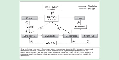
However, the pathophysiology of acute inflammation-related anemia (e.g., trauma, surgery) is somehow different. In this setting, inflammatory responses are mediated mainly by IL-6 and IL-8 (with transient contribution of TNF-α and IL-1 in some visceral surgeries, such as gastrointestinal or cardiac procedures), whereas IFN-γ plasma levels are undetectable or within the normal range Citation[3–5]. Therefore, in most of these conditions the two major mechanisms leading to anemia are perioperative or traumatic blood loss and blunted erythropoiesis due to decreased iron availability, whereas EPO levels are normal or near-to-normal Citation[5].
Hepcidin, a 25-amino acid peptide produced mainly by hepatocytes in response IL-6 levels, plays a major role in dysregulation of iron homeostasis during inflammation. Once synthesised, hepcidin is secreted into the bloodstream and interacts with ferroportin 1 (the only know iron exporting protein) at enterocyte basolateral membrane, hepatocytes and macrophages . The binding of hepcidin to ferroportin 1 causes internalization and lysosomal degradation of the carrier protein. Thus, hepcidin regulates the rate of iron absorption by villous enterocytes and the rate of iron recirculation from macrophages and hepatocytes, resulting in hypoferremia. In addition, inflammatory mediators increased divalent metal transporter 1 (IFN-γ), transferrin receptor expression (IL-10) and ferritin synthesis (TNF-α, IL-1, IL-6, IL-10) in macrophages leading to increased iron storage Citation[1,2].
It is important to recall that transferrin-bound iron is the primary iron source for erythropoiesis, entering the erythroblast by a process involving transferrin receptor-mediated endocytosis. This iron may be obtained by absorption of dietary iron and/or mobilization of iron stores at macrophages and liver Citation[1,6]. The amount of iron required for daily renewal of red blood cells (20–30 mg) is provided mostly by recycling the iron from senescent erythrocyte at macrophages. Therefore, as daily absorption (1–2 mg) just balances daily loss, internal turnover of iron is essential to meet the bone marrow requirements for erythropoiesis Citation[1,6].
Thus, during Anemia of Chronic Disease (ACD), inhibition of intestinal absorption and reticuloendothelial sequestration of iron result in decreased iron availability for the bone marrow, referred to as iron-restricted erythropoiesis or FID. This is characterized by low serum iron and decreased transferrin saturation (TSAT), in the face of adequate body iron stores defined by the presence of stainable iron in the bone marrow and/or a serum ferritin value within or above normal limits. Finally, when persisting decreased iron absorption and/or chronic blood loss are present, FID may evolve to absolute iron deficiency (FID+ID).
Therefore, treatment of ACD should rest on three fundamental pillars: the correction of the causing disease (if possible), the administration erythropoiesis-stimulating agents (which is rather restricted nowadays, especially for kidney disease or cancer associated anemia) and iron supplementation.
Regarding iron supplementation, it has been shown that individuals suffering from FID+ID have significantly lower hepcidin levels than those with FID without ID, and are able to absorb some dietary iron from the gut and mobilize some iron from macrophages Citation[3]. Thus, hepcidin levels may be useful in differentiating between FID and FID+ID and have been also shown useful in predicting non-responsiveness to oral iron salts in patients with IDA Citation[7]. As for individuals resistant to conventional oral iron therapy, the use of iv. iron formulations has been long time recommended. However, as it will be presented during this course, recent reports strongly suggest that liposomal oral iron formulation, which have an absorption mechanism different to iron salts, may represent an efficacious alternative to iv. iron Citation[8].
Monoclonal anti-hepcidin antibodies (12B9m) and spiegelmers (Lexatepid), the use of isocitrate or the administration of vitamin D supplements for treating ACD are in early stages of study Citation[9]. Finally, it must be stressed that the use allogeneic blood transfusion should only be indicated in poorly tolerated and severe anemias.
Regarding iv. iron, there are several formulations available. The efficacy of iv. iron is directly related to the amount of iron administered, but differences in core size and carbohydrate chemistry determine pharmacological and biological differences between the different iron complexes. These differences include clearance after injection, iron release in vitro, early evidence of iron bioactivity in vivo, and maximum tolerated dose and rate of infusion, as well as effects on oxidative markers, propensity for inducing hypophosphatemia, and propensity to cause transient proteinuria following administration Citation[2]. However, comparative cost-efficacy of these formulations has rarely been attempted.
As anemia is one of the most frequent extra-intestinal manifestations of inflammatory bowel diseases (IBD), for which iv. iron provides a faster Hb increase and iron store repletion, with low rates of treatment discontinuation, we comparatively estimated the cost-efficacy of an 8-week treatment course, pooling data of four different iv. iron compounds from five recent studies Citation[10–14]. The efficacy of iron sucrose (IS), ferric carboxymaltose (FCM), iron isomaltoside-1000 (MNF) given as infusion (I) or as bolus (B), and low-molecular-weight iron dextran (LMWID) was estimated as the difference (ΔHb g/dL) Hb values at week 8 and at baseline. Cost calculation per patient was performed from a Spanish perspective, taking into account: the cost of IVI; direct hospital costs (personnel, infusion material, infusion and observation time); and indirect hospital costs (the general functioning costs) Citation[15]. To correct for a possible effect of the different IVI doses administered, the cost per Δ1 g/dl Hb was also calculated.
At week 8, ΔHb was 2.2 g/dl for IS, 3.1 g/dl for FCM, 2.6 g/dl for MNF, and 2.0 for LMWID. Mean IVI doses were 1130, 1390, 885, and 949 mg, respectively, and ΔHb/g IVI were 1.9, 2.2, 2.9, and 2.1 g/dl, respectively. As depicted in , mean treatment cost both per patient and per Δ1 g/dl Hb were higher for IS when compared to FCM, MNF and LMWID. Therefore, the four iv. iron formulations were efficacious at correcting anemia, with ΔHb showing an apparent dose-response pattern. However, FCM, MNF and LMWID allow for giving up to 1000–1500 mg in a single session, thus facilitating patient management and reducing treatment costs when compared to IS. Head-to-head prospective cost-efficacy comparisons of these iv. iron formulations are, as well as with newer oral iron products (e.g., liposomal iron), are needed.
Table 1. Comparative cost-efficacy of different iv. iron formulation for treating IBD-associated anemia.
References
- Weiss G, Goodnough LT. Anemia of chronic disease. N Engl J Med 2005;352:1011-23.
- Muñoz M, Garcia-Erce JA, Remacha AF. Disorders of iron metabolism. Part II: iron deficiency and iron overload. J Clin Pathol 2011;64:287-96
- Theurl I, Aigner E, Theurl M, et al. Regulation of iron homeostasis in anemia of chronic disease and iron deficiency anemia: diagnostic and therapeutic implications. Blood 2009;113:5277-86.
- Muñoz M, García-Vallejo JJ, Sempere JM, et al. Acute phase response in patients undergoing lumbar spinal surgery: modulation by perioperative treatment with naproxen and famotidine. Eur Spine J 2004;13:367-73.
- Van Iperen CE, Kraaijenahgen RJ, et al. Iron metabolism and erythropoiesis after surgery. Br J Surg 1998;85:41-5.
- Muñoz M, Garcia-Erce JA, Remacha AF. Disorders of iron metabolism. Part 1: molecular basis of iron homoeostasis. J Clin Pathol 2011;64:281-6.
- Bregman DB, Morris D, Koch TA, et al. Hepcidin levels predict nonresponsiveness to oral iron therapy in patients with iron deficiency anemia. Am J Hematol 2013;88:97-101.
- Yuan L, Geng L, Ge L, et al. Effect of iron liposomes on anemia of inflammation. Int J Pharm 2013;454:82-9.
- Ganz T. Systemic iron homeostasis. Physiol Rev 2013;93:1721-41.
- Lindgren S, Wikman O, Befrits R, et al. Intravenous iron sucrose is superior to oral iron sulphate for correcting anaemia and restoring iron stores in IBD patients: A randomized, controlled, evaluator-blind, multicentre study. Scand J Gastroenterol 2009;44:838-45.
- Evstatiev R, Marteau P, Iqbal T, et al. FERGIcor, a randomized controlled trial on ferric carboxymaltose for iron deficiency anemia in inflammatory bowel disease. Gastroenterology 2011;141:846-53.e1-2.
- Kulnigg S, Stoinov S, Simanenkov V, et al. A novel intravenous iron formulation for treatment of anemia in inflammatory bowel disease: the ferric carboxymaltose (FERINJECT) randomized controlled trial. Am J Gastroenterol 2008;103:1182-92.
- Reinisch W, Staun M, Tandon RK, et al. A randomized, open-label, non-inferiority study of intravenous iron isomaltoside 1,000 (Monofer) compared with oral iron for treatment of anemia in IBD (PROCEED). Am J Gastroenterol 2013;108:1877-88.
- Khalil A, Goodhand JR, Wahed M, et al. Efficacy and tolerability of intravenous iron dextran and oral iron in inflammatory bowel disease: a case-matched study in clinical practice. Eur J Gastroenterol Hepatol 2011;23:1029-35.
- Calvet X, Ruíz MÀ, Dosal A, et al. Cost-minimization analysis favours intravenous ferric carboxymaltose over ferric sucrose for the ambulatory treatment of severe iron deficiency. PLOS One 2012;7:e45604.
Anemia may adversely affect patients with chronic diseases in several ways. In particular, anemia in cancer patients is associated with significant decreases in health-related quality of life (HRQOL) and may have a negative impact on prognosis. Alleviating anemia with erythropoiesis stimulating agents (ESA) improves energy, activity and overall QOL, particularly among patients with mild-to-moderate anemia, and helps patient cope with active treatments. However, research suggests that anemia is still under-recognized and under-treated. This may be partly due to the limitations of current ESA therapy, which includes a large percentage of patients who do not respond to this treatment, the need for frequent dosing and the relatively slow time to response. Adequate patient selection for treatment with ESA or other drugs/procedure is of pivotal importance in this setting.
Dysregulations of iron metabolism causing iron deficiency represent a major cause of anemia of chronic diseases (ACD). Also, data from the dialysis and cancer populations has clearly shown that an important factor that seriously limits response to ESA is functional iron deficiency (FID), which is an imbalance between iron needs in the erythropoietic marrow and iron supply. FID may be either pre-existing or occurrs during ESA therapy, when red cells are produced at a rate that outstrips labile iron availability. As a consequence, iron supplementation may still be required to achieve or maintain an optimal response to ESA. In anemic cancer patients, iron deficiency has to be investigated by dosing transferrin saturation, a parameter that is modestly influenced by inflammation. Ferritin, in contrast, belongs to the group of acute phase proteins and often does not reflect iron stores in cancer, due to its interdependence with inflammatory reactions. The iron regulatory peptide, hepcidin, is the key factor underlining the occurrence of iron dysregulation in the anemia of chronic diseases (ACD), including cancer. Hepcidin is up-regulated in ACD, resulting in the inhibition of iron transport across cell membranes, which decreases the accessibility of storage iron and gastrointestinal absorption of dietary iron, leading to an increased frequency of iron-restricted erythropoiesis, especially during therapy with ESA. Hepcidin dysregulation may well represent the mechanism by which oral iron supplementation has been reported ineffective in cancer patients.
Prospective trials published over the last decade demonstrate that anaemic patients with cancer undergoing chemotherapy and receiving ESA respond better, without additional toxicity, when parenteral iron is administered. Such benefit is more relevant when FID is present at baseline but appear to be independent of baseline iron variables in one large study. This issue is clinically relevant because appropriate iron supplementation, apart from allowing more patients to benefit from ESA therapy, may represent a strategy to improve the cost–effectiveness of ESA in oncology, as it has occurred in nephrology. However, the use of iron supplementation during treatment with ESA is not rigorously pursued in anemic patients with cancer as it is in chronic kidney disease. This underuse is likely to be related to: the false perception that cancer patients do not have decreased iron stores (as measured by serum ferritin) and therefore thought not to require iron supplementation during ESA therapy; the often misinterpreted incidence and clinical nature of serious adverse events of intravenous iron; the lack of studies demonstrating the efficacy of traditional oral iron agents to favour response to ESA.
Novel iron preparations capable of increasing iron absorption and bioavailability, including those carried by a liposome membrane, may well facilitate a more widespread use of iron supplementation in cancer anemia. A randomized study in this setting is required.
References
- Cella D, Dobrez D, Glaspy J. Control of cancer-related anemia with erythropoietic agents: a review of evidence for improved quality of life and clinical outcomes. Ann Oncol 2003;14:511-9.
- Cella D, Stone AA. Health-related quality of life measurement in oncology: Advances and opportunities. Am Psychol 2015;70:175-85.
- Sankaran VG, Weiss MJ. Anemia: progress in molecular mechanisms and therapies. Nat Med 2015;21:221-30.
- Tessitore N, Solero GP, Lippi G, et al. The role of iron status markers in predicting response to intravenous iron in hemodialysis patients on maintenance erythropoietin. Nephrol Dial Transplant 2001;16:1416–23.
- Pedrazzoli P, Farris A, Del Prete S, et al. Randomized trial of intravenous iron supplementation in patients with chemotherapy-related anemia without iron deficiency treated with darbepoetin alfa. J Clin Oncol 2008;25:1619-25.
- Ludwig H, Müldür E, Endler G, Hübl W. Prevalence of iron deficiency across different tumors and its association with poor performance status, disease status and anemia. Ann Oncol 2013;24:1886-92.
- Auerbach M. Should intravenous iron be the standard of care in oncology? J Clin Oncol 2008;25:1579-81.
- Petrelli F, Borgonovo K, Cabiddu M, et al. Addition of iron to erythropoiesis-stimulating agents in cancer patients: a meta-analysis of randomized trials. J Cancer Res Clin Oncol 2012;138:179-87.
- Pedrazzoli P, Rosti G, Secondino S, Siena S. Iron supplementation and erythropoiesis-stimulatory agents in the treatment of cancer anemia. Cancer 2009;115:1169-73.
- Yuan L, Geng L, Ge L, et al. Effect of iron liposomes on anemia of inflammation. Int J Pharm 2013;454:82-9.
Introduction
Anemia is a common manifestation of cancer patients and is usually associated with advanced disease, malnutrition and poor prognosis. It is one of the reasons for fatigue, delay/reduction and change in dose intensity of cancer treatments, poor activity of radiation therapy due to reduced oxygen effect, increase use of blood transfusions, and finally of rise of financial burden in oncology setting. Rapid correction of hemoglobin levels (Hb) is necessary for patients wellbeing and needs iron plus or minus erythropoiesis-stimulating agents (ESAs) and eventually, if they do not permit correction of Hb, red blood cells transfusions (RBC). Usually, ESAs associated with parenteral iron are appropriated to treat moderate (grade [G]2) anemia, conversely more severe and symptomatic grade of anemia (G3–4) needs prompt transfusions. ESAs significantly reduced the use of RBC transfusions (relative risk [RR] 0.65, 95% CI: 0.62-0.68) according to a 2012 Cochrane meta-analysis Citation[1]. The risk of venous thromboembolism was increased in patients receiving ESAs (RR 1.52, 95% CI: 1.34-1.74). Current data cover chemotherapy-related form of anemia where parenteral iron has showed a better and more rapid response of ESAs agents according to a meta-analysis of randomized trials Citation[2]. In general, the availability of iron can limit the Hb response following treatment with ESAs in patients with cancer-related anemia as well as in those with renal failure. Nevertheless, these are the best markers of iron stores that are currently available, and serum iron, transferrin saturation, and serum ferritin should be evaluated at baseline in all anemic patients who are being considered for an ESA and in patients who fail to respond to ESA therapy within 6–8 weeks. Iron should be given during ESA therapy if necessary, in order to maintain a transferrin saturation of ≥20% and a serum ferritin level of ≥100 ng/mL. The reason of failure of oral iron formulation in cancer anemic patients seems related to the hepcidin protein, an acute phase protein produced primarily by the liver. Several observation suggest that hepcidin, and perhaps other regulatory molecules produced in the liver Citation[3–5], plays a major role as a negative regulator of intestinal iron absorption and iron release from macrophages Citation[6,7], by interacting with, and inactivating, the iron export protein ferroportin Citation[8]. If chemotherapy and radiotherapy are the main reasons for cancer-associated anemia, several other agents or conditions in cancer patients can interfere with erythropoiesis. A direct effect of neoplasia (bleeding), reduced oral intake, increase of RBC destruction (hemolytic anemia) are common cause of reduced Hb levels. Molecular targeted agents as monoclonal antibodies (e.g anti HER2, EGFR or VEGF agents) or small molecules (multi-target) tyrosine kinase inhibitors (e.g sunitinib or sorafenib) are able to interfere with specific pathway leading to a reduced/altered erythro- or myelopoiesis as reflected by high rate G1–2 leucopenia and/or anemia in randomized trials. Unfortunately, ESAs are not recommended for the treatment of anemia that is unrelated to chemotherapy in patients with malignancy. Large trials with ESAs in patients not receiving chemotherapy or radiotherapy, in fact, showed that their use is not useful and may potentially be detrimental for patient's outcome Citation[9,10]. However, data regarding patients treated with targeted agents for solid tumors were ever be reported sistematically, and the treatment of these emergent forms of anemia is unknown.
Etiology of anemia in cancer patients
The causative role of anemia with molecular agents is partially unknown, but the general opinion is that they can interfere with myelo- and/or erythropoiesis in bone marrow. If other etiologies cannot be excluded (microangiopathic hemolytic anemia or macrocytic anemia associated with sunitinib therapy Citation[11–17]) the main reason seems related to the interference with the FLT-3 pathway. Fms-like tyrosine kinase 3 (known as FLT-3 or CD 135) is a cytokine receptor that belongs to the receptor tyrosine kinase class III. CD135 is the receptor for the cytokine Flt3 ligand (FLT3L). It is expressed on the surface of many hematopoietic progenitor cells. Signaling of FLT3 is necessary for the proper development of hematopoietic stem cells and progenitor cells. An old study in rabbit showed that hematopoietic recovery occurred after total body irradiation if protected by FLT-3 ligand, and suggests a radioprotective clinical potential of FLT3 receptor Citation[18].
Incidence of anemia with targeted therapies
Analyzing data from large randomised trials that included common labelled targeted agents used for treatment of solid tumours an high rate of G1-2 anemia events were found and published in a systematic review and meta-analysis randomized trials by Barni et al. Citation[19]. Overall, the addition of targeted therapies to standard treatment (chemotherapy or placebo/best supportive care) increased the risk for all grades of anemia by 7%. The relative risk for all grades (incidence, 44%) and grades 1–2 (incidence, 38.9%) of anemia was higher with biological therapies alone but not when combined with chemotherapy. The risk was significant for erlotinib, trastuzumab and sunitinib. Bevacizumab was associated with a lower risk for anemia, as for a protective effect. Anti-epidermal growth factor receptor, anti-human epidermal growth factor receptor 2, anti-vascular endothelial growth factor receptors, and tyrosine kinase inhibitors predicted RRs of 1.24, 1.20, 0.82, and 1.33, respectively, and all of these values were significant. Risk was largely increased with agents targeting multiple pathways as VEGFR, RET, cKIT, FLT-3, CSF-1R that likely interfere with myelo-and erythropoiesis as previously described above. Even mTOR inhibitors exhibited an increased risk of anemia though not significant due to small number of included studies. Risk of anemia was independent of the underlying disease and was associated only with oral multitarget tyrosine kinase inhibitors due to their pleiotropic effect on receptor for growth factor expressed on hematopoietic cells. As other similar analysis showed Citation[20,21] the meta-analysis showed a risk-lowering effect of bevacizumab on anemia adverse events, but this results is hard to be explained on the basis of biological effect of bevacizumab. The hypothesis of targeting FLT-3 and its downstream pathway is confirmed by the different toxicity profiles of sunitinib and pazopanib, the last targeting VEGFR1,2,3, PDGFR and c-KIT but not RET, an exquisite target of sunitinib. The value of Hb, fell down the 2 weeks after the cycle of sunitinib (4 week on–2 week off schedule), conversely the anemia levels during pazopanib therapy remained quite steady during treatment, and largely above the lower normal limit in the COMPARZ study Citation[22]
Therapy considerations
Treatment of anemia with targeted agents is commonly not conventional. In fact, ESAs are not labeled for this indication and blood transfusions are needed only for lower Hb levels (G3–4 anemia or symptomatic anemia). However, a preemptive strategy can be suggested due to high risk of fatigue observed with these agents that could worsen anemia symptoms Citation[23]. Published guidelines regarding management of anemia with sunitinib suggest that G3/4 anemia usually does not require relevant dose modification; however, because of concern about the potential toxicities and angiogenesis triggering, the use of ESAs should be cautioned Citation[24]. Baseline evaluation of nutritional status, iron balance, B12, and folate deficiency should be performed. Chronic bleeding must be promptly recognized and treated (e.g., with palliative radiotherapy for example), and treatment not started in presence of risk of bleeding. Seldom blood transfusion is needed before starting treatment and iron supplementation can be started. In particular liposomal iron is an attractive way of iron administration in cancer patients. Pyrophosphate liposomal iron (Sideral forte®) is composed of protected iron and vitamin C, useful in case of deficiency or increased requirements. The iron, included is uniquely coated using a liposomial technology that allows the molecule to pass through the stomach, avoiding any gastrointestinal irritation, to be directly absorbed through the lining of the digestive tract. In particular, liposomal iron has been showed to be effective in a similar way to iv. iron when used in association with epoetin alfa in patients with refractory anemia. In particular, a clinical feasibility study in cancer patients treated with molecular agents seems be urgently needed in medical oncology to verify safety and efficacy in anemic patients with metastatic tumors.
Conclusions & future perspectives
In conclusion, mild anemia is a common event in patients treated with targeted therapies for solid tumors (up to 40-50% of patients showing anemia adverse event mainly of low grade). Early treatment of this hematological toxicity is of paramount importance due to deterioration of quality of life; increase of fatigue and cost saving with transfusion prevention. Due to lack of a standardized treatment, not labeling of ESAs agents and largely unknown cause of anemia, the treatment is a challenge. Use of the liposomial iron in patients treated with tyrosine kinase inhibitors could be an appealing way to treat anemia associated with these agents that could rise up to 50% with sunitinib. A prospective observational study was launched in 2014 at Oncology Unit of Treviglio Hospital with pyrophosphate liposomal iron (1 tablet QD for 3 months) for patients with mild (G1: Hb level 10-12 g/dL) anemia before starting chemotherapy for solid tumors. In the absence of proper guidelines preemptive use of iron, schedule changing (e.g with sunitinib for example), correction of Hb level before starting with treatment, and short dose interruption/reduction of these drugs should be implemented to treat hematological toxicities associated with these treatments.
References
- Tonia T, Mettler A, Robert N, et al. Erythropoietin or darbepoetin for patients with cancer. Cochrane Database Syst Rev 2012;12:CD003407.
- Petrelli F, Borgonovo K, Cabiddu M, et al. Addition of iron to erythropoiesis-stimulating agents in cancer patients: a meta-analysis of randomized trials. J Cancer Res Clin Oncol 2012;138(2):179-87.
- Lin L, Valore EV, Nemeth E, et al. Iron transferrin regulates hepcidin synthesis in primary hepatocyte culture through hemojuvelin and BMP2/4. Blood 2007;110(6):2182-9.
- Muckenthaler M, Roy CN, Custodio AO, et al. Regulatory defects in liver and intestine implicate abnormal hepcidin and Cybrd1 expression in mouse hemochromatosis. Nat Genet 2003;34(1):102-7.
- Meynard D, Babitt JL, Lin HY. The liver: conductor of systemic iron balance. Blood 2014;123(2):168-76.
- Ganz T.Hepcidin, a key regulator of iron metabolism and mediator of anemia of inflammation. Blood 2003;102(3):783-8.
- Frazer DM, Inglis HR, Wilkins SJ, et al. Delayed hepcidin response explains the lag period in iron absorption following a stimulus to increase erythropoiesis. Gut 2004;53(10):1509-15.
- Nemeth E, Tuttle MS, Powelson J, et al. Hepcidin regulates cellular iron efflux by binding to ferroportin and inducingits internalization. Science 2004;306(5704):2090-3.
- Smith RE Jr, Aapro MS, Ludwig H, et al. Darbepoetin alpha for the treatment of anemia in patients with active cancer not receiving chemotherapy or radiotherapy: results of a phase III, multicenter, randomized, double-blind, placebo-controlled study. J Clin Oncol 2008;26(7):1040-50.
- Wright JR, Ung YC, Julian JA, et al. Randomized, double-blind, placebo-controlled trial of erythropoietin in non-small-cell lung cancer with disease-related anemia. J Clin Oncol 2007;25(9):1027-32.
- Talebi TN, Stefanovic A, Merchan J, et al. Sunitinib-induced microangiopathic hemolytic anemia with fatal outcome. Am J Ther 2012;19(4):e143-5.
- Bevacizumab + sunitinib: microangiopathic haemolytic anemia. A serious drug interaction between 2 cancer drugs. Prescrire Int 2009;18(102):165.
- Price J, Shaarbaf R, Wood L. Sunitinib causes macrocytosis in patients with advanced renal cell carcinoma. Curr Oncol 2010;17(2):30-3.
- Rini BI, Choueiri TK, Elson P, et al. Sunitinib-induced macrocytosis in patients with metastatic renal cell carcinoma. Cancer 2008;113(6):1309-14.
- Jain R, Mathew P, Wood CG, et al. Sunitinib-induced acute hemolysis without hypertension: a case report. Clin Genitourin Cancer 2008;6(2):122-3.
- Schallier D, Trullemans F, Fontaine C, et al. Tyrosine kinase inhibitor-induced macrocytosis. Anticancer Res 2009;29(12):5225-8.
- Billemont B, Izzedine H, Rixe O. Macrocytosis due to treatment with sunitinib. N Engl J Med 2007;357(13):1351-2.
- Gratwohl A, John L, Baldomero H, et al. FLT-3 ligand provides hematopoietic protection from total body irradiation in rabbits. Blood 1998;92(3):765-9.
- Barni S, Cabiddu M, Guarneri P, et al. The risk for anemia with targeted therapies for solid tumors. Oncologist 2012;17(5):715-24.
- Schutz FA, Je Y, Azzi GR, et al. Bevacizumab increases the risk of arterial ischemia: a large study in cancer patients with a focus on different subgroup outcomes. Ann Oncol 2011;22(6):1404-12.
- Sher A, Wu S. Anti-vascular endothelial growth factor antibody bevacizumab reduced the risk of anemia associated with chemotherapy-A meta-analysis. Acta Oncol 2011;50(7):997-1005.
- Motzer RJ, Hutson TE, Cella D, et al. Pazopanib versus sunitinib in metastatic renal-cell carcinoma. N Engl J Med 2013;369(8):722-31.
- Santoni M, Conti A, Massari F, et al. Treatment-related fatigue with sorafenib, sunitinib and pazopanib in patients with advanced solid tumors: an up-to-date review and meta-analysis of clinical trials. Int J Cancer 2015;136(1):1-10.
- Kollmannsberger C, Bjarnason G, Burnett P, et al. Sunitinib in metastatic renal cell carcinoma: recommendations for management of noncardiovascular toxicities. Oncologist 2011;16(5):543-53.
- Funakoshi T, Latif A, Galsky MD. Risk of hematologic toxicities in cancer patients treated with sunitinib: a systematic review and meta-analysis. Cancer Treat Rev 2013;39(7):818-30.
Introduction
Myelodysplastic syndromes are clonal neoplastic diseases. The cases of anemia in said syndromes are partly due to ineffective haematopoiesis and partly to a functional iron deficit. This is already known with regard to cancer-related anemia and has led several authors to supplement erythropoietin with intravenous iron in order to treat anemia in cancer patients undergoing chemotherapy with good results. These results have also been confirmed in patients with cancer-related anemia with lymphoproliferative disorders who are not undergoing chemotherapy or blood transfusion. Moreover, in cancer patients receiving chemotherapy, intravenous iron supplementation appears to be superior to typical oral iron supplements with ferrous sulfate or organic compounds.
The objective of this study is to verify the efficacy and cost of therapy in the groups treated with intravenous iron, oral liposomal iron and no iron supplement in patients with refractory anemia who are receiving erythropoietin alpha.
Patients and methods
This is a retrospective study. We considered 44 patients with refractory anemia. All patients had received 1 ampoule of 40,000 IU of subcutaneous erythropoietin alpha/week, 7.5 mg of oral calcium levofolinate/day, vitamin B12: 400 mg orally/day. 20 patients (group A) received 62.5 mg of intravenous sodium ferric gluconate/day in 100 ml of normal saline solution under continuous infusion for 1 hour, on the day the patient received erythropoietin. Another 20 patients (group B) received 14 mg of oral liposomal iron (Sideral®), 2 capsules/day throughout the treatment period with erythropoietin. Four patients (group C) received no iron supplementation. The characteristics of the patients are listed in . The median follow-up was 21 months (range 2–36).
Table 1. Demographic characteristics.
The costs of treating each patient were also determined, in consideration of the more frequent expense items, as listed in . These costs are a national average, referring to the costs provided by the various regional health services and, as regard the cost of one lost working day, the costs provided by the National Institute of Social Security.
Table 2. Details of costs per patient.
The average treatment costs in each group were calculated as follows. For each patient, the overall cost of patient treatment during the entire follow-up period was calculated, using the costs listed in . This cost was then divided by the months of the patient’s follow-up, in order to give an average monthly patient treatment cost. This provided an estimate of the costs, independent of the precise cost of the drug, but tied to the final outcome (efficacy) of the therapeutic strategy used during the observation period. In fact, by using this type of calculation, very costly, but very effective drugs that require few administrations during the observation period would have an average monthly cost in line with less costly, but less effective, drugs which require more frequent administrations. Subsequently, for each treatment group, the median was calculated of the monthly cost averages of each patient in a given group. This provided a summary figure for each treatment group that is sufficiently independent of the clinical outcome of each patient. There was no iron, B12 or folate deficiency documented in the lab exam of any patient included in the study.
Results
The results are summarised in . The number of transfusions required by month of follow-up was lower in the group treated with liposomal iron compared to the group treated with iv. iron and the one which did not receive any iron at all. The response to treatment with erythropoietin in the groups that received iron supplementation was around double that of the group that did not receive iron supplementation. Side effects were more serious and common in the group treated with iv. iron (1 headache, 3 hypotension and 1 urticaria), compared to the other two groups. The need for transfusions and the number of transfusions/month were higher in the groups receiving intravenous iron or not receiving supplementation compared to the group taking liposomal iron. The speed of initial response to erythropoietin was greater in the group treated with intravenous iron, while the frequency of administration of maintenance doses of erythropoietin was lower in the group treated with liposomal iron, due to this group reaching normal hemoglobin values earlier. In addition, the median working days lost per month were much higher in the groups treated with intravenous iron or that did not receive iron, compared to the group treated with liposomal iron. The loss of response to erythropoietin was in proportion greater in the group that did not receive iron supplementation, while the need for re-treatment with iron was greater in the group that received intravenous iron. The group that did not receive iron supplementation had a greater cost per month of treatment than the groups supplemented with iron intravenously or with liposomal iron. The latter group had the lowest cost of all. This was due to the fact that the costs of blood tests, working days lost and transfusions received were much higher in the group not supplemented with iron than in the groups receiving iron supplementation. The group supplemented with liposomal iron, however, had a significantly lower cost per follow-up month than the other two groups, especially because it required less expenditure for medical and nursing support in day hospital or clinic, fewer transfusions, fewer lost working days (2 hours per month) and a lower quantity of erythropoietin than the other two treatment groups.
Table 3. Summary of main results.
Table 4. Median monthly costs in Euros for each treatment group.
Discussion
Certainly, the data from this study present interpretation difficulties, as they are limited by the retrospective nature of the study and the small number of patients in each treatment group, especially in the group that did not receive iron therapy (data available only for 4 patients). The low caseload is partly due to the rarity of the disorders in question, myelodysplastic syndromes, which have an incidence of around 5 cases/100,000 population/year, which drops to around 1 case/100,000 population/year in the case of refractory anemia. However, there are data worthy of attention and consideration.
Iron supplementation in erythropoietin therapy seems to be effective in myelodysplastic syndromes and solid tumours treated with or without chemotherapy4-6 or in indolent lymphoma not treated with chemotherapy. In addition, as shown by Henry, in cancer patients being treated with chemotherapy, intravenous iron supplementation of erythropoietin results in a superior response to anemia than seen without supplementation or with oral supplementation with organic iron compounds or iron sulphate. The data in this study show that things probably change when using liposomial iron. In fact, even if patients who need transfusion are the same in the group supplemented with intravenous iron and liposomal iron, the number of transfusions required per month of follow-up is significantly less in the group treated with liposomal iron. Side effects are significantly more common and potentially dangerous in the group with iv. iron. Nothing comparable was reported in the group supplemented with liposomal iron. This also explains the recent warning issued by the EMA and the Italian Medicines Agency (AIFA), which advised strongly against and limited the use of iv. iron, given the potentially fatal side effects. Supplementation with iv. iron generates a faster response to erythropoietin and the need for lower maintenance doses when compared to those receiving no supplementation. These data are coherent with what is already acknowledged in the literature. However, providing supplementation with liposomal iron leads to the need for lower maintenance doses than with iv. iron supplementation. This is also confirmed by the low expenditure on erythropoietin recorded in the group treated with liposomal iron. This could be interpreted in several ways. In fact, as has been known for some time, neoplastic disorders have an inflammatory status. During inflammation, the liver produces an acute-phase protein, hepcidin, that inhibits the membrane protein ferroportin, which facilitates hepatic absorption from the intestine and its transportation from macrophage and tissue deposits. Liposomal iron may not follow these usual routes of absorption and may be absorbed by direct fusion with the cell membranes and be subsequently released into circulation or could be absorbed as dietary chylomicrons. If this happens, the effects of liposomal iron should be comparable with those of intravenous iron. In this study, liposomal iron supplementation of erythropoietin seems to generate a greater stimulus for erythropoiesis, which leads to greater clinical efficacy and less use of erythropoietin. This suggests that there is some other effect linked to the liposomal formulation. In fact, it is already known that the liposome could in itself have an anti-inflammatory action or at least facilitate the absorption of the iron in chronic inflammation, as demonstrated by some recent trials. Therefore, by eliminating the inflammatory component from anemia, it could increase the bioavailability of iron deposits with a consequent greater efficacy of haematopoiesis. In the clinical context this translates into a lower need for erythropoietin and transfusions and, financially, less expenditure on these two items. In addition, the group treated with iv. iron required administration of the iron in hospital, with a greater cost in medical and nursing care in day hospitals or clinics and, consequently, the cost of more lost working days. Given that liposomal iron does not require all of this, the monthly cost in the group with such supplementation is significantly lower. In all countries with public health services, this inevitably also translates into lower social costs.
Conclusion
Supplementation with liposomal iron (Sideral®) in refractory anemia treated with erythropoietin seems to be safe, feasible, cost-effective and not inferior to iv. iron supplementation.
Acknowledgement: Prof. Manuel Muñoz Gómez
Introduction
The prevalence of pre-operative anemia may be high among surgical patients, depending on the patients' co-morbidities, gender, age and the underlying pathology for which they require surgery Citation[1]. This preoperative anemia is quite common (20 to 50% depending on conditions) and is an independent risk factor for morbidity and mortality Citation[2,3]. The hemoglobin (Hb) level is an independent transfusional risk factor Citation[4] and there is a relationship dose-dependent between postoperative complications and the allogeneic blood transfusion (ABT) Citation[1,3,5]. The procedures in orthopedic and trauma surgery (OST), vascular or oncological surgery can cause significant blood loss and provoke a postoperative acute anemia, or aggravate previous preoperative anemia, which often requires ABT.
The clinical, financial and logistical disadvantages of ABT have promoted the development of generically known as Patient Blood Management programs (PBM) multidisciplinary and multimodal programs whose aim is to reduce or eliminate the need for ABT and improve clinical outcome Citation[3,6,7]. These programs are supported by the application of four groups of perioperative measures: use of “restrictive” transfusion criteria (administer the minimum effective dose guided by clinical signs o symptoms); stimulation of erythropoiesis (diagnosing and treating the perioperative anemia); reducing bleeding (improving the hemostasis and avoiding the hyperfibrinolysis); and autologous blood transfusion Citation[3].
The objective of this lecture is to briefly review the efficacy, safety and recommendations of one of the PBM pillar: the stimulation of erythropoiesis. Standard operating procedures, multimodal strategies and protocols are needed to improve the Hb perioperative levels to avoid (or to reduce to minimum) ABT and to achieve the best clinical outcome.
Patient blood management Citation[3,6,7]
PBM is an evidence-based approach to optimizing the care of patients who could or might need transfusion. A focus on improved patient outcomes and economic and operational pressures are leading key industry thinkers to examine appropriate blood usage with new interest. Hospitals are eager to improve patient safety and clinical outcomes, while also reducing the need for allogeneic blood components. PBM programs can achieve these goals by reducing variation in transfusion practice and managing patients with nontransfusion – and, if appropriate, transfusion – treatment modalities Citation[7].
Assessment and management of preoperative patients involve maximizing Hb levels to prevent anemia and optimizing coagulation function to limit bleeding. Starting with the primary care physician, the health-care team supporting medical and presurgical patients should focus efforts on determining whether there is a reason to suspect any medical conditions that might predispose the patient to transfusion Citation[7].
As any leading organization in the education of health-care professionals about blood management and utilization review, the scientific societies related with ABT (like the American Association of Blood Banks) must offer resources that address the various aspects of PBM, helping members achieve their goals of optimizing patient outcomes, preventing unnecessary blood usage and auditing physician compliance with established criteria for transfusion Citation[3,6,7].
Perioperative stimulation of erythropoiesis
Preoperative anemia
Using the WHO criteria, preoperative anemia is present in many surgical patient Citation[2]. Preoperative anemia is recorderd in up to 30% of orthopedic patients, up to 50% in digestive cancer, and up to 66% in hip fracture Citation[1–5]. Pre-operative anemia has been linked to post-operative infections, poorer physical functioning and recovery, decreased quality of life, and increased length of hospital stay and mortality Citation[1–6].
It is well known that the preoperative Hb level is the main independent risk factor for receiving ABT Citation[4,5]. Blood transfusion is also associated dose-dependent with an increased morbidity and perioperative mortality Citation[1,3–6]. Postoperative anemia is more frequent (up to 90%) and must be corrected, which does not necessarily imply that shall be done by the administration of ABT (only if clinically is needed: ‘restrictive transfusion criteria’) Citation[8].
Decisions to transfuse should be based on assessment of an individual patient including their underlying cause of anemia. Blood transfusion has become a routine medical response despite cheaper and safer alternatives in some settings. We must not transfuse red blood cells for iron deficiency without hemodynamic instability. Pre-operative patients with iron deficiency and patients with chronic iron deficiency without hemodynamic instability (even with low Hb levels) should be given oral and/or intravenous iron Citation[9]. There is high quality evidence that demonstrates a lack of benefit and, in some cases, harm to patients transfused to achieve an arbitrary transfusion threshold. If necessary, transfuse only the minimum number of units required instead of a liberal transfusion strategy Citation[9,10]. This is the first cornerstone of appropriate PBM.
Perioperative stimulation of erythropoiesis is the second fundamental pillar of PBM program. Normal erythropoiesis needs a healthy bone marrow with an adequate supply of various nutrients (iron, vitamins C, B1, B6, B12 and folic acid), and hormones (erythropoietin, thyroid hormones and steroids). In the absence of information on other haematinics, only the possible benefit of oral and iv. iron administration to reduce transfusion rate has been studied Citation[7].
Diagnosis and treatment of peri-operative anemia
Patients scheduled for any major surgery should investigate the presence of preoperative anemia at least 30 days before surgery, for differential diagnosis and appropriate therapy, if needed (GRADE 1C) Citation[8,11]. Faced with an unexpected anemia, an elective surgical procedure should be postponed until it has been properly classified and treated.
Usually the presence of anemia is diagnosed if Hb level is under 13 g/dl in men or under 12 g/dl in women. Perhaps we need a different definition in surgical patients and a higher Hb level objective. Women have a lower tidal volume than men, while blood loss in these surgical procedures is similar for both genders Citation[3].
Therefore, for women scheduled for major surgery, like arthroplasties, ‘anemia definition’ should be at least the same as for male patients; that is, Hb <13 g/dl. Some authors Citation[3] invite to change the term ‘preoperative anemia’ by ‘sub-optimal level of preoperative Hb’ when it is <13 g/dl. Consequently, the aim of preoperative treatment should be to optimize to reach a level Hb ≥13 g/dl (closer to 14 g/dl) and minimize the risk of transfusion Citation[3].
In the case of postoperative anemia, the goal of treatment is to achieve safe Hb levels that avoid any transfusion and the correction of anemia in the shortest time, to facilitate functional recovery and enhance the quality of life. In this period should pay attention to drug interactions that may cause or worsen anemia Citation[3].
Another important aspect, and frequently overlooked, is the diagnosis of hematinics deficiencies without anemia (iron deficit, B12 vitamin or folate deficiency), since its correction is crucial to optimize preoperative Hb levels, especially in case of patients under treatment with recombinant erythropoietin (rHU-EPO), and to ensure and accelerate the recovery of postoperative anemia (GRADE 1C) Citation[11].
Iron therapy
Indications
Some studies have shown that, for patients presenting with iron deficiency and iron-deficiency anemia, administration of oral iron (ferrous salts 100–200 mg/day for 4–6 weeks) improves presurgical Hb levels, reduces transfusion rates and, in some cases, shortens the time spent in hospital Citation[3,4,8,12–14].
If there is poor absorption or poor tolerance of oral iron or an accelerated response to treatment is required, pre-operative intravenous iron supplementation, starting 3–4 weeks prior to the scheduled procedure, increases Hb levels and/or corrects anemia and reduces ABT requirements Citation[1,3,4,8]. The intramuscular route for iron administration is not recommended Citation[8].
As for patients presenting with slight anemia (Hb between 10 and 13 g/dl), but without iron deficiency and/or with clinical or laboratory signs of inflammation, pre-operative administration of rHU-EPO has been proven to increase effective and safety Hb levels and reduce the rate of ABT Citation[1,3,4,8]. The minimum effective dose of ESA for this indication is presently unknown, but it has been shown that most patients attain the target Hb level with only one or two doses Citation[1,3,4,8]. All patients under treatment with rHU-EPO must receive an adequate source of iron (and vitamins) to ensure a nice response and avoid reactive thrombocytosis.
Evidence and recommendations
The European Society of Anaesthesia (ESA) guidelines recommends treating iron deficiency by administration of oral or intravenous iron (GRADE 1B) Citation[11], but this recommendation must be tempered by the severity of the anemia, the type of surgery and the time available to treat it. Faced with a preoperative iron deficiency anemia, whenever possible and the necessary time, consider the use of oral iron for its low cost and easy administration (GRADE 2B) Citation[11].
Table 1. Preoperative correction of anemia of “Management of severe perioperative bleeding ESA Guidelines”.
The update of Seville´s document Citation[8] suggests the preoperative administration of oral iron to improve preoperative Hb levels and/or reduce transfusion rate (Grade 2B) . In anaemic colon cancer patients, the preoperative administration of oral iron (ferrous salts), starting 14–30 days prior to surgery, improved the level of Hb and decreased ABT Citation[12,13]. In patients scheduled for total knee or hip arthroplasty, the administration of oral iron, together with a restrictive transfusion protocol, improved Hb levels, reduced transfusion rates and, in some cases, the length of hospital stay Citation[14].
Table 2. Summary of recommendations related to anemia of the Spanish Update Consensus Document on alternatives to reduce allogeneic blood transfusion (in descending order of strength).
However, sometimes, either by malabsorption, contraindication, poor tolerance or limited time avaible before surgery, is fully justified the use of intravenous iron instead classical oral iron, with which the medullary response and repletion deposits will be faster (1–2 weeks) (GRADE 2B) Citation[11]. In anemic patients scheduled for surgery, the administration of iv iron increase the Hb levels, the anemia was mostly corrected and reduced ABT needs Citation[1,3,8].
Commentary
Although the orthodox view is that early pre-operative anemia assessment (at least one month before surgery) Citation[15], classification and management is preferred, data from more pragmatic approaches suggest that anemia treatment (mainly with iron) should always be attempted in any major surgical procedures, as any time may be a good time for patients to benefit from it Citation[5].
The PBM multimodal programs must be well defined, adapted to the means of each hospital, the characteristics of our patients and the experience of the health professionals involved. This is the right way to reach the objective of conducting major surgical procedures without the use of ABT, without complications and reducing costs.
Reference
- Muñoz M, García-Erce JA, Cuenca J, Bisbe E, Naveira E; AWGE (Spanish Anemia Working Group). On the role of iron therapy for reducing allogeneic blood transfusion in orthopaedic surgery. Blood Transfus 2012;10(1):8-22
- Spahn DR. Anemia and patient blood management in hip and knee surgery. A systematic review of the literature. Anesthesiology 2010; 113:482-95.
- Canillas F, Gómez-Ramírez S, García-Erce JA, Pavía-Molina J, Gómez-Luque A, Muñoz M. “Patient blood management” in orthopaedic surgery. Rev Esp Cir Ortop Traumatol 2015;59(3):137-49
- García-Erce JA, Cuenca J, Solano VM. [Predictive factors for transfusion requirements in patients over 65 years old with subcapital hip fracture]. Med Clin (Barc) 2003;120(5):161-6
- Muñoz M, Gómez-Ramírez S, García-Erce JA. Implementing Patient Blood Management in major orthopaedic procedures: orthodoxy or pragmatism? Blood Transfus 2014;12(2):146-9
- García Erce JA, Peral García AI. New paradigms in patient blood management in surgery. Cir Esp 2015;93(1):59-61.
- Patient BloodManagement. (http://www.aabb.org/pbm/Pages/default.aspx) ( last access March 2015)
- Leal-Noval SR, Muñoz M, Asuero M, Contreras E, García-Erce JA, Llau JV, Moral V, Páramo JA, Quintana M; Spanish Expert Panel on Alternatives to Allogeneic Blood Transfusion. Spanish Consensus Statement on alternatives to allogeneic blood transfusion: the 2013 update of the “Seville Document”. Blood Transfus 2013;11(4):585-610.
- Canadian Society of Hematology. Five Things Physicians and Patients Should Question. http://www.choosingwiselycanada.org/recommendations/hematology/ ( last access March 2015)
- American Association of Blood Banks. Five Things Physicians and Patients Should Question. http://www.choosingwisely.org/doctor-patient-lists/american-association-of-blood-banks/ ( last access March 2015)
- Kozek-Langenecker SA, Afshari A, Albaladejo P, Santullano CA, De Robertis E, Filipescu DC, et al. Management of severe perioperative bleeding: guidelines from the European Society of Anaesthesiology. Eur J Anaesthesiol 2013 Jun;30(6):270-382.
- Lidder PG, Sanders G, Whitehead E, et al. Pre-operative oral iron supplementation reduces blood transfusion in colorectal surgery - a prospective, randomised, controlled trial. Ann R Coll Surg Engl 2007; 89: 418-21.
- Okuyama M, Ikeda K, Shibata T, et al. Preoperative iron supplementation and intraoperative transfusion during colorectal cancer surgery. Surg Today 2005;35:36-40.
- Cuenca J, García-Erce JA, Martínez F, Cardona R, Pérez-Serrano L, Muñoz M. Preoperative haematinics and transfusion protocol reduce the need for transfusion after total knee replacement. Int J Surg 2007;5(2):89-94.
- Goodnough LT, Maniatis A, Earnshaw P, Benoni G, Beris P, Bisbe E, et al. Detection, evaluation, and management of preoperative anemia in the elective orthopaedic surgical patient: NATA guidelines. Br J Anaesth 2011;106:13-22.
Purpose
Recombinant human erythropoietin (ESA) is the standard of care for patients with chemotherapy related anemia. Functional iron deficiency may impair response to erythropoiesis-stimulating agents in iron-replete patients with chemotherapy-associated anemia.
Intravenous (iv.) iron improves hemoglobin (Hb) response with chemotherapy-associated anemia. The concomitant use of oral iron as a supplement to ESA is controversial. In different -other–some studies iv. iron produces a significantly greater increase in Hb and Hb response compared with oral iron. Our study evaluated the safety and efficacy of liposomal iron versus iv. iron to increase hemoglobin in anemic cancer patients receiving chemotherapy and darbepoietin alfa.
Patients and methods
This prospective multicentric, randomized study enrolled 64 patients with chemotherapy-related anemia (Hb > 8 g/dl <10 g/dl; serum ferritin >100 ng/ml or transferring saturation >15%) scheduled to receive chemotherapy and ESA. All patients received darbepoetin alfa 500 mcg once every 3 weeks and were randomly assigned to receive 8 weeks of ferric gluconate 125 mg intravenously (iv.) every weeks (weekly) or oral liposomal iron (Sideral® Forte) 30 mg once a day (daily). The primary endpoint of this study was to demonstrate the non inferiority of oral liposomal iron in improving Hb response compared to intravenous iron.
The Hb response was defined as the Hb increase ≥ 2 g/dl from baseline or achieving Hb ≥ 12g/dl. Safety profile, red blood cell transfusion and quality of life was also evaluated.
Results
There was no difference in the Hb response rate between the two treatment arms. 71% of iv. iron-treated patients achieved an erythropoietic response compared with 70% who received oral iron. Chi squared equals 0.014 with 1 degree of freedom. The two-tailed P value equals 0.9060. By conventional criteria, this difference is considered to be not statistically significant. There were also no differences in the proportion of patients requiring red cell transfusions, changes in quality of life. Liposomal oral iron was very well tolerated.
Conclusion
In cancer patients with chemotherapy-related anemia receiving darbepoietin alfa, liposomal oral iron (Sideral® Forte) provides similar increase in Hb and Hb response (efficacy) with better tolerability and more convenient administration than iv. iron.
Iron deficiency is common in patients with chronic kidney disease (CKD), particularly in those requiring hemodialysis (HD) Citation[1].
Factors predisposing to iron deficiency in CKD patients include increased blood losses, increased iron demands from erythropoiesis-stimulating agents (ESA) therapy, decreased duodenal iron absorption or impaired iron release from tissue stores. Increased blood losses are due to frequent blood drawing for routine lab tests, gastrointestinal or other bleeding causes (as a result of uremic platelet dysfunction), or recurrent blood losses in hemodialysis circuits in HD patients (an average of 1–2 g of iron losses per year in HD patients). Impaired iron absorption may be due to antacids, phosphate binders and increased hepcidin levels. Hepcidin excess in CKD, due to its reduced renal clearance and/or inflammation, contributes to the impaired dietary iron absorption and release from tissue stores Citation[1]. All these mechanisms lead to iron-restricted erythropoiesis, which aggravates the anemia due to insufficient erythropoietin production associated with CKD.
Thus, iron supplementation is one of the cornerstones of anemia therapy in CKD patients in order to optimize erythropoiesis, to minimize the use of ESA and to improve ESA responsiveness. In fact, iron deficiency (either absolute or functional) is the most common cause of ESA resistance in CKD Citation[2].
Iron supplementation can be currently given either orally or intravenously (iv.), to treat and prevent the development of iron deficiency in CKD patients. Oral ferrous (Fe2+) iron preparations, such as ferrous sulfate, ferrous gluconate, and ferrous fumarate have traditionally been used to treat iron deficiency and are often effective in non-dialysis CKD patients. They are inexpensive, readily available, do not require an iv. access, and do not have serious adverse events. However, the use of oral iron salts among non-dialysis CKD patients may be limited by the gastrointestinal (GI) side effects, and the reduced intestinal absorption with current available iron formulations. GI side-effects are the most commonly reported adverse events associated with these compounds and include nausea, flatulence, abdominal pain, diarrhea, constipation, and black or tarry stools, which can reduce patient’s compliance with oral ferrous iron-based therapy. GI symptoms associated with oral ferrous salts are likely due to: the doses recommended in CKD patients (200 mg of elemental iron/day) are high and a large proportion of the administered iron is not absorbed and subsequently undergoes free radical generation through iron-induced redox cycling in the gut lumen and at the mucosal surface which can promote inflammation and changes in the microbiota composition or metabolism. Ferrous oral iron negatively impacts the colonic microbiota, promoting the presence of potentially pathogenic bacteria at the expense of beneficial bacteria Citation[3,4]. This can be especially relevant in CKD patients, since recent studies have shown the close relationship between the kidney and the GI tract in CKD patients through: 1) the production and accumulation of uremic toxins derived from increased bacterial fermentation of protein and other nitrogen-containing substances in the GI tract, and 2) the translocation of endotoxins and other bacteria-derived products from the gut lumen into the bloodstream, due to alterations in the intestinal epithelial barrier and changes of the intestinal microbiota associated with the uremic milieu. These changes may trigger chronic inflammation, increase cardiovascular risk and worsen uremic toxicity Citation[5]. It is tempting to speculate that an increased iron availability with oral iron supplements stimulates the proliferation of intestinal bacteria and further increases the production of microbial-derived uremic toxins and bacterial translocation. Furthermore, there have been also concerns over ‘available’ iron in the colon as a risk factor for inflammatory signalling and colorectal carcinogenesis Citation[6].
Thus, there is a need for long-term studies that assess the safety of oral ferrous salts in this population, as well as of newer oral iron formulations with better intestinal absorption (not limited by hepcidin), higher bioavailability, and better GI tolerance.
A new generation of iron-based phosphate binders are being introduced in the clinical setting. Both ferric citrate and sucroferric oxyhydroxide are at different stages of regulatory approval and have demonstrated the efficacy and safety for the treatment of hyperphosphatemia in CKD and dialysis patients in randomized controlled trials. Iron from ferric citrate is more readily absorbed than that from sucroferric oxyhydroxide. Thus, ferric citrate may be more suitable for chronic treatment of hyperphosphatemia in CKD patients requiring iron supplements, although its use may have to be limited in time because of potential for iron overload in patients not needing iron or not receiving ESA. In contrast, sucroferric oxyhydroxide may be a better election for hyperphosphatemic CKD patients not requiring iron supplements Citation[7].
Intravenous iron therapy is an alternative for CKD patients intolerant to or with an inadequate response to oral iron. Several randomized prospective studies have compared parenteral and oral ferrous salts in non-dialysis CKD patients. These studies yielded inconsistent results concerning the relative efficacy of oral iron versus iv. iron therapy. A recent meta-analysis of the Cochrane Collaboration showed an increase in hemoglobin levels, ferritin concentration, and transferrin saturation index associated with iv. compared with oral iron among non-dialysis CKD patients, although the effect was smaller than in dialysis patients Citation[8]. In fact, the KDIGO guidelines in 2012 stated that, for non-dialysis CKD patients, a clearly defined benefit of iv. iron therapy was not supported by evidence at the time the guidelines appeared Citation[9]; and they recommended that either oral iron therapy or iv. iron therapy can be given in non-dialysis CKD patients. However, more recent studies with newer iv. iron formulations suggest a better efficacy of iv. iron administration over the oral route Citation[10]. However, the oral route may be preferred in these patients in order to preserve the veins of the arm for a possible future vascular access for HD and a lower risk of severe adverse events.
In peritoneal dialysis patients several studies support the concept that iv. iron is more effective than oral iron and that oral iron may be poorly absorbed and accordingly less effective in these patients Citation[8]. Furthermore, current oral iron salts are frequently associated with constipation, which may be an additional problem for the adequacy of the dialysis technique. Thus, new oral iron compounds with better bioavailability, and improved GI tolerance may also be an attractive alternative in this setting.
Most HD patients require iv. iron because of the higher iron requirements (1–2 g per year) that cannot be compensated by oral iron administration. The Cochrane meta-analysis, showed an increase in hemoglobin levels, ferritin concentration, and transferrin saturation index associated with iv. compared with oral iron among HD patients. Furthermore, there was a significant reduction in ESA dose in patients requiring dialysis with iv. iron Citation[8]. However, iv. iron formulations are not devoid of limitations. They require an iv. access (important issue in non-dialysis CKD and peritoneal dialysis patients) and they have to be administered in a hospital facility. Furthermore, iv. iron formulations are associated with adverse events that can be potentially serious. iv. iron has been associated with an increased risk of hypotensive episodes and hypersensitivity reactions as compared with oral iron Citation[8]. Labile (free) iron is often detectable after iv. iron administration, induces oxidative stress and is toxic to cells. Potential risks associated with iv. iron administration, include iron overload, increased oxidative stress and endothelial dysfunction, accelerated progression of cardiovascular disease, higher risk of infection by promoting bacterial growth and virulence and impairing host defense; as well as, impaired insulin production and higher insulin resistance, among others Citation[1,11,12]. Thus limiting its uncritical use in these patients. Although data in favour of maintenance rather than intermittent iron dosing is limited, recent data support the use of maintenance iv. iron versus intermittent iron bolus, in order to reduce haemoglobin variability during ESA therapy and because of a lower risk of infection. Studies investigating the effect of iron supplementation on mortality of HD patients have yielded conflicting results. Whereas some studies found a higher mortality rate in patients treated with high iron doses, other studies could not confirm these observations. Unfortunately, long-term safety data on the effect of iron supplementation on such important clinical endpoints are lacking from prospective randomized controlled trials.
Therefore, in all CKD patients the risks of iv. iron administration must be weighed against any potential clinical benefits that are expected Citation[9].
There are several intravenous iron preparations for the treatment of iron deficiency in CKD, such as iron dextrans, iron sucrose, ferric gluconate, ferric carboxymaltose, iron isomaltoside-1000 and ferumoxytol. These compounds have different molecular weights and physiochemical properties, with different degradation kinetics and ability to release ‘free’ iron into the circulation. The new iron formulations (ferric carboxymaltose, iron isomaltoside-1000 or ferumoxytol) bind iron more avidly, minimizing the release of labile iron, thus allowing larger dose infusions.
References
- Zumbrennen-Bullough K, Babitt JL. The iron cycle in chronic kidney disease (CKD): from genetics and experimental models to CKD patients. Nephrol Dial Transplant 2014;29:263-73
- Kalantar-Zadeh K, Lee GH, Miller JE, et al. Predictors of hyporesponsiveness to erythropoiesis-stimulating agents in hemodialysis patients. Am J Kidney Dis 2009;53:823–34.
- Cancelo-Hidalgo MJ, Castelo-Branco C, Palacios S, et al. Tolerability of different oral iron supplements: a systematic review. Curr Med Res Opin 2013;29:291–303
- Kortman GA, Raffatellu M, Swinkels DW, et al. Nutritional iron turned inside out: intestinal stress from a gut microbial perspective. FEMS Microbiol Rev 2014;38:1202-34
- Sabatino A, Regolisti G, Brusasco I, et al. Alterations of intestinal barrier and microbiota in chronic kidney disease. Nephrol Dial Transplant. 2014 Sep 4. In press
- Radulescu S, Brookes MJ, Salgueiro P, et al. Luminal iron levels govern intestinal tumorigenesis after apc loss in vivo. Cell Rep 2012;2:270–82
- Nastou D, Fernández-Fernández B, Elewa U, et al. Next-generation phosphate binders: focus on iron-based binders. Drugs 2014;74:863-77
- Albaramki J, Hodson EM, Craig JC, et al. Parenteral versus oral iron therapy for adults and children with chronic kidney disease. Cochrane Database Syst Rev 2012;1:CD007857
- KDIGO clinical practice guidelines for anemia in chronic kidney disease. Kidney Int 2012;Suppl 2:279-335
- Macdougall IC, Bock AH, Carrera F, et al. FIND-CKD Study Investigators. FIND-CKD: a randomized trial of intravenous ferric carboxymaltose versus oral iron in patients with chronic kidney disease and iron deficiency anemia. Nephrol Dial Transplant. 2014; 29: 2075-84
- Litton E, Xiao J, Ho KM. Safety and efficacy of intravenous iron therapy in reducing requirement for allogeneic blood transfusion: systematic review and meta-analysis of randomised clinical trials. BMJ 2013; 347:f4822
- Fishbane S, Mathew A, Vaziri ND. Iron toxicity: relevance for dialysis patients. Nephrol Dial Transplant. 2014;29: 255-9
Background
European Medicines Agency (EMA) has recommended measures to be taken to manage and minimize the risk of hypersensitivity reactions to all intravenous iron (FeIV) drugs available across Europe. The aim of this survey was to analyze the effects on FeIV clinical management after the introduction of this recommendations among hemodialysis centers (HDC) in Lombardy Region.
Materials and methods
A questionnaire was sent to all 117 hemodialysis centers (HDC) in Lombardy where there are currently 4990 patients on haemodialysis treatment. Items concern HDC characteristics (hospital: HC and peripheral: CAL presence of intensive care unit (ICU), emergency trained staff), and their organization: iron molecules and their organization: and their organization: iv. administration modalities, side effects, and variations in clinical practice on FeIV therapy between 2013 and 2014 (ΔFeIV). Linear regression model was used to analyze the raw ‘focus’ effects of HDC types on outcome and a forward stepwise procedure to assess the confounding impact by percentage variation (Δ%) of the effect.
Results
Survey response rate was 73.5%. Overall FeIV therapy was used in 69.1% (range 11%-100%) of the patients after EMA's recommendation were reduced by 12.6% (Δ-FeIV%). No severe adverse reactions were reported. Differing from CAL, HC had a larger number of ICU (97.2 vs 20%, OR = 128.8, P < 0.001), emergency trained staff (97.2 vs 61.2%, OR = 22.2, P < 0.001) and facilities (91.7 vs 58%, OR = 7.8, P < 0.001). Linear regression model demonstrated a significant raw ‘focus’ effect of HDC types on Δ-FeIV% (=19.6, P < 0.001). Non-significant association were found in ICU-adjusted model (=6.7, P = 0.199) and all-confounding adjusted model (=5.6, P = 0.337).
Conclusions
The absence of serious hypersensitivity reactions confirms the safety of FeIV products and under-utilization of the FeIV therapy was detected. EMA’s recommendation was followed by a drop in FeIV therapy prescriptions occurred in CAL compared to HC. This was true only for CAL without the presence of ICU. This survey identified a sector where it can achieve an improvement to prevent treatment disparities.
Introduction
Both absolute and functional iron deficiencies frequently complicate anemia in patients with non dialysis-dependent chronic kidney disease (ND-CKD). The optimum route of administration of iron is controversial in ND-CKD patients: although oral iron is less expensive, easier to administer, and may be safer, it can be compromised by gastrointestinal side effects that can result in poor patient compliance and suboptimal iron absorption. The purpose of this study is to determine if liposomal iron, a preparation of ferric pyrophosphate encapsulated in a phospholipid membrane is as effective as endovenous (ev) iron in the treatment of iron-deficiency anemia for ND-CKD patients.
Methods
This was an open label, phase IV, prospective randomized controlled trial May 2013. A total of 99 patients with CKD 3–5 stage not on dialysis (eGFR <60 ml/min/1.73 m2) and anemia secondary to absolute or functional iron deficiency (hemoglobin ≤ 12g/dl; ferritin ≤ 100ng/ml or ferritin between 100 and 300 ng/ml with TSAT ≤ 25%) were enrolled. If the participant was being treated with an erythropoiesis stimulating agent (ESA), the dose medication had to be on fixed dose for the last three months prior to study enrollment. The eligible patients were enrolled and randomized in a 1:2 ratio to receive, respectively, intravenous iron (a total dose of 1000 mg of iron gluconate divided into weekly administrations of 125mg diluted in 250 ml normal saline) or oral iron (pyrophosphate liposomal iron 30 mg/day plus ascorbic acid 70mg/day, Sideral® Forte). We determined the full panel of hematologic and iron at enrollment (T0) and months 1 (T1), 2 (T2), 3 (T3) and 4 (T4) after initiating therapy The primary outcome variable was change from baseline (CFB) in hemoglobin (Hb) values at any time point of the study in the two groups.
Results
Both groups had similar baseline characteristics. Three patients in oral group and only one in iron group received ESA therapy. Hb concentrations increased from baseline in both groups at T1, T2, T3 but were higher at every study time point during ev treatment (Figure 1); Hb decreased when oral iron was stopped, remained stable after the suspension of ev iron. In the oral group, only at T3 the CFB in Hb was significant (0.6±0.67 g/dl, p = 0.05); in the ev group, the CFB was significantly (p <0.05) at T1 (0.6 ±0.91 g/dl), T2 (0.89 ± 1.09 g/dl), T3 (0.92 ±1.0 g/dl) and T4 (0.93 ±0.94 g/dll). CFB in Hb were different at T1 (P<0.003) e T2 (P<0.027) in the two groups, it became similar at T3 (p = ns) and different again at T4 (P < 0.0001). In comparison to oral iron, ev iron achieved greater and significant improvements in ferritin and TSAT. No side effects were reported in oral group; the most common side effect reported with ev iron was hypotension.
Conclusions
Three months of therapy with liposomal iron (Sideral® Forte) are required to have increments of Hb similar to those obtained after administration of a total dose of 1000 mg of ev gluconate iron.
Background
In chronic kidney disease (CKD) the anemic condition depends on insufficient production of Erythropoietin (Epo). The association between iron therapy and Epo is a fundamental step for anemia correction. Oral iron is poorly absorbed and is usually supplemented in conservative CKD patients; however, oral iron has several side effects, as intravenous iron therapy can cause allergic events. Recently, has been developed an oral liposomal iron preparation (Sideral® Forte) that showed to have a lower incidence of gastrointestinal side effects.
Objectives
To evaluate the effectiveness of treatment with liposomal iron compared to intravenous iron.
Design
The protocol had a duration of 9 months divided as follows: first period (iv1) during which intravenous iron was administered, second period (os) where liposomal oral iron replaced iv iron and a third period (iv2) in which all patients resumed iv iron therapy.
Patients
Ten anemic CKD patients in hemodialysis since, at least, three months were enrolled.
Measurements
Hemoglobin (Hb), Ferritin, Transferrin saturation (TSAT), Reactive C Protein (CRP), Albumin, and weekly consumption of Epo were evaluated.
Results and conclusions
The os compared to the iv1 period, showed a significant increase in terms of Hb concentration and TSAT and a significant decrease regarding CRP values and weekly consumption of Epo. While comparison between iv2 and os period showed a significant reduction of Hb and a significant increase in the weekly consumption of Epo and increase of CRP. In conclusion liposomal iron (Sideral® Forte) seems to be a valid alternative to intravenous iron therapy.
Figure 1. Graphical representation for hematochemical parameters of all patients at the end of the three different treatment periods (iv1, os, iv2). (A) Hemoglobin variations during the study (Hb). (B) Transferrin Saturation (TSAT). (C) C-reactive protein (CRP). D: Erythropoietin weekly consumption (Epo).
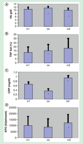
Background
Liposome has a described anti-inflammatory effect and transports its content directly in the bloodstream, beyond gastric and enteric wall.
Aim
Aim of this study is to verify if liposomal iron is most effective than iron sulfate in correction of anemia of chronic inflammatory disease of young women.
Patients and methods
In group A, 9 patients (4 with systemic erythematous lupus, 3 with mixed connectivitis, 2 with rheumatic fibromyalgia), median age 32 years (R27-42), Hb 8.5 g/dl (R8-10), saturation of iron binding capacity < 20%, with a median ferritin level of 100 ng/ml (R90-250), ESR 35 mm/1st hour (R22-95), CRP 18 mg/I (R12-24), normal B12 and folate, received liposomial iron (Sideral® Forte) 60 mg/day orally for 3 months. In group B 12 patients (6 with systemic erythematosus lupus, 3 with mixed connectivities, 3 with rheumatic fibromyalgia), median age 38 years (R29-45), Hb 9 g/dl (R8-9.5), saturation of iron binding capacity < 20%, with a median ferritin level of 120 ng/ml (R80-190), ESR 33 mm/1st hour (R20-87), CRP 15 mg/I (R13-27), normal B12 and folate, received iron sulfate 210 mg/day orally for 3 months.
Results
After treatment, group A showed a median hemoglobin level of 11.5 g/dl (R10.5-12), a median ferritin level of 260 ng/ml (R 190-280), a ESR decrease to a median value of 8 mm/1st hour (R 3-10) and a median CRP 3 mg/I (R2-4). After treatment, group B showed a median hemoglobin level of 9.5 g/dl (R8-9.5), a median ferritin level of 100 ng/ml (R 90-180), and ESR and CRP don’t showed any improvement. 4 patients showed epigastralgia, 2 stypsis, 5 diarrohea.
Conclusion
Liposomal iron (Sideral®) is most safe, effective, well tolerated, effective than iron sulfate in increase hemoglobin level and reduce inflammatory markers in correction of anemia of chronic inflammatory disease of young women.
Oral Communications
Background
Anemia is a common manifestation of neoplastic disease, is linked to adverse prognosis, symptoms such as fatigue and can require supportive therapy as blood transfusions or use of erythropoiesis-stimulating agents (ESAs). Oral liposomal iron (Sideral® Forte) is an oral formulation with good bioavailability and tolerability, and may improve anemia similar to intravenous iron, with or without ESAs. Treating early mild grade anemia could prevent a fall in hemoglobin (Hb) level below concentrations that could require the use of ESAs/transfusions.
Objective
A prospective observational study was started in mild anemic cancer patients treated with oral liposomal iron (30 mg/day), with the primary aim to evaluate iron parameters during treatment and the rate of patients with Hb drop below 10 g/dl.
Methods
Patients with solid tumors and planned to start on chemotherapy, with pre-existing grade (G)1 anemia (Hb levels range 10–12 g/dl and transferrin saturation (TSAT) 15–50% before chemotherapy) were included. Only patients, who have not started chemo or radiotherapy treatments, were included. Any use of ESA agents or other iron formulations was not allowed. A continued treatment of 3 months was performed and iron parameters were tested at 6 and 12 weeks. The planned recruitment is of 80 patients.
Results
Up to March 2015, 10 patients were enrolled (n = 6 and n = 4 in advanced and adjuvant setting, respectively). In three cases, treatment lasted less than 12 weeks. Among them, one patient stopped liposomal iron for chemotherapy-related vomiting after few days. Concomitant chemotherapy was platinum based in 60% of patients and fluorouracil or anthracycline based in the remaining 40%. At baseline, medium Hb level was 11.28 g/dl. After 6 (n=10) and 12 (n=7) weeks, medium Hb levels were 11.09 and 11.30 g/dl, respectively. Each patient had G3 nausea, G2 vomiting, and G2 diarrhea unrelated to the studied drug. TSAT increased from 12.45 to 20.8 and 20.7% at 6 and 12 weeks, respectively. No patient was transfused.
Conclusion
In a cohort of 10 patients with G1 anemia at the beginning of cytotoxic therapy, administration of liposomal iron (Sideral® Forte) for 3 months maintained Hb above level that requires supportive therapy and may potentially worsen cancer-related symptoms. Its use could be considered as a prophylactic measure to prevent transfusions/ESAs in cancer patients treated with chemotherapy and pre-existing mild anemia.
Background
Anemia is one of the most common hematological issues in oncology patients. The etiology is multifactorial: chronic inflammatory status, type of neoplasm, dyserythropoiesis caused by cytotoxic treatments, possible blood loss, hemolysis, bone marrow infiltration by neoplastic cells and nutritional deficiencies. Most chemotherapeutic drugs exert their cytotoxic effect through the inhibition of DNA synthesis and replication. Toxic effects often involve also the healthy tissues with the highest proliferative index and, among them, the hematopoietic one. As widely demonstrated in other studies, the use of a best supportive care for the treatment of anemia during chemotherapy improves the response and compliance of the patient.
Objective
The aim of this study is to assess the benefits of the continuation of oral liposomal iron administration (Sideral® Forte, 1 capsule/day for 8 weeks) to 10 anemic patients affected by lymphoma in the follow-up after chemotherapy, in terms of quality of life improvement, oxygen saturation (sO2) increase, heart rate reduction, in correlation with the increase of hemoglobin (HB) and ferritin levels in blood.
Methods
In this study, 10 anemic patients were examined: 4 with Hodgkin lymphoma (3F:1M) and 6 with non-Hodgkin lymphoma (3M:3F), who underwent oral administration of Sideral® Forte during chemotherapy and continued to be treated with Sideral® Forte 1 capsule/day for 8 weeks during follow-up.
The parameters assessed at the beginning and at the end of the study were:
Hb (normal range in adult males: 14–18 g/dl, in adult females: 12–16 g/dl);
sO2 (normal range: 95–98%);
Heart rate (normal range: 60–80 HR at rest);
Ferritin (normal range in adult males: 20–300 ng/ml, in adult females: 12–150 ng/ml);
Quality of Life (QoL) FACT Questionnaire- An Version 04 Modified Fatigue Scale (normal range: 0–28).
Results
After the administration of Sideral Forte 1 capsule/day for 8 weeks, patients enrolled in this study showed a mean increase of Hb levels of 1,26 g/dl (9.96–11.22, p = 0.0309), of ferritin levels of 49.946 ng/mL (42.839–92.785, p = 0.2105) of sO2 of 1.1% (95.2–96,3%, p = 0.3365) and a mean decrease of HR of −2 (85.4–83,4, p = 0.3843). Furthermore, we evaluated the symptom “fatigue” through the QoL FACT Questionnaire (An version 04 Modified Fatigue Scale, see ) at week 0 and at week 8. After 8 weeks a mean decrease of fatigue of 8.8 (20.4–11.6, p = 0.0088) was observed ().
Figure 1. The histogram explains the efficacy of Sideral® Forte showing a mean increase of Hb levels of 1.26 g/dl (9.96–11.22 g/dl, p = 0.0309), ferritin levels of 49.946 ng/mL (42.839–92.785 ng/ml, p = 0.2105), of sO2 of 1.1% (95.2–96.3%, p = 0.3365) and a mean decrease of HR of −2 (85.4–83,4 HR, p = 0.3843).
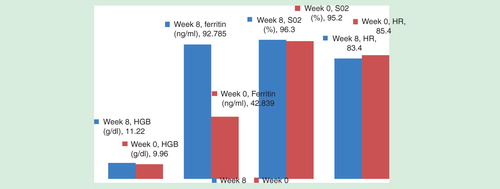
Table 1. Quality of Life FACT Questionnaire- An Version 04 Modified Fatigue Scale (normal range: 0–28).
Figure 2. The histogram shows an improvement of the quality of life after 8 weeks of treatment with Sideral® Forte (20.4–11.6, p = 0.0088).
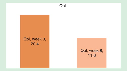
Conclusions
The oral administration of liposomal iron (Sideral Forte, 1 capsule/day for 8 weeks) to anemic patients with lymphoma in the follow-up after chemotherapy reduces fatigue, improves cardiac and respiratory functions and quality of life.
Background
This study aims to investigate the efficacy and tolerability of liposomal oral iron in comparison to intravenous iron gluconate in CKD patients undergoing chronic hemodialysis. Liposomal iron is a new iron formulation with high bioavailability and a low incidence of side effects and is highly tolerated. The use of intravenous iron in hemodialysis patients treated in an out-hospital setting is still under debate.
Methods
Twelve chronic HD patients undergoing regular intravenous iron therapy and ESAs treatment were randomized 1:1 to receive iron liposomal iron (Sideral®, PharmaNutra) or to continue iron gluconate intravenous treatment for three months. Oral liposomal iron was administered in a comparable weekly dosage (from 30 to 180 mg/week) The primary end point was to evaluate the effects of the two different treatments on Hb levels; the iron status, compliance and adverse effects were also evaluated. All patients received alfa EPO and iron and ESAs dose were not changed during the study period.
Results
No significant variations were observed in the two groups at the follow-up time. Hemoglobin levels varied from 12.03 ± 1.8 g/dl to 12.57 ± 2.1 g/dl in the intravenous group and from 12.68±2.3 g/dl to 12.66 g/dl (p = n.s.) in the oral group. Iron saturation index varied from 27.6 to 30.8% in the iron group and from 24 to 21% in the oral group (p = n.s.). Oral iron was highly tolerated.
Conclusions
Oral liposomal iron (Sideral® Forte) is a safe and efficacious alternative to iron gluconate therapy in chronic hemodialysis patients. Further studies are needed to investigate iron liposomal effects in severely inflamed patients.
Backgrounds
Iron supplementation, together with a gluten-free diet, is essential for the treatment of anemia in subject affected by celiac disease. Liposomal iron is a preparation of ferric pyrophosphate carried within a phospholipidic membrane. Compared to other oral formulations, it is well absorbed from the gut and demonstrates high bioavailability together with a lower incidence of side effects.
Objectives
The aim of the study was to evaluate the effectiveness of the oral liposomal iron treatment (30 mg/day – Sideral® Forte) compared to ferrous sulfate (105 mg/day) in celiac patients showing iron deficiency anemia occurring during the follow-up of celiac disease. The degree and timing of improvement of hemoglobin level (Hb, g/dl), total iron-binding capacity (TIBC, μg/dl) and transferrin saturation (TSAT%) of the two treatment groups (group 1: liposomal iron and group 2: ferrous sulfate) were evaluated.
Methods
In the last 12 months, 24 Type 1 diabetes mellitus patients (T1DM) aged 31.2 ± 14.7 years (M ± SD), attending our outpatient clinic (regional HUB-SPOKE network) were monitored. The mean time from T1DM diagnosis (in all cases the onset was before celiac disease) was 5,08 ± 11.9 vs 4.91 ± 9.1 in group 1 and 2, respectively. The mean age at the onset of the celiac disease was 29.5 ± 17.4 years versus 18.67 ± 10.9 years. All patient biopsies showed celiac disease stage III (b and c) according to the Marsh–Oberhuber classification. The patients were divided into two groups: 12 patients were treated with oral liposomal iron, while 12 with ferrous sulfate. Hb, TIBC and TSAT values were measured after 4 weeks of treatment for both groups.
Results
After 4 weeks of treatment, hemoglobin levels were significantly higher compared to baseline in the liposomal group (1.27 ± 0.34 g/dl vs 0.82 ± 0.39 g/dl, F = 0,81, t = 2.98, P < 0.007, IC = 95% 0.13–0.76). No significant change in TSAT value at baseline was observed between the two groups (P >0,05). However, changes in TSAT values (ΔTSAT) after treatment (T1) were significantly higher in the liposomal group (TSAT(T1) = 23.6% ± 4.27 vs 18.7% ± 5.33, F = 0.7, t = 2.4, P < 0.02, IC = 95% 0.77–8.9) (ΔTSAT = 23.6% ± 4.67 vs 7.2% ± 5.33, F = 2.1 t = 2.09, P < 0.04, IC = 95% 0.51–10,4).
Conclusions
In this preliminary study, liposomal iron (Sideral® Forte) supplementation was shown to rapidly achieve higher hemoglobin levels in Type 1 diabetic patients with celiac disease showing iron deficiency anemia during follow-up and gluten-free diet. The efficacy of this therapeutic approach is important when iron deficiency is present even under unknown gluten-free diet compliance, giving the possibility to improve iron deposits in condition of potential gut mucosal damage.
Posters
Background
CKD patients on regular hemodialysis (HD) have a reduced erythropoietin response to anemia. Treatment with erythropoiesis-stimulating agents (ESA) is generally effective, but results in a substantial increase in the iron demand. Indeed, most of ESA-treated individuals require iron supplementation. In HD population, iron is almost exclusively administered intravenously, as for practical use and because most studies have shown an advantage of intravenous injection over oral iron supplementation. Nevertheless, the enhanced body iron stores, in prolonged or inappropriate doses, may lead, in the end, to an excess of tissue iron accumulation, manifested by hyperferritinemia, an expression of unavailable iron. Oral iron supplementation in HD population (mainly with iron sulfate) is often ineffective due to the reduced absorption and gastrointestinal side effects that reduce patient compliance. The liposomal iron pyrophosphate, carried within a phospholipidic membrane, has a pattern of better bioavailability and tolerance.
Objectives
The aim of this study is to analyze the efficacy of continued oral iron supplementation with liposomal iron pyrophosphate in HD patients, after suspension of intravenous iron, evaluating the effects on ferritin, anemia and the ESA doses needed to maintain hemoglobin (Hb) within the target range. We also evaluated C-reactive protein (CRP) values to exclude the inference of inflammation.
Methods
To this purpose, we studied eight CKD patients on a regular three times a week HD, treated with ESA and intravenous iron supplementation (sodium iron gluconate, FERLIXIT®, 31.25–125 mg/week). The patients showed hyperferritinemia (1066–3822 ng/ml) and all of them had Hb values within the target range (12.0 ± 0.8 g/dl), a slightly increased serum iron level (108 ± 38 mg/dl) and transferrin saturation (TSAT% ) (53.5 ± 18.9% ). CRP was normal. At time 0, the intravenous iron therapy was stopped and the oral iron supplementation was introduced using liposomal iron pyrophosphate (SIDERAL® Forte), at a dose of 30 mg/day. On a monthly basis was evaluated Hb, hematocrit (Ht), ferritin and serum iron levels, TSAT% and CRP, monitoring ESA doses needed to obtain Hb values within the target range. All data are expressed as mean ± SD and statistical significance evaluated by paired t-test.
Results
The results of this study are preliminary, as they refer to the first 3 months of the liposomal iron treatment. One patient was excluded due to the relapsing of tumor pathology. The seven remaining patients showed a sharp significant decrease in ferritin values after the first month (2094 ± 1112 to 1663 ± 942 ng/ml, p < 0.001), with no further decrease after 3 months of treatment (1569 ± 967 ng/ml). Hb levels remained steadily within the target range (12.2 ± 0.4 g/dl), while it was noticed a slight but not significant decrease in serum iron (89 ± 18 mg/dl vs 108 ± 38 mg/dl) and in TSAT% (45.0 ± 10.1% vs. 53.5 ± 18.9%) within normal values. The EPO dose was reduced but not significantly from 4286 ± 4821 to 1500 ± 1414 IU/week (p < 0.06). No changes were observed for CRP values.
Conclusions
The substitution of intravenous iron supplementation with liposomal iron pyrophosphate (SIDERAL Forte) in CKD patients on HD with hyperferritinemia was effective in significantly lowering the ferritin levels, while keeping Hb levels, blood iron and TSAT% within the target ranges. These data could be related to a better iron utilization without the risk of tissue iron overload and a better compliance and tolerance of the patients to the liposomal iron. The results were obtained despite an evident ESA dose reduction, which it was, however, not significant.
Background
Hemorrhoids are fibromuscular and vascular cushions physiologically present in the inside lining of the rectum and they are important in the control of continence and closure of the anus. Abnormal dilatation of the vascular vessels within these cushions determines a haemorrhoidal disease characterized by symptoms such pain, incontinence, prolapses and haemorrhages varying from hematochezia to proctorrhagia. This disease affects both sexes equally and it has an incidence of about 13–36%, while surgical removal is required in 10% of cases. Hemorrhoidal disease occurs more frequently in patients aged 45–65 years and iron deficiency anemia (IDA) is often very severe, thus complementary iron therapy is essential.
Aims and methods
In order to evaluate the efficacy of liposomal iron, between October 2014 and January 2015, 9 patients aged 27–45 years, who showed IDA (hemoglobin < 9.5 g/dl; serum iron < 30 μg/dl) due hemorrhoids were treated with liposomal iron (Sideral Forte®) 2 cps/day for 3 months associated with phlebotonic drugs. Hemoglobin (Hb), serum iron, ferritin and transferrin saturation (TSAT) were measured at T0 and at 1 (T1), 2 (T2) and 3 (T3) months.
Results
The patients showed a significant increase in all the blood parameters tested already after 1 month of iron supplementation . After 3 months of supplementation, the increase in serum iron level in women of fertile age is significantly lower compared to the men group () possibly due to monthly menstruations. Despite the use of a high dose of liposomal iron (60 mg/day), no gastrointestinal side effects were reported by the patients, demonstrating the high tolerability of liposomal iron.
Table 1. Women aged 27–41 years (n = 4).
Table 2. Men aged 31–45 years (n = 5).
Table 3. Both sexes patients aged 27–45 years (n = 9).
Conclusions
The use of liposomal iron (Sideral® Forte) is an effective and safe treatment of IDA in patients with haemorrhoidal disease.
Intestinal malabsorption is a syndrome characterized by an abnormal absorption of food nutrients. Different forms of this syndrome are known (generalized, partial and selective), and they may be due to gastrointestinal diseases. The generalized malabsorption is characterized by steatorrhea (fecal fat >7 g/day with the 100 g fat diet) and diarrhea (fecal volume >250 g/day), which allow to distinguish a serious generalized malabsorption secondary to acinar pancreatic diseases from milder forms secondary to biliary diseases. In contrast, the partial malabsorption is characterized by nutritional deficiency (iron, folate, calcium, B12 vitamin, bile acids) related to the extent of damage to the wall of the small intestine. Finally, in the selective malabsorption, there is a specific biochemical defect of the enterocytes.
Celiac disease (CD) is an autoimmune multiple organ disease with production of autoantibodies, which cause intestinal damage due to the ingestion of gluten. If a potential celiac disease is suspected, it is indicated to perform test for a specific autoantibodies (antitransglutaminase and endomysial antibody). In case of positive titration, an endoscopic examination of the upper digestive tract (esophagogastroduodenoscopy [EGD]) is generally performed. Despite the incidence of celiac disease is about 1% in the general population, it is one of the major causes of malabsorption.
The non-celiac gluten sensitivity (NCSG) is a disorder, first described in 1980, that is characterized by intestinal and extra-intestinal symptoms correlated to the ingestion of gluten in non-celiac and patients not allergic to wheat.
Materials and methods
For the purpose of this study, 45 patients have been recruited (m/f 15/30; age: 51.6 ± 16.4 year; BMI: 29.3 ± 3.2 kg/m2; waist: 99.1 ± 5.6 cm) and monitored in a clinic setting for gastrointestinal symptoms such as for the irritable bowel syndrome (IBS) (abdominal pain, bloating, diarrhea or constipation) and systemic symptoms (brain fog, headache, fatigue, myalgia and arthralgia, dermatitis, depression, anemia) that may be refractory to the therapy given from the homecare provider.
Subsequent to the enrolment and physical examination, blood tests (blood count, creatinine, erythrocyte sedimentation rate (ESR), C-reactive protein (CRP), serum iron, ferritin, thyroid-stimulating hormone antibody (TRAb), free triiodothyronine (fT3), free thyroxine (fT4), anti-thyroid peroxidase (TPO) antibody, thyroglobulin (hTg) antibody, anti-thyrotropin receptor (TSH receptor) antibody, IgA and total IgG, anti-transglutaminase antibody, gliadin antibody, calprotectin, serum calcium, folic acid, vitamin B12, Helicobacter pylori serum and faecal antibody) and diagnostic procedures (ECG, upper abdomen and thyroid ultrasound, EGD) were performed on each patients.
The diagnosis of gluten enteropathy (EGD) was determined on the basis of histopathological data (according to modified Marsh criteria) in association with the titration celiac autoantibody outcome.
From the results of the blood test and diagnostic procedures performed at recruitment (T0), 12 patients were excluded for major diseases (5 chronic renal failure, GFR < 45 mg/dl per day; 4 dysmenorrhea (flow > 7 days), 1 neoplasia and 1 rheumatoid arthritis). Therefore, 34 patients were supplemented with a combination of therapy and gluten-free diet for 90 days (T1). After a full blood count test at 90 days from the beginning of the therapy, patients with a persisting condition of iron deficiency anemia (IDA) (Hb: <11.0 g/dl, MCV 78fl; RDW 11.5%) were supplemented with iron liposomal treatment (Sideral Forte-1cps twice a day for 15 days then 1 cps a day for 75 days) (T2).
Results
Table 1. Anthropometric measure in recruited patients.
Table 2. Anthropometric, hematologic measures and autoimmunity response in CD patients.
Table 3. Anthropometric, hematologic measures and autoimmunity response in NCSG patients.
Table 4. Liposomal iron supplementation in NCSG patients.
Conclusions
The diagnosis of gluten enteropathy, in the presence of hypochromic microcytic anemia is for the physician a challenge due to multiple clinical symptoms found in the general population. The history and physical examinations do not usually emphasize gastrointestinal symptoms as well as anthropometry and blood analytes. However, a significant inverse correlation exits between the severity of the damage of the villi and anemia, a condition that appears to be related to intestinal malabsorption.
Our observational experience has identified that in the presence of Marsh III type with autoantibody positivity, undertaking a gluten-free diet leads to a small but significant even if marginal improvement of the iron status. Furthermore, it was observed that association of oral liposomal iron supplementation to the gluten-free diet significantly improve Hb, ferritin and serum iron levels, possibly due to the high bioavailability of the drug.
In patients with SG (autoantibody positivity and villi damage [Marsh I–II]) the improvement of Hb, ferritin and serum iron was significant when the gluten-free diet was associated with an oral liposomal iron supplementation.
The liposomal iron is an oral preparation containing ferric pyrophosphate conveyed within a specific phospholipid membrane. The liposomal iron technology (Sideral® Forte), compared to other oral formulations, has a high gastrointestinal absorption and high bioavailability responsible for the reduced side effects.
Background
Tumor hypoxia is linked to tumor progression, development of treatment resistance and thus poor prognosis. Since anemia is a major factor in causing tumor hypoxia, the association between blood hemoglobin concentration (cHb) and tumor oxygenation status is a field of continuous clinical research Citation[1]. Head and neck cancer patients treated with radiotherapy or radio-chemotherapy have often a poor nutritional intake and development of mucositis that may increase anemia incidence. In this report, the effect of oral liposomal iron supplementation was described as improving hemoglobin levels in an old patient with local advanced glottic–supraglottic cancer treated with adjuvant radiotherapy after total laryngectomy and bilateral nodal dissection.
Case report
A 78-year-old woman, with a long smoking history and local advanced squamous cell carcinoma of the glottic–supraglottic region, underwent total laryngectomy and bilateral nodal dissection (pT4N2aMx, AJCC 7th. Edition). Six weeks after surgery, the patient started an adjuvant radiotherapy treatment aiming to minimize the locoregional recurrence risk, especially considering that the post-surgical Magnetic Resonance Imaging (MRI) showed a suspected upper jugulo-digastric positive node. Due to the clinical frailty of the patient (ECOG 2 after surgery), the multidisciplinary board decided to optimize tumor control probability through the use of simultaneous integrated boost (SIB) technique on the suspected nodal volume without the addition of chemotherapy or immunotherapy (cetuximab) to the adjuvant treatment. 160 cGy in 33 fractions were dispensed to the neck nodes at levels IIa, IIb, IIIa, IIIb, IV, V and VI (Delphian nodes), 200 cGy in 33 fractions to nodes at levels II, III and to the laryngeal surgical bed, while 225 cGy in 33 fractions to the MRI positive node at level IIb left chain. During the first 4 weeks of treatment with these dosage scheme, a reduction trend in hemoglobin levels (lower value = 8.7 g/dl) was observed in conjunction with the development of a more severe mucositis (Grade 2–3 CTCAE toxicity scale). It was also observed a decrease in mean corpuscular volume values (MCV, lower value = 43 fl) and mean corpuscular hemoglobin values (MCH, lower value = 16 pg/cell) suggesting an anemia by reduced iron intake confirmed by ferritin levels ≤100 ng/ml and transferrin saturation ≤25%. The clinicians started an oral supplementation with liposomal iron (30 mg/2 times a day) [Sideral Forte, Pharmanutra S.P.A.] for all the remaining period of radiotherapy treatment (about 28 days). The patient was monitored for the entire treatment and followed-up for 1 month after the end of the therapy. The hemoglobin value at the end of treatment was significantly higher (12.4 g/dl) and there was also observed an improvement in mucositis level (no more than grade 2 on the CTCAE toxicity scale) and in the deglutitory function of the patient. No gastrointestinal symptoms were described and thus an increase of the standard treatment of 20 mg/day of omeprazole (patient treated also with corticosteroid) was not needed. A PET/CT scan 3 months after the end of the radiation treatment demonstrated a complete metabolic response with excellent locoregional control of the disease.
Discussion
Correct and prompt management of nutritional deficiency in patients with head and neck cancer is of paramount importance to improve their quality of life and mitigate the acute side effects caused by the treatment. Recent studies, including large retrospective analyses, have demonstrated the dramatic adverse impact of anemia on the locoregional tumor control and survival Citation[2]. This case report underlines the efficiency of oral liposomal iron (Sideral® Forte) supplementation in improving the anemic status in a patient with reduced iron intake due to the pathological alteration of the irradiated mucosae and to the poor deglutitory function. Moreover, an important clinical observation as a reduction in the mucositis score was also noticed, suggesting an active role of the hemoglobin oxygen in the repair of the mucosal cells damaged by the irradiation.
References
- Vaupel P, Mayer A, Höckel M. Impact of hemoglobin levels on tumor oxygenation: the higher, the better? Strahlenther Onkol Organ Dtsch Röntgenges Al. febbraio 2006;182(2):63–71.
- Kumar P. Impact of anemia in patients with head and neck cancer. The Oncologist. 2000;5 Suppl 2:13–8.
Background
Chemotherapy-associated anemia (CAA) is a common symptom in patients with lymphoproliferative diseases and is associated with red blood cells (RBC) transfusions requirement and decreased quality of life (QOL). Erythropoiesis-stimulating agents (ESAs) and iron supplementation are used to avoid or reduce blood transfusion. Many trials demonstrated that intravenous (iv.) iron significantly increases hematopoietic response rate and decreases blood transfusions rate both in trials with and without ESAs. Iron deficiency is a common reason for a lack of response to ESAs. Even among patients with normal or increased total body iron stores, functional iron deficiency (lack of bioavailable iron) as a result of inflammation-related hepcidin production and pathologic sequestration of iron inside the macrophages can restrict erythropoiesis and impair response to ESAs treatment. Intravenous iron administration might overcome functional iron deficiency by providing elemental iron in a readily bioavailable form. Some trials suggest that oral liposomal iron is an optimal alternative to iv. iron supplementation.
Objectives
To evaluate the efficacy and tolerability of liposomal iron associated with darbopoietin in CAA in patients with lymphoma in our institution.
Methods
We retrospectively analyzed 21 patients, treated in our institution in 2014, affected by lymphoproliferative diseases, with chemotherapy-related anemia. They received darbopoietin 150 IU weekly plus oral liposomal iron (Sideral® Forte, Pharmanutra Spa) 30 mg once daily. We examined iron status, hemoglobin (Hb) values before chemotherapy, when they began ESAs, at 4 and 8 weeks, RBC transfusions and characteristic of the disease. Characteristic of patients are summarized in .
Table 1. Characteristics of analysed patients.
Results
We reported a medium increase of Hb during the 8 weeks of treatment of 1,73 g/dl and a medium increase of 0,98 g/dl compared to Hb level before chemotherapy. Eight patients required RBC transfusion and this was more frequent in patients with bone marrow lymphomatous infiltration. No serious adverse events were reported. 50% of patients had a functional iron deficiency.
Conclusions
This experience confirmed that also in lymphoma patients with CAA, oral liposomial iron (Sideral® Forte, Pharmanutra Spa) associated with darbopoietin appears to be safe, well tolerated and effective in increasing hemoglobin level, decreasing the need for transfusions and improving QOL.
Background
Anemia is the most common hematological problem in elderly patients and its prevalence increases every 10 years of age in both sexes affecting in total about 13% of patients over 70 years old (Salive ME et al. Anemia and hemoglobin levels in older persons: relationship with age, gender, and health status. J Am Geriatr Soc. 1992; 40: 489-96.). Iron deficiency anemia combined with posthemorrhagic anemia represents 20–40% of the causes of anemia in the elderly (Joosten E et al. Prevalence and causes of anemia in a hospitalized geriatric population. Gerontol 1992; 38: 111–7). In elderly patients, the iron deficiency is often related to a daily intake of gastrolesive drugs, gastrointestinal ulcers, diverticulitis, colorectal cancer and in some cases to inadequate diet or absorption deficit. Low levels of serum ferritin, high total iron binding capacity, high levels of serum transferrin and low saturation of it, high levels of soluble transferrin receptor and absence of medullary iron deposits are characteristic signs of iron deficiency (Smith DL. Anemia in the elderly. Am Fam Physician 2000; 62: 1565–72.). In mild and moderate forms, the first-line treatment is represented by oral ferrous iron salts therapy (Cook JD Diagnosis and management of iron-deficiency anemia. Best Pract Res Clin Haematol 2005; 18: 319–332.). However, the gastrointestinal side effects of this therapy frequently include nausea, heartburn, abdominal pain, diarrhea and flatulence. The high incidence of these side effects causes a low therapeutic adherence of up to 50% of patients and a consequent therapeutic failure (Cancelo-Hidalgo MJ, et al. tolerability of different oral iron supplements: a systematic review. Curr Med Res Opin 2013; 29: 291–303). Liposomal iron is a new formula of oral iron with a high gastrointestinal absorption and bioavailability with few side effects, highly effective in the treatment of iron-deficiency anemia. (Pisani et al. Effect of oral versus intravenous liposomal iron iron for treatment of iron deficiency anemia in CKD patients: a randomized trial. Nephrol Dial Transplant. 2014 November 13).
Objectives
Preliminary data on the tolerability of the iron supplementation with protected liposome iron in elderly patients with iron-deficiency anemia, clinical complexity and polypharmacy are presented in this study.
Methods
A retrospective analysis was performed on twenty elderly patients with iron deficiency anemia of various origins with clinical complexity and polypharmacy undergoing iron supplementation with protected liposomal iron (Sideral®, Pharmanutra). Patients were assessed at least after 4 weeks from the beginning of the treatment and the incidence of side effects and adherence to therapy was reported.
Patients’ characteristics
See in text.
Table 1. Characteristics of analysed patient.
Results
Patients were monitored as outpatients at least 4 weeks after the first visit and they were evaluated also for side effects due to the intake of liposomal iron. After 2 months from the liposomal iron prescription, the patients were contacted to check treatment adherence. Only two patients have reported gastrointestinal symptoms during the first week of therapy. One patient discontinued his therapy after a month due to problems unrelated to the drug. The total therapeutic adherence was 95%.
Conclusions
The protected liposomal iron (Sideral®) is well tolerated in elderly polypharmacy patients affected by complex clinical disease, allowing a better therapeutic adherence than other iron formulas.
Background and Objectives
Liposomal iron is effective also in dialysis patients. A very first experience report on the changes that occurred between guidelines, formulations, advances in the understanding of the pathogenic mechanisms of iron resistance.
Case study report
The first time liposomal iron (Sideral®) was administered to a dialysis patient in our facility was in 2008. The patient, a woman intolerant to iron in both the oral and intravenous formulations (sodium ferric gluconate), was started on liposomal iron (Sideral®). Despite the iron deficiency, she was treated with high doses of epoetin alfa (30000 IU/week). Yet, she did not reach a hemoglobin level recommended by K-DOQI guidelines. Sideral® 14 mg, the only formulation available at the time, was proposed to the patient, who showed excellent tolerability. The therapy was given for 90 days, for a total cumulative iron dose of 1260 mg, while maintaining weekly values of erythropoietin at 30,000 IU. After 2 months of treatment, hemoglobin value increased to 13 mg/dl as well as serum iron, while there was an initial decrease in ferritin level. After 3 months of therapy, hemoglobin remained unchanged, while iron and transferrin saturation were further increased until ferritin values finally increased.
Conclusions
Since 2008, liposomal iron showed well tolerability and efficacy and has become a valuable tool to treat not only patients with intolerance to other iron formulations, but also patients at any stage of kidney failure or on dialysis. This is made possible as liposomal iron follows absorption pathways that are not inhibited by hepcidin. Nowadays, a new formulation with higher iron content (30 mg per capsule) is also available. Moreover, liposomal iron (Sideral®) can replace intravenous iron with two advantages: reduce allergic risks and preserve superficial veins by avoiding the use of parenteral infusion, an important issue for CKD patients. Thus, a good beginning bodes well.
Table 1. Characteristics of analysed patients.
Background
Intravenous iron support simultaneous to erythropoietin (EPO) administration improves hemoglobin response in myelodysplastic patients. There are many evidences that iron, useful for bacterial growth, might increase risk of infection.
Objectives
The aim of this study is to verify incidence of number of febrile episodes in low-risk myelodysplastic patients supported with iron.
Methods
A multicenter retrospective study was performed. Between July 2008 and December 2014, 107 patients affected by low-risk refractory anemia were studied. Median follow-up was 24 months (R12–60). 20 patients had no support, 27 EPO support, 30 EPO + liposomal iron 14mg (Sideral®) 2 capsules orally/day for 3 months, 15 EPO + iron sulfate (525 mg, 2 tablets orally/day for 3 months) and 15 EPO + iv. sodium ferric gluconate (62.5mg iv. in NS100 ml in 1 h/day for 5 day/month). Statistical analysis was performed by Chi square test and Fisher exact test.
Results
In the group with no support, median packed red blood cells unit (PRBCU) transfused was 0.2/month (R0–0.5). Median number of febrile episodes/year was 1.5 (R0–2). In the group supported with EPO alone median PRBCU transfused was 0.4/month (R0-0.7). Median number of febrile episodes/year was 2 (R0-2). In the group supported with EPO + iron sulfate median PRBCU transfused was 0.3/month (R0-0.6). Median number of febrile episodes/year was 3 (R0-3). In the group supported with iv. sodium ferric gluconate median PRBCU transfused was 1.5 /month (R1–3). Median number of febrile episodes/year was 6 (R0–9). In the group supported with liposomal iron, median PRBCU transfused was 0.2/month (R0–1). Median number of febrile episodes/year was 1 (R0–2).
Conclusion
Number of febrile episodes does not appear to correlate to basal neutrophil count or hemoglobin level reached after 3 months of treatment. Number of febrile episodes was higher in the group with higher transfusion requirement and in the group treated with iv. sodium ferric gluconate (p = 0.02). It can be hypothesised that liposomal iron support provides a reduced amount of nontransferrin bound iron that might block bacterial growth. However, these data need confirmation on a larger cohort of patients.
Background
Fatigue is the most invalidating symptom in neoplastic disease and it is frequently linked to an iron deficiency. In inflammatory diseases as myelodysplastic syndromes fatigue might be linked to a functional iron deficiency with elevated ferritin level and a saturation of total iron binding capacity < 20%.
Objectives
The aim of this study is to verify whether liposomal iron support in myelodysplastic syndromes as refractory anemia improves fatigue perception in patients with a saturation of total iron binding capacity < 20%.
Methods
Between June 2011 and December 2014, 20 patients affected by refractory anemia were studied. Median follow-up was 12 months (R10-24). Patients were randomized 1:1 and in group A median age was 60 years (R65-70), M/F: 8/2. In group B median age was 66 years (R60-75), M/F: 6/4. Karyotype was normal in group A and B patients. Median level of haemoglobin was 9 g/dL in group A (R8.5-11) and 8.8 g/dL (R8.5-11.5) in group B. to Group A received alpha erythropoietin 40,000 IU sc/week + calcium levofolinate 7.5 mg/day orally + Vitamin B12: 400 mg/day orally. Group B received liposomal iron 14 mg (Sideral®), 1 capsule orally/day + alpha erythropoietin 40000 IU sc/week + calcium levofolinate 7.5 mg/day orally + Vitamin B12: 400 mg/day orally. Fatigue was measured using the Modified Fatigue Impact Scale (FISC - Fisk 1994).
Results
Patients in group A reached a median hemoglobin level of 11.5 g/dl and after 3 month of therapy referred a median FISC score of 74 (R65-80). Patients in group B reached a median hemoglobin level of 12.5 g/dl and after 3 months of therapy referred a median FISC score of 54 (R42-68).
Conclusion
Liposomal iron (Sideral®) support improves fatigue perception in patients with refractory anemia. This study needs confirmation on a lager cohort of patients.
Background
In iron deficiency anemia, support with intravenous iron allows a faster anemia correction and a faster ferritin increase than with iron sulfate. Frequently, iron sulfate and intravenous iron generate adverse events as hypotension, rash, shock, abdominal pain, constipation or diarrhea. Thus, high doses of oral iron frequently are poorly tolerated because of these adverse events.
Objectives
The aim of this study was to verify whether high doses of oral liposomal iron are safe, cost-effective and well tolerated as standard doses of intravenous ferric gluconate in patients with iron deficiency anemia.
Methods
Two groups of patients (randomized 1:1) with iron deficiency anemia without other relevant comorbidities were considered in this study. Group A had a M/F ratio of 2/3, 7 patients had hemorrhagic gastritis, 3 hemorrhagic enteric bleeding angiodysplasia, 10 hypermenorrhaea. Median level of hemoglobin (Hb) was 8 g/dl (R 7-10), median ferritin level was 10 ng/ml (R 3–20), with normal level of CRP or ESR. Group A received liposomal iron (Sideral® Forte) 30 mg 4 capsules/day. Group B had a M/F ratio of 1/3, 9 patients had hemorrhagic gastritis, 1 hemorrhagic enteric bleeding angiodysplasia, 10 hypermenorrhea. Median level of Hb was 8.5 g/dl (R 8-9.5), median ferritin level was 8 ng/ml (R 2–18), with normal level of CRP or ESR. Group B received iv. sodium ferric gluconate 62.5 mg iv. in NS 100 ml 3 h per day. The median treatment costs in each group were calculated considering the monthly global treatment cost for each patients in the treatment period. This provided an estimate of the total costs, which do not consider only the cost of the drug, but tied to the final outcome (efficacy) of the therapeutic strategy used during the observational period.
Results
In group A, 1 g/dl Hb increase was observed after a median of 8 days (R 7–12) and a target Hb level of 12 g/dl was achieved in a median time of 4 weeks (R 2-4) with a median monthly cost of €110 (R 92–162). 6 patients (30%) showed adverse events (3 abdominal pain, 3 diarrhea). In group B, 1 g Hb increase was observed after a median of 7 days (R 6-10) and a target Hb level of 12 g/dL was achieved in a median time of 3.5 weeks (R 1.5-4) with a median monthly cost of €326 (R 250-360). 4 patients (20%) showed adverse events (2 hypotension, 2 rash and headache).
Conclusion
High dose of oral liposomal iron (Sideral® Forte) support is safe, fast, well tolerated and cost-effective as intravenous iron in sideropenic anemia. This study needs confirmation on a larger cohort of patients.
Background
Liposome has a described anti-inflammatory effect and transports its content, beyond gastric and enteric wall, directly into the bloodstream.
Objectives
The aim of this study is to verify whether liposomal iron is more effective than iron sulfate in the correction of anemia of chronic inflammatory disease of young women.
Methods
In the group A, 9 patients (4 with systemic erythematous lupus, 3 with mixed connectivitis and 2 with rheumatic fibromyalgia), median age 32 years (R27–42), Hb 8.5 g/dl (R8-10), saturation of iron binding capacity <20%, with a median ferritin level of 100 ng/ml (R90-250), ESR 35 mm/1st hour (R22-95), CRP 18 mg/I (R12-24), normal B12 and folate, received liposomial iron 60 mg/day (Sideral® Forte, 2 cps/day) orally for 3 months. In the group B12 patients (6 with systemic erythematous lupus, 3 with mixed connectivitis and 3 with rheumatic fibromyalgia), median age 38 years (R29-45), Hb 9 g/dl (R8-9.5), saturation of iron binding capacity < 20%, with a median ferritin level of 120 ng/ml (R80-190), ESR 33 mm/1st hour (R20-87), CRP 15 mg/I (R13-27), normal B12 and folate, received iron sulfate 210 mg/day orally for 3 months.
Results
After treatment, the group A showed a median hemoglobin level of 11.5 g/dl (R10.5–12), a median ferritin level of 260 ng/ml (R 190–280), a ESR decrease to a median value of 8 mm/1st hour (R 3–10) and a median CRP 3 mg/I (R2–4). After treatment, the group B showed a median hemoglobin level of 9.5 g/dl (R8-9.5), a median ferritin level of 100 ng/ml (R 90–180), and ESR and CRP did not show any improvement. 4 patients showed abdominal pain, 2 constipation and 5 diarrhea.
Conclusion
Liposomal iron (Sideral® Forte) is more safe, effective, well tolerated and effective than iron sulfate in increasing hemoglobin level and reducing inflammatory markers to correct anemia of chronic inflammatory disease in young women.
Background
Myelodysplastic syndromes (MDS) represent a heterogeneous group of hematologic neoplasms typical of the elderly, characterized by morphologic dysplasia, aberrant hematopoiesis and peripheral blood refractory cytopenias. Among these, anemia is the most frequent one, occurring approximately in 80-90% of the patients at diagnosis, and is often associated with many potentially debilitating symptoms including fatigue, depression, reduced cognitive capacity, dyspnea. For patients with low-risk MDS (typically with IPSS low or intermediate-low) the main objective of treatment is to control and improve symptoms associated with cytopenias and in particular to reduce the transfusion requirements and transfusion side effects. The advent of hematopoietic growth factors, such as recombinant erythropoietin (EPO), has greatly improved survival and quality of life in MDS patients. Moreover, studies in patients with chronic renal insufficiency largely demonstrated that iron supplementation can improve responses to EPO treatment. Despite patients with MDS have often high serum ferritin levels and iron overloading, sometimes refractory anemia is accompanied by low or normal iron reserves. In these patients iron supplementation may increase hemoglobin (Hb) responses to EPO.
Purpose
In this case report we aimed at studying whether iron supplementation could improve hemoglobin response to EPO treatment.
Material and methods
We report the case of a patient diagnosed with low-risk MDS and treated with EPO and iron supplements for severe anemia.
Results
A 76 year old man was referred to our institution for sever and symptomatic anemia (Hb < 8 g/dl). Medical history comprised moderate chronic renal insufficiency and a thalassemia trait. Complete blood counts showed microcytic anemia (Hb 7.7 g/dl and MCV 60 fl) with normal platelets (110×106/l) and leucocytes (4.7×10⁹/l, neutrophils 3.2×10⁹/L) values. Serum creatinin was 3,75 mg/dl (normal range <1.1 mg/dl), endogen EPO level was lower than 500 UI/µl and iron metabolism showed serum ferritin within normal range (50 mg/dl), but reduced transferrin saturation levels (approximately 14%). There were no signs of hemolytic anemia since reticulocyte counts, LDH, haptoglobin and bilirubin levels were within range of normality. The patient had been treated in the past 2 years with darbepoietin, at the dosage of 40 µg/week , prescribed by the nephrologist, with no improvement in Hb values. Moreover, 1 year in advance, the patient was treated with iv. iron infusions without any apparent improvement of blood counts. We therefore performed a bone marrow trephine that showed the presence of erythroid dysplasia, abnormal maturation of myeloid cells without an increase of blast cells, and no cytogenetic abnormalities. We diagnosed the patient with MDS, such as refractory anemia (RA), characterized by a low International Prognostic Scoring System (IPSS). At diagnosis, the patient was transfusion dependent with a transfusion need of 4 packed red blood cell (PRBC) units per month. Two months after MDS diagnosis, we started a treatment with rEPO alpha, at the dosage of 40,000 U biweekly, plus an oral liposomal iron supplementation (Sideral® Forte). Four weeks later, Hb levels increased from 7.7 to 9 g/dl. In the following months, the transfusion need dramatically decreased to 2 PRBC every 3 months, and 1 year later, the patient became transfusion independent. Current Hb levels range between 10 and 11 g/dl, the patient is free of symptoms due to anemia and performance status is good.
Conclusions
This case report brings further strength to the importance of providing an adequate iron intake to support efficacious hematopoiesis. In fact, the patient object of this case report did not achieve a hematologic response unless we combined both rEPO and oral liposomal iron supplementation. Treatment with erythropoiesis stimulating agents (ESA) may be a cause itself of iron deficiency. ESAs require iron for stimulating effective erythropoiesis and it has been estimated that 1 g of iron is needed to raise the hemoglobin level from 8 to 11–12 g/dl. Therefore, in MDS patients with no iron overload or reduced iron deposit, it is important to remember that iron supplementation may improve or trigger hematologic responses in those subjects with no previous benefits from ESAs alone.
Background
As a result of possible deficiency of methoxy polyethylene glycol epoetin beta (CERA) in the national territory, AIFA, according to an agreement with EMA, elaborated a document inviting prescribers to switch patients in therapy with different CERA's doses to any erythropoiesis stimulating agent (ESA), for the treatment of anemia associated with chronic kidney disease(CDK) This recommendation emphasized the need to monitor hemoglobin levels (Hb) and the parameters of safety and efficacy.
Purpose
To evaluate variations of efficacy (Hb levels) and safety (immunological reaction) of a new treatment, in patient with CKD after switching from CERA to epoetin zeta (EPO zeta), as per international and national guidelines. To keep the same Hb level obtained before the shift. To compare the cost differences of the two ESA.
Materials and methods
Preliminary observational study (April–September 2012) was carried on CDK patients in dialysis care at the Department of Nephrology. The patients enrolled were treated with some of the doses of CERA indicated in the Recommendation at least during 10 months. We evaluated ESA dosage, Hb level and dosage/kg.
Result
The study included 12 patients (7 men and 5 women) with mean age 56, 64 years(range 40–75). All patients were treated with EPO zeta (average initial dose 6500UI/Kg/week); after monthly monitoring Hb levels, the initial dose of EPO zeta was increased on 7.69% (average dose 7000 UI/kg/week) and 3 months later, median Hb level observed was 11.28 g/dl. Statistical analysis showed no significant differences between CERA and EPO zeta in term of Hb level (p=0.408). No adverse events due to treatment were registered; no iron supplementation was provided. The use of EPO zeta resulted in saving 250 euros per month/patient versus CERA treatment.
Conclusion
After switch of CERA therapy, the use of EPO zeta appears effective and safe for CDK patient’s treatment. Data showed the need to increase the dose of EPO zeta to maintain steady Hb level. Despite the implementation of consumption, the use of this biosimilar could contribute to containing the pharmaceutical costs.
Background
On the 25/10/2013 AIFA (Italian Medicine Agency) released a safety communication stating that ‘intravenous iron containing drugs may only be given where emergency personnel and equipment are immediately available to treat the potentially life-threatening allergic reactions that can occur with treatment’. This communication banned the administration of intravenous iron drugs in facilities that do not have a resuscitation unit. Due to this limitation, the administration of intravenous iron therapy to patients undergoing their hemodialysis treatment at a supported outpatient center was no longer allowed.
Objectives
To evaluate an alternative iron administration therapy in hemodialysis patients in a supported outpatient center using oral iron treatment that may have good gastric tolerability associated with low risk side effects.
Methods
28 patients treated in a supported outpatient hemodialysis center were included. In order to ensure full patient compliance, 2 capsules of Sideral® Forte were administered during every dialysis treatment for a total of 6 capsules per week. Blood values of hemoglobin, hematocrit, red blood cell volume, reticulocytes, C-reactive protein, sideremia, transferrin and ferritin were monitored at the beginning and after 4 months of treatment.
Results
A significant reduction in the value of hemoglobin and hematocrit of 0.5 g/dl and 1.4%, respectively was observed. No modification in the dose of erythropoietin was needed. No significant variation in the other parameters was observed.
Conclusions
The administration of Sideral® Forte for 4 months resulted in a decline in hemoglobin and hematocrit levels without reducing the iron deposit of the hemodialysis patients. We suggest that Sideral® Forte can be a valid alternative to the intravenous iron therapy in supported outpatient centers where intravenous iron therapy is no longer allowed. Administering higher daily dose of the liposomal iron and increasing the erythropoietin dose should be considered to optimize the therapy.
Background
In the last decade, a new class of anticancer drugs targeting the angiogenesis was developed. These compounds, antagonizing the vascular endothelial growth factor activity (anti-VEGF), are ‘tyrosine kinase inhibitors’ (i.e. sunitinib, sorafenib, regorafenib, axitinib, pazopanib) and ‘anti-VEGF-receptor’ (i.e. bevacizumab); other new antiangiogenetic drugs (AAG) are under investigation. Their employment is currently approved for a number of neoplasms, comprising colorectal, lung, breast, kidney and liver cancer. Although the toxicity profile has been well investigated and new toxicities previously unreported with other anticancer drugs have emerged, anemia represents as well a potential side effect. Its incidence may vary according to each drug, ranging between ‘very common’ (i.e. >1/10) for sunitinib and regorafenib, ‘common’ (i.e. >1/100 to 1/10) for sorafenib and axitinib and ‘not reported in SPC’ for pazopanib.
Objectives
The aim of this paper is to evaluate the incidence of anemia in a series of AAG-treated patients (pts) at Treviglio-Caravaggio Hospital (Italy) between March 2012 and March 2015. Hemoglobin (Hb) levels were recorded before starting AAG, and at 12 and 16 weeks after its introduction. Anemia was defined according to local laboratory normal limits ( < 11.5 g/dL in females, < 13.0 g/dl in males), and its grading according to the NIH Common Terminology Criteria for Adverse Events. Those patients receiving also a chemotherapy regimen or presenting other evident causes of anemia were excluded from the study.
Results
Forty-one consecutive AAG-treated patients were evaluated (median age = 67 years, mean age = 66.6 years, age range = 49–84 years; male/female = 27/14). Thirteen patients receiving bevacizumab were excluded from the study because of concomitant chemotherapy. The remaining 28 evaluable patients were treated with: sorafenib (N=11), sunitinib (N=10), axitinib (N=3), pazopanib (N=2), and regorafenib (N=2). Sites of neoplasms were: kidney (N=12), liver (N=8), colon (N= 2), kidney plus colon (N=5), liver plus colon (N=1). Before starting AAG, anemia was already present in 10 patients (36%), being of Grade (G) 1 in 9 patients, and G2 in 1. After AAG introduction, a further decrease of Hb was observed in 4 patients (3 of G1 and 1 of G2). Among the 18 patients without anemia before AAG, a decrease of Hb > 1 g/dl after AAG introduction was observed in 6 of them, but only 4 patients (22%) developed anemia (G1 in all cases). When considering all patients, independent of basal status, anemia was observed in 32% (G1 in 8 patientsand G2 in 1), but no patient received additional treatment for anemia and no dose reduction or AAG withdrawal due to anemia were performed. Of note, in both patients receiving pazopanib an increase of Hb levels (+1.8 g/dl and +3.3 g/dl, respectively) was observed.
Conclusions
Despite the small number of evaluated patients, our data appears to confirm literature reports. The low incidence of anemia > grade 2, the absence of AAG withdrawal due to anemia, and the number of possible management strategies suggest that anemia itself does not represent a ‘limiting’ adverse event for AAG-treated patients. Moreover, iron based therapy (i.e. liposomial iron) should be widely considered in these pts, since in most of them the use of erythropoietin is not indicated.
Background
Iron deficiency anemia (IDA) is a public health problem that affects about 2 billion people, as the WHO Global Database on Anemia in 2008 estimates. IDA is more prevalent in children and young adults because of the physiological high iron requirements. A diet with an insufficient amount of iron is usually the most frequent cause of IDA, but also the regular consumption of milk in infants down-regulates the iron uptake. In addition, calcium and casein phosphopeptide directly inhibit iron absorption. In adolescents, additional causes of anemia are celiac disease, food intolerance, Helicobacter pylori (HP) infection, gastroesophageal reflux disease and inflammatory bowel disease (IBD).
Objectives
The aim of this retrospective study is to analyze the incidence and the characteristics of bowel diseases in patients younger than 20 years with unexplained iron deficiency.
Methods
From January 1997 to July 2011, 122 pediatric patients (age <20 years) with unexplained iron deficiency were observed in our center. Iron deficiency was defined by serum ferritin and transferrin saturation levels according to the CDC. IDA was defined by low levels of hemoglobin (Hb), according to the age and sex groups. Blood cells count and iron assessment were performed at planned intervals: first visit, 7–10 days, 3–4 weeks and during the follow-up until the resolution of IDA. Hb, Medium corpuscular volume (MCV), serum iron and ferritin levels were evaluated. Screening for gastrointestinal diseases was performed during the observational period. Fisher exact test (qualitative) and Wilcoxon or Kruskal–Wallis test (quantitative) were used for statistical analysis. The significance level was set at p= 0.05. This study is carried out in agreement with the Declaration of Helsinki.
Results
Among 122 patients (67 males and 55 females, median age 4.7 years), 26 (21.3%) were < 2 years old, 42 (34.4%) were between 2 and 6 years old, 8 (6.5%) were between 6 and 10 years old and 46 (37.7%) were >10 years old. At baseline, 84 out of the 122 patients (68.9%) presented anemia. Hemoglobin level, MCV and ferritin values at the first observation are reported in figure 1(a, b, c). Clinical history noticed an insufficient dietary intake of iron in 13 out of the 26 (50%) patients, all of them younger than 2 years. Clinical symptoms suggested a screening for gastrointestinal diseases in 36 patients (29.5%): six patients showed no evidence of gastrointestinal involvement, while 30 patients were found positive to the screening (types of gastrointestinal pathology according to the age and sex are shown in ). Oral ferrous iron was started in 28/30 patients (93%) and 12 of them (43%), who developed gastrointestinal discomfort and diarrhea, were switched to oral liposomal iron (Sideral®). Six out of 7 patients with HP infection started concomitant therapy for HP eradication, reaching normal range value for Hb, but not for serum ferritin after 3 months. Fifteen patients with food intolerances were managed with dietary restrictions. During follow-up, all patients with gastrointestinal diseases achieved Hb and serum ferritin values within the normal range, while MCV values remained lower than normal in 67% of the patients regardless of the compound used.
Table 1. Type of gastrointrestinal disease considered with number of patients.
Conclusions
In our experience, 25% of children and adolescents with unexplained iron deficiency suffer from gastrointestinal diseases. Detailed clinical history was important to identify those patients with unexplained iron deficiency who may benefit from a screening for gastrointestinal diseases. Food intolerances are prevalent in children <6 years and sporadic in those older than 10 years, while HP infection is prevalent in patients >10 years. No case of IBD was found. Suitable treatment for the various gastrointestinal disease combined with iron supplementation is needed for the treatment of IDA. Oral liposomal iron (Sideral®) showed efficacy in patients with gastrointestinal diseases, who were intolerant to the oral ferrous compounds.
Figure 1. (A) Hemoglobin levels according to age. No statistical difference was found between the different age groups. (B) MCV values according to age. MCV values were significantly lower in children aged >2-6 years and in those older than 10 years. (C) Ferritin values according to the age. Ferritin values were significantly lower in children older than 10 years.
Background
Iron-deficiency anemia is the most frequent anemia (low red blood cell or hemoglobin levels) caused by an insufficient dietary intake, an altered absorption of iron, and/or iron loss from bleeding which can originate from a wide range of sources such as the intestinal, uterine or urinary tract. Iron deficiency causes approximately half of all anemia cases worldwide and affects women more often than men. World estimates of iron deficiency occurrences are somewhat vague, but the true number probably exceeds one billion people. Anemia is sometimes treatable, but certain types of anemia may be a lifelong condition. In case of a simple diet-related cause, the use of oral iron supplement is usually able to overcome anemia. In many other circumstances, such as inflammatory anemia or gastroresected patients, the conventional oral iron therapies usually fail. The main problem of the conventional therapy with ferrous sulfate or other oral bivalent drugs is the occurrence of side effects. Moreover, the long-term treatment required several months induce the patients to a low compliance and in some cases intravenous treatment is needed. Parenteral iron therapy involves risks of fever, chills, backache, myalgia, dizziness, syncope, rash, and for some preparations, anaphylactic shock. The total incidence of adverse events is much lower compared with bivalent oral tablets and blood transfusions, but, however, parental iron therapy clearly appears more dangerous and serious. Liposomal iron is a preparation of ferric pyrophosphate carried within a phospholipidic membrane. Compared to other oral formulations, it is well absorbed from the gut and it demonstrates high bioavailability combined with a lower incidence of side effects.
Objectives
Considering the liposomal iron a promising new strategy for iron therapy, a study was conducted to determine whether liposomal iron is able not only to correct anemia, but also to replace the intravenous iron treatment.
Methods
In this randomized, open-label trial, 18 patients (13 females, 5 men), mean age 56.16 years, with different diseases were included. All patients were eligible for the iv. treatment due to a very low hemoglobin range (7.8 ≤ Hb ≤ 11.8 g/dl) and to a low ferritin levels (average ferritin 16.4 ng/ml). All patients received oral liposomial iron (Sideral® Forte 30 mg/day), and they were divided in three groups based on the treatment time: A (20 days, n=2), B (40 days, n=8), C (60 days, n=8). All the patients who showed a low response at the 40 days of treatment continued the therapy for further 20 days (not depending on the cause of the disease). The primary endpoint was to evaluate the effects of the treatments measuring Hb levels, compliance and adverse effects.
Results
By the end of this study, all the patients enrolled completed the treatment and no side effect was reported. As shown below (), in the group
A the average of Hb improvement was +1 g/dl (range 0.6-1.4 g/dl).
B the average of Hb improvement was +1,7 g/dl (range 0.6-3.3 g/dl).
C the average of Hb improvement was +2 g/dl (range 0.4-5.5 g/dl).
Figure 1. Average Hb g/dl improvement.
Figure 2. Response Hb(t0)/Hb(t1).
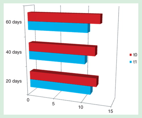
Conclusions
All the patients enrolled in this study have completed the treatment without side effects that are often common to other oral therapies. All of the patients presented a significant increase in hemoglobin level by the end of the study, showing a good response and a high compliance to the treatment. We could assess that the new liposomal iron is a valid alternative to the iv. standard drugs.
Group A
Group B
Group C