Abstract
Vogt–Koyanagi–Harada (VKH) disease is usually defined as a bilateral chronic granulomatous panuveitis that may be associated with CNS, auditory and integumentary manifestations. An autoimmune reaction against melanocyte antigens in genetically predisposed patients without history of ocular trauma is presumed to cause the disease. Features of ocular and extraocular involvement vary according to disease phase, and the condition can present with disease features limited to intraocular inflammation without associated systemic manifestations. Exudative retinal detachment is the most specific feature to acute VKH disease. Ancillary tests (mainly fluorescein angiography) may help in establishing the definitive diagnosis. On the other hand, sunset glow fundus is typical to chronic VKH disease. Complications are more likely to occur in the chronic recurrent phase. The mainstay of treatment for acute VKH relies on prompt systemic corticosteroid therapy initiated as a high dose and then tapered gradually. The duration of corticosteroid therapy is recommended to be at least 6 months. Other immunosuppressants may be given as second-line or, in selected cases, as first-line therapy. Early diagnosis with rapid initiation of corticosteroids reduces recurrence and thus improves visual prognosis.
Medscape: Continuing Medical Education Online
This activity has been planned and implemented in accordance with the Essential Areas and policies of the Accreditation Council for Continuing Medical Education through the joint sponsorship of Medscape, LLC and Expert Reviews Ltd. Medscape, LLC is accredited by the ACCME to provide continuing medical education for physicians.
Medscape, LLC designates this Journal-based CME activity for a maximum of 1 AMA PRA Category 1 Credit(s)™. Physicians should claim only the credit commensurate with the extent of their participation in the activity.
All other clinicians completing this activity will be issued a certificate of participation. To participate in this journal CME activity: (1) review the learning objectives and author disclosures; (2) study the education content; (3) take the post-test with a 70% minimum passing score and complete the evaluation at www.medscape.org/journal/expertimmunology; (4) view/print certificate.
Release date: 13 December 2012; Expiration date: 13 December 2013
Learning objectives
Upon completion of this activity, participants should be able to:
• Analyze the pathophysiology and epidemiology of VKH disease
• Assess the clinical course of VKH disease
• Evaluate diagnostic tools for VKH disease
• Distinguish first-line therapy for acute VKH disease
Financial & competing interests disclosure
EDITOR
Elisa Manzotti
Publisher, Future Science Group, London, UK
Disclosure: Elisa Manzotti has disclosed no relevant financial relationships.
CME AUTHOR
Charles P Vega, MD
Health Sciences Clinical Professor; Residency Director, Department of Family Medicine, University of California, Irvine, CA, USA.
Disclosure: Charles P Vega, MD, has disclosed no relevant financial relationships.
AUTHORS
Sonia Attia, MD
Department of Ophthalmology, Fattouma Bourguiba University Hospital, Monastir, Tunisia; Faculty of Medicine and University of Monastir, Tunisia
Disclosure: Sonia Attia, MD, has disclosed no relevant financial relationships.
Sana Khochtali, MD
Department of Ophthalmology, Fattouma Bourguiba University Hospital, Monastir, Tunisia; Faculty of Medicine and University of Monastir, Tunisia
Disclosure: Sana Khochtali, MD, has disclosed no relevant financial relationships.
Rim Kahloun, MD
Department of Ophthalmology, Fattouma Bourguiba University Hospital, Monastir, Tunisia; Faculty of Medicine and University of Monastir, Tunisia
Disclosure: Rim Kahloun, MD, has disclosed no relevant financial relationships.
Sonia Zaouali, MD
Department of Ophthalmology, Fattouma Bourguiba University Hospital, Monastir, Tunisia; Faculty of Medicine and University of Monastir, Tunisia
Disclosure: Sonia Zaouali, MD, has disclosed no relevant financial relationships.
Moncef Khairallah, MD
Department of Ophthalmology, Fattouma Bourguiba University Hospital, Monastir, Tunisia; Faculty of Medicine and University of Monastir, Tunisia
Disclosure: Moncef Khairallah, MD, has disclosed no relevant financial relationships.
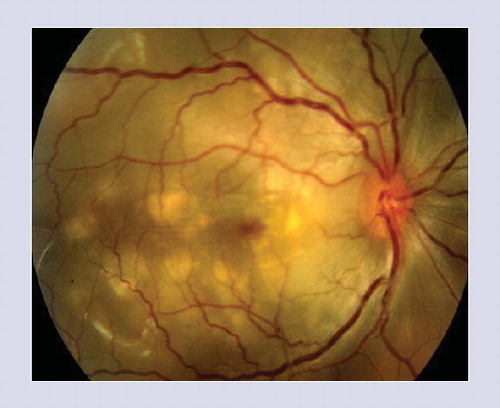
Note the presence of associated exudative retinal detachment, retinal folds and optic disc hyperemia.
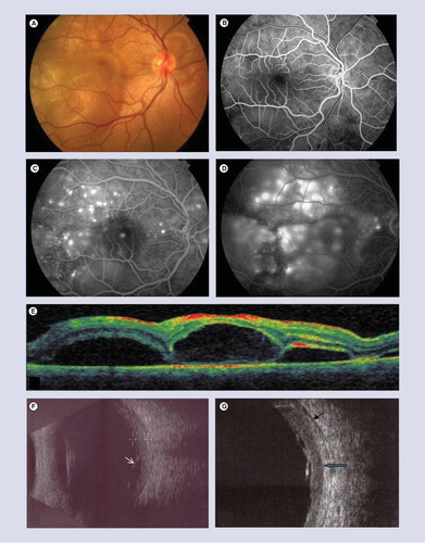
(A) Fundus photography shows bilateral exudative retinal detachment involving the posterior pole with associated retinal and choroidal folds. (B) Early-phase fluorescein angiogram shows areas of delayed choriocapillaris filling. (C) Mid-phase fluorescein angiogram shows multiple pinpoints that enlarge with pooling of dye in subretinal space in the late-phase (D). (E) Optical coherence tomography shows exudative retinal detachment with subretinal septa dividing the subretinal space into several compartments. (F) 10-MHz ultrasonography of the same patient shows diffuse-low to medium-reflective choroidal thickening most marked in the posterior fundus and associated exudative retinal detachment (white arrow). (G) 20-MHz ultrasonography shows better definition of sclero-choroidal limit (blue arrow) and episcleral space (black arrow) with more accurate measurement of choroidal thickening.
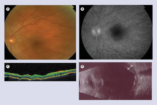
(A) Fundus photograph of left eye shows optic disc edema and retinal and choroidal folds. There is no evident exudative retinal detachment. (B) Early-phase fluorescein angiography shows hypofluorescent striae and optic disc hyperfluorescence. (C) Optical coherence tomography of the same eye shows multifocal folds of the retinal pigment epithelium and Bruch’s membrane. (D) 10-MHz ultrasonography shows diffuse medium reflective choroidal thickening (white arrow).
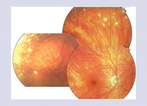
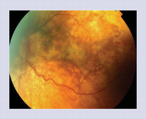
Note the presence of nummular, round, well-limited chorioretinal depigmented scars located in the midperiphery and focal hyperpigmentation in the foveal area.
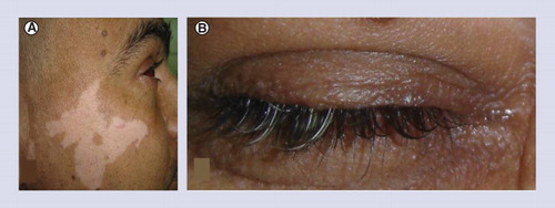
(A) Vitiligo on the face and (B) poliosis
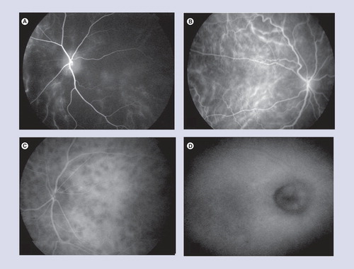
(A) Early-phase indocyanine green angiograms shows a marked choriocapillaris filling delay and (B) choroidal vascular fuzziness due to perivascular leakage. (C) Intermediate-phase indocyanine green angiography shows numerous hypofluorescent dark dots. (D) Late-phase indocyanine green angiography shows optic disc hyperfluorescence.
Vogt–Koyanagi–Harada (VKH) disease commonly affects pigmented races and people of a certain genetic predisposition. It is often defined as a bilateral, chronic granulomatous panuveitis frequently associated with CNS, auditory and integumentary manifestations Citation[1,2]. It was individualized after the former descriptions of the disease by Vogt in 1906 Citation[3], Harada in 1926 Citation[4] and Koyanagi in 1929 Citation[5]. The most accepted origin of the disease is a T-cell mediated autoimmunity reaction against melanocytes Citation[1]. The course of the disease has been divided into four phases: prodromal phase, acute uveitic phase, chronic convalescent phase and chronic recurrent phase Citation[1,2]. In the early acute uveitic phase, ocular involvement typically manifests as diffuse choroiditis with exudative retinal detachment (ERD). Later on, anterior chamber inflammation develops in association with posterior uveitis, resulting in bilateral panuveitis. During the chronic phase, typical sunset glow fundus (SSGF) occurs. Associated systemic manifestations, when present, are helpful in establishing the definitive diagnosis of VKH disease. Fluorescein angiography (FA) provides important diagnostic clues in the acute uveitic phase. Other ancillary tests, mainly optical coherence tomography (OCT), indocyanine green angiography (ICGA) and ultrasonography show typical manifestations and are helpful for the diagnosis, follow-up and monitoring of treatment.
Treatment modalities mainly include systemic corticosteroids and immunosuppressive agents. The most common complications include cataract, glaucoma, choroidal neovascularization and subretinal fibrosis. Vision loss may also result from pigmentary changes secondary to previous inflammation and ERD.
The primary aim of this article was to review current data on the clinical characteristics, diagnosis, management and prognosis of VKH disease.
Epidemiology
VKH disease is a globally distributed condition that has a predilection for dark-skinned individuals such as Asians, Native Americans, Hispanics, Asian Indians and those with Middle Eastern heritage. Black African people are less commonly affected, indicating that VKH disease is associated with genetics rather than with the amount of skin pigmentation Citation[1,2]. In Saudi Arabia, VKH disease accounts for 19.4% of all uveitis cases Citation[6]. It represents 9.7% of uveitis cases in Japan Citation[7], 7.4% in Tunisia Citation[8] and only approximately 1–4% in the USA Citation[9]. The disease usually occurs in patients between the ages of 20 and 50 years, but children may also be affected Citation[10,11]. Two to 15% of VKH patients are less than 14 years of age Citation[8,12–14]. The disease may also affect elderly patients Citation[12,13]. Women are often more likely to be affected than men. The reported proportions of affected women to men are 47.9% in Saudi Arabia Citation[6], 50% in Singapore Citation[15], 65.3% in Tunisia Citation[8] and 71.1% in Turkey Citation[13].
Pathogenesis & pathology
Although the precise etiology of VKH disease is unknown, several studies have suggested that a T-cell mediated autoimmune reaction against one or more antigens found on or associated with melanocytes may play a major role in the disease Citation[1]. The antigenic target of melanocytes often seems to be tyrosinase or tyrosinase-related proteins Citation[16–18].
There is a genetic susceptibility and the disease has been described in twins Citation[19–21]. Data from previous studies showed a strong relationship between VKH disease and the human leukocyte antigens DR4 and Dw53 (HLA-DR4 and HLA-Dw53) in many populations Citation[22–27]. Among the various polymorphic variations in the β-chain of the DR4 haplotype, the HLA-DRB1*0405 allele has been considered the most significant one Citation[28,29]. Specific killer cell immunoglobulin-like receptor (KIR) genes, HLA-C alleles, CTLA-4 genetic polymorphisms, as well as a decreased expression of CD18 and AKNA transcription factors may also increase the susceptibility to VKH disease Citation[30–33].
Low levels of advanced glycation end products, decreased 1.25-dihydroxyvitamin D3 levels, high levels of osteopontin and high leptin levels in the serum have been implicated in the development of VKH disease Citation[34–37]. Besides these, infectious factors may possibly trigger VKH disease, including Epstein–Barr virus reactivation Citation[38] and cytomegalovirus, whose antigens showed a cross-reaction with tyrosinase peptides by T cells from VKH disease patients Citation[39]. VKH disease has been described in association with other systemic diseases, with a presumed autoimmune mechanism including diabetes mellitus, celiac disease, psoriasis, thyroid disease and rheumatoid arthritis Citation[1,40,41].
The typical histopathological features seen in the early phase of VKH disease include granulomatous inflammation that primarily involves the choroid with milder inflammatory infiltrate involving the iris and ciliary body and subsequent ERD. The choroid is characterized by a diffuse lymphocytic infiltration interrupted at multiple sites with collections of epithelioid cells and few multinucleated giant cells, which contain pigment with no apparent choroidal necrosis. The choriocapillaris is spared from inflammatory cell infiltration Citation[42]. The choroidal infiltration is made of T lymphocytes which exhibit the markers of helper (CD4+) and suppressor/cytotoxic cells, along with Class II MHC molecules. The ocular infiltrating CD4+ T cells from patients with VKH disease recognize human melanocyte antigens Citation[17]. Many cytokines could be involved in the pathogenesis of the inflammatory process, predominantly IL-2 and IFN-γ Citation[43]. Besides, IL-7, IL-21 and IL-23 stimulate CD4+ T cells to produce IL-17, which in turn promotes the development of uveitis in VKH disease Citation[44–47]. In active VKH patients, increased IL-17 may also result from a decreased IL-27 expression Citation[48]. By contrast, IL-10 and TGF-β cytokines are associated with the resolution of active inflammation Citation[49].
The SSGF appearance in the convalescent phase results from the disappearance of choroidal melanocytes. In chronic VKH disease, the peripheral fundus scars correspond to focal chorioretinal atrophy with loss of retinal pigment epithelium (RPE). Focal areas of hyperpigmentation in depigmented fundi are the consequence of RPE proliferation. In fact, chronic and recurrent inflammation in the choroid, as noted in VKH disease, often stimulates retinal pigment epithelial cells to proliferate Citation[50,51].
Clinical features
VKH disease typically consists of four consecutive phases: prodromal phase, acute phase, chronic convalescent phase and chronic recurrent phase Citation[52,53].
Prodromal phase
The prodromal phase, which lasts for only a few days before ocular manifestations, is characterized by neurologic and auditory manifestations. Prodromal symptoms often start suddenly, and consist of meningeal irritation. This aseptic meningitis may manifest with meningismus, headache, tinnitus, a flu-like illness and nausea Citation[52–54]. Patients complain of peri-orbital pain in more than a third of cases, and occasionally photophobia with tearing may follow within 1–2 days Citation[52].
The auditory disturbances include a dysacusis that is often not perceived by the patient Citation[55] and occasionally mild vestibular syndrome with vertigo occurs Citation[56]. Other more specific, but rare, neurologic manifestations can occur, including cranial nerve palsies and cerebellar or psychiatric signs Citation[52,57,58]. These prodromal signs may be mild, and diagnosis is only made exceptionally during the prodromal phase.
Acute phase of the disease
This phase usually occurs several days after the prodromal phase, may take several weeks and is characterized by the occurrence of acute uveitis Citation[52,53]. Mostly, the patient complains of a sudden blurred vision in both eyes. The visual acuity (VA) varies between less than 20/200 and more than 20/40 Citation[8,13,59]. At presentation, ocular involvement usually occurs simultaneously in both eyes (70–95%) Citation[8,52]. Unilateral ocular involvement is rare and patients may not notice second eye involvement for a period of 1–10 days Citation[52,60,61].
Bilateral posterior uveitis is the most typical feature of VKH disease. It is characterized by a diffuse granulomatous choroiditis, manifesting with ERD, vitritis and optic disc swelling. Occasionally anterior uveitis is associated with posterior segment inflammation, so bilateral panuveitis is the clinical presentation Citation[52,53].
ERD, present in over 85% of cases, is typically multifocal, may be either localized to the posterior pole and/or the midperiphery Citation[62–64]. Early on, these are discrete with shallow elevations of the neural retina, resulting in occult ERD Citation[58]. In advanced cases, these can become bullous or be extended to the far periphery, and occasionally form an extensive ERD, simulating rhegmatogenous retinal detachment. It is important to emphasize that bilateral ERD with vitreous inflammation is highly suggestive of VKH disease. In an ethnically and geographically diverse group of patients with nontraumatic bilateral acute uveitis, ERD was found to be a highly specific symptom of this entity Citation[65]. Optic disc hyperemia or significant optic disc swelling is common, occurring in up to 87% of patients Citation[66,67]. It may be difficult to distinguish from bilateral optic neuritis Citation[68]. Moreover, it has been reported that the occurrence of disc swelling in VKH was significantly correlated with age and disc morphology, rather than the severity of inflammation. Some VKH patients with disc swelling develop visual field defects from optic disc involvement Citation[69].
Fundus examination may show deep, round, white-yellowish lesions, which are variable in size and localized in the posterior pole or sometimes in the midperiphery Citation[52,53]. Retinal and/or choroidal folds in the posterior pole may precede or accompany ERD Citation[70]. Patients may present early, with optic disc edema and retinal and/or choroidal folds . This clinical presentation may be considered to be a ‘pre-exudative’ stage, and should suggest a diagnosis of VKH disease Citation[71].
Several ancillary tests including FA, ICGA, ocular ultrasonography and OCT are recommended to characterize and analyze posterior segment changes associated with acute VKH disease (see section ‘ancillary tests’).
Anterior uveitis occurs later in the course of acute VKH disease. It is often nongranulomatous with usually mild-to-moderate flare. Tonic pupils are also common in the presenting phase. As a result of the inflammation extension to the anterior segment, anterior uveitis is lacking in patients who are seen early. However, severe anterior uveitis, occasionally granulomatous, frequently accompanies the posterior segment involvement in patients seen late in the disease’s course.
Anterior chamber shallowing secondary to ciliary body edema and cilio-choroidal detachment, which is easily revealed by biomicroscopic ultrasound, is not uncommon Citation[72–74]. Anterior chamber shallowing results from swelling of the ciliary body that may displace the lens-iris diaphragm forward Citation[43,73,74], leading to elevated intraocular pressure and episodes of angle closure that may reveal VKH disease Citation[75]. Therefore, VKH disease should be considered in the differential diagnosis of unilateral or bilateral acute angle closure glaucoma in darkly pigmented young patients Citation[52].
Chronic convalescent phase
The convalescent phase occurs several weeks after the acute phase and may take up to four months. It is characterized by a variable development of choroid depigmentation and teguments changes Citation[52,53].
Ocular depigmentation
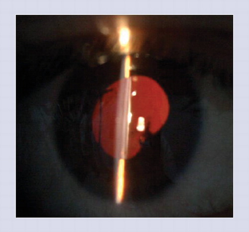
Depigmentation of the choroid resulting from loss of melanocytes produces the typical orange-red ‘sunset glow’ appearance of the fundus . It may be suspected when examining the red reflex Citation[52,64]. Associated optic disc pallor is commonly observed . SSGF, occurring 2–3 months after the acute uveitic phase, is useful for retrospective diagnosis of VKH disease. This sign was found to be very specific to this entity in the setting of chronic nontraumatic bilateral uveitis Citation[65]. In addition to SSGF, fundus examination shows nummular chorioretinal depigmented, round, well-limited lesions in the midperiphery Citation[52,53]. These depigmented lesions have been considered to be Dalén–Fuchs nodules, but there is no histopathologic evidence to support such a consideration. Other fundus lesions may occur including focal or linear RPE migration/clumping Citation[52,53] or subretinal fibrosis . Pseudotumoral RPE proliferation has also been reported in one case of VKH disease at the chronic convalescent phase Citation[76].
The limbus can also become depigmented (Suguira sign). This perilimbal vitiligo may occur 1 month following the acute phase of the disease Citation[77]. It is found more frequently in the Japanese population (85%) than in other populations Citation[8,52,53]. Moreover, peripheral iris depigmentation may also be observed in patients with VKH disease during the chronic convalescent phase Citation[78].
Integumentary manifestations
Poliosis of the eyebrows or eyelashes, alopecia and vitiligo occur during the chronic convalescent phase as a characteristic, but not specific, finding of VKH disease Citation[52]. The vitiligo often has a symmetric distribution over the head, eyelids, trunk and especially over the sacrum. Cutaneous manifestations are more frequent in Japanese than in the Hispanic populations Citation[52]. On the other hand, early initiation of corticosteroids in the acute uveitic phase might prevent or delay the onset of integumentary signs Citation[8,15,53]. Besides, reversal of poliosis, vitiligo and alopecia in patients with VKH has been reported; it has been suggested that this could reflect the restoration of the normal immune homeostasis Citation[79].
Chronic recurrent phase
This phase may interrupt the convalescent phase with anterior uveitis, which can be recurrent, chronic or both. In fact, some patients do not develop inflammation recurrences Citation[80], whereas other patients present with chronic intraocular inflammation relapsing after several weeks to many months Citation[37,56,57]. The rate of ocular complications has been reported to occur proportionally to the number of recurrences of inflammation and to the duration of the disease Citation[9,52,81]. Delay in initiation of treatment with corticosteroids in the acute stage of the disease leads to recurrence Citation[52,53]. In addition, most vision-threatening complications occur in the chronic recurrent phase of VKH disease Citation[52,53,80].
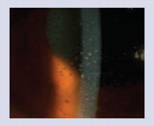
The chronic recurrent phase is characterized by typically acute episodes of anterior uveitis that are often resistant to systemic steroid therapy Citation[52,58]. Recurrent granulomatous anterior uveitis, with cells and flare in the anterior chamber and mutton-fat keratic precipitates on the corneal endothelium, is the typical feature of this recurrent chronic phase . Iris nodules, developing on a background of atrophic iris stroma, can also be seen in the chronic recurrent phase of the disease Citation[52,82]. Focal pigment atrophy of the iris may also take place and gonioscopy may reveal pigment pearls in the angle Citation[52]. Bilateral peripheral transilluminating iris depigmentation and atrophy with sometimes severe ocular hypotony may occur Citation[78]. Recurrent posterior uveitis with vitritis, ERD and optic disc swelling would be distinctly uncommon during this phase Citation[52,53,58]. However, choroidal inflammation relapses may occur, particularly during the first 6 months, usually caused by rapid corticosteroid tapering Citation[83,84]. Furthermore, recurrence of choroidal involvement with a concomitant recurrent anterior uveitis may be subclinical and revealed only by ICGA Citation[85].
Ancillary tests
Fluorescein angiography
FA in the acute phase characteristically reveals patchy choroidal filling due to delayed choroidal perfusion in early frames and numerous punctate hyperfluorescent dots at the level of RPE. These dots gradually enlarge and stain surrounding subretinal and sub-RPE fluid, and the dye clearly outlines the full extent of neurosensory detachments and stains it in a fairly homogenous fashion . Optic disc hyperfluorescence and leakage are usually seen with late pooling of fluorescein and late choroidal hyperfluorescence Citation[52,86–88].
Choroidal folds, radiating from the optic disc to the periphery (some even passing through the macula horizontally or vertically) and similar to the large retinal vessels in number and shape, show alternate hyperfluorescent and hypofluorescent bands on FA Citation[70].
When the exudative lesions of VKH disease have resolved, FA generally demonstrates areas of window defects and blockage by areas of damaged RPE. In some eyes, choroidal neovascularization and subretinal fibrosis can occur as late complications.
Indocyanine green angiography
ICGA findings in the acute phase includes: patchy filling delay of choriocapillaries in the very early angiographic phase and filling delay of the large choroidal arteries, perivascular leakage of individual vessels in the early phase, fewer choroidal vessels in the posterior as well as peripheral fundus. Other findings include diffuse leaking fuzzy vessels in the intermediate phase, diffuse choroidal hyperfluorescence in the late phase and hypofluorescent dark dots that appear at the intermediate phase and either become isofluorescent or remain hypofluorescent in the late phase of the angiogram. These dark dots probably represent partial or full-thickness granuloma Citation[89–92]. Other findings may occur, including reduction in the number of large choroidal vessels, disc hyperfluorescence indicating severe papillitis, hyperfluorescent pinpoints in the area of ERD and choroidal folds showing hypofluorescence or normal fluorescence at the early stage and turning hyperfluorescent at the late stage Citation[70]. The quantitative evaluation of choroidal dye filling velocity in patients with VKH disease showed that the acute phase choroidal model’s time-constant values in VKH disease were significantly longer, when compared with controls, suggesting choroidal circulation disturbance. Furthermore, the choroidal model’s time-constant values shortened significantly during the recovery phase, suggesting improvement of choroidal circulation Citation[93].
In the chronic phase, hypofluorescent lesions corresponding to chorioretinal atrophy, as well as signs similar to those observed at the early phase but less severe, may be observed Citation[71,88].
ICGA helps to determine disease activity in the chronic or convalescent stages of VKH disease when signs of active choroidal inflammation begin to show, including dark spots, choroidal vessel fuzziness and diffuse late hyperfluorescence. These signs show significant decrease after systemic corticosteroid therapy, confirming their inflammatory character Citation[29,66,94,95]. Thus, the ICGA is the test of choice for monitoring by watching for early relapses related to a too-fast tapering of corticosteroids Citation[71].
Optical coherence tomography
In the acute phase, OCT reveals ERD with subretinal septa that divide the subretinal space into several compartments . These septa are composed of an inflammatory product such as fibrin that develops on the RPE and then disappears completely after steroid therapy. The presence of dot reflex in the subretinal space argues in favor of the presence of a subretinal fluid rich in proteins. The fibrin membrane is pushed upward by further leakage from the RPE, leading to its partial detachment from the RPE and, as a consequence, development of septa. Subretinal septa caused multilobular dye pooling in ERD associated with acute VKH disease Citation[87,96–100].
OCT also shows multifocal folds of RPE and the Bruch’s membrane (BM) with undulations and bumps on the RPE surface related to choroidal folds , however, the number of folds was larger than on FA and ICGA Citation[70,101]. Gupta et al., using spectral-domain OCT, suggested that these folds are not true and only represent the undulations of the RPE that are seen clinically as choroidal striations Citation[101]. Spectral domain OCT and high-penetration OCT showed markedly thickened choroid, possibly related not only to inflammatory infiltration but also to increased exudation that decreased quickly with corticosteroid treatment Citation[101,102]. Choroidal thickness decreased over time after treatment and increased markedly again in eyes with recurrent disease Citation[102]. Spectral-domain OCT also shows a loss of focal hyper-reflectivity in the inner choroid in both acute and convalescent stages Citation[103]. When comparing VKH patients in acute and convalescent stages with control subjects, enhanced depth imaging spectral-domain OCT shows significant loss of focal hyper-reflectivity in the inner choroid in patients with VKH disease Citation[104].
In the chronic phase, spectral-domain OCT shows thickening of RPE/BM layer corresponding to pigmented scars or areas of pigmentation, thinning of RPE/BM layer ± loss of outer retinal layers corresponding to atrophic scars, mild irregularities on top of RPE/BM layer or involvement of the outer retina, as well as marked atrophy of neural retina, RPE and choroid Citation[105].
In summary, OCT is useful in evaluating acute VKH disease and monitoring response to treatment. It may also be useful in the evaluation of chronic VKH disease.
Ultrasonography
B-scan ultrasonography is a noninvasive, useful diagnostic tool and can be of a great help in the diagnosis of acute VKH disease, especially when visualization of the fundus is poor or when the clinical presentation is atypical Citation[106].
Conventional ultrasonography (10-MHz) in patients with acute VKH, shows low to medium reflective diffuse choroidal thickening, which is mostly marked in the peripapillary area and extending to the equatorial region , confirming the pathology findings in this entity Citation[106]. Measurement of choroidal thickening is an important parameter for the diagnosis of acute VKH disease , especially in patients with ‘pre-exudative’ stage. Moreover, ultrasonography confirms the presence of ERD and may reveal a prominent optic disc, related to optic disc swelling and fine vitreous opacities Citation[8,106]. High-resolution ultrasonography (20-MHz) is helpful in detecting subclinical ERD, allows a more reproducible and accurate choroidal thickening measurement and better defined episcleral space than with the 10-MHz probe Citation[8]. Furthermore, ultrasonography may be useful in monitoring response to treatment Citation[8].
Other
Ultrasound biomicroscopy
Biomicroscopic ultrasound is a useful tool in the evaluation of VKH disease activity in the anterior chamber and in monitoring its clinical course. Cross-sectional imaging at the active phase may show a shallow anterior chamber, ciliochoroidal detachment, a thickened and swollen ciliary body and low internal reflectivity of the ciliary stroma with ciliary processes being unclear, suprachoroidal effusion, circumferential supraciliary fluid, anterior rotation and anterior bowing of the iris Citation[72,107,108]. These symptoms disappear after corticosteroid therapy. In the remission phase, the internal reflection becomes homogenous and the ciliary process becomes clearly delineated Citation[74].
Laser flare meter
Data about changes of aqueous flare and cells in eyes with VKH disease are scarce. Only one study shows that the breakdown of the blood–aqueous barrier associated with recurrent VKH disease was more severe and longstanding than that observed in acute VKH disease Citation[109].
Autofluorescence
In acute VKH disease, fundus autofluorescence (FAF) shows hyperautofluorescence in the macula coupled with hypoautofluorescence in the areas of ERD Citation[110,111]. The abnormal FAF returns to normal at 6 months after corticosteroid treatment.
In patients with chronic VKH disease, FAF shows different patterns, such as decreased autofluorescence signal corresponding with peripapillary atrophy, multiple nummular and/or irregular atrophic and pigmented scars, increased autofluorescence signal corresponding to patches, strands or irregular areas of pigmentation, cystoid macular edema and normal retina and autofluorescence signal similar to background of normal areas corresponding to SSGF, disciform scar and sectoral chorioretinal atrophy Citation[105].
Electroretinography
Full-field electroretinography analysis demonstrated diffusely diminished amplitudes in both scotopic and photopic phases in patients in the chronic phase. The alterations of full-field electroretinography may explain why patients with good VA have progressive loss of visual field Citation[112].
Multifocal electroretinography (mfERG) in patients with VKH disease shows a severe decrease in VA and diffuse mfERG abnormalities before treatment, reflecting severe damage of macular function that may result from RPE changes after ERD. 12 months after treatment, mfERG shows delayed and limited recovery of macular function. mfERG may be a useful tool for therapy monitoring in patients with VKH disease Citation[113].
Lumbar puncture
Lumbar puncture is a helpful additional test in cases of atypical VKH disease. This test is rarely used in clinical practice when history, clinical and angiographic findings argue in favor of a diagnosis Citation[114].
Lumbar puncture shows cerebrospinal fluid (CSF) pleocytosis (lymphocytosis) in 80% of patients within 1 week and in 97% of patients within 3 weeks of the onset of inflammation. The CSF pleocytosis is transient and resolves within 8 weeks, even in patients who develop recurrences of intraocular inflammation Citation[52].
A recent study showed that profiles of surface markers (CD3+, CD4+ and CD4+ CD45RO+) were identical for CSF and aqueous humor. However, these markers appear to be different from those for peripheral blood cells. These findings suggest that CSF may reproduce the active local immunological reactions in sites affected by VKH disease (mainly uvea and meninges) Citation[115].
The frequency of CSF pleocytosis and the number of cells in the CSF may influence the development of SSGF in VKH disease Citation[64].
MRI
MRI is rarely performed. It shows choroidal thickening to be highly raised after gadolinium injection Citation[52,116,117]. However, MRI is useful in cases of neurologic involvement by showing hypersignal lesions, particularly in the periventricular area of the cerebellum Citation[118].
Audiogram
A deafness of perception in the acute frequencies (low and average in the initial stages) more or less symmetrical with an average loss of 30 dB is the main audiogram finding in the acute phase of disease. This is generally reversible within 2–3 months Citation[55]. This dysacusis is often not perceived by the patient, so an audiogram is essential in case of any suspicion of VKH disease Citation[52]. Deafness may be reversed following early corticosteroid therapy Citation[119].
Diagnostic criteria
The diagnosis of VKH disease is primarily based on clinical features. Several criteria have been proposed to clarify the diagnostic approach, including the American Uveitis Society (AUS) criteria Citation[9] and the Sugiura’s Criteria Citation[120]. Owing to the close similarities in clinical findings between VKH disease and sympathetic ophthalmia (SO), exclusion of a history of penetrating ocular trauma or ocular surgery preceding the onset of uveitis is the constant criteria Citation[121].
The AUS criteria fail to clearly separate the acute phase from the chronic phase and seem to be inadequate in establishing diagnosis in the initial phase of the disease Citation[9]. Chronology of clinical manifestations is not considered. FA and ultrasonographic data, which are substantial criteria for diagnosis, are not taken into account by the AUS criteria. Sugiura’s criteria are seldom used outside Japan because, according to Japanese literature, CSF analysis is mandatory Citation[120]. In 2001, the International Nomenclature Committee proposed the Revised Diagnostic Criteria Citation[122], which classifies the disease into three categories: complete, incomplete and probable VKH (Box 1). The VKH Committee’s new criteria allow the use of both FA and ultrasonography for the detection of early manifestations of the disease in equivocal cases. These new revised diagnostic criteria make up the deficiencies of AUS criteria. However, classification of the disease into three categories (complete, incomplete and probable) might have a low clinical relevance and may well lead to confusion, as the chronologic type of clinical manifestations is not visibly considered. In fact, complete categorisation of VKH disease reveals a delayed diagnosis and not a definite diagnosis. Furthermore, ICGA data Citation[87,94] and those of OCT Citation[87,96] were not considered by these revised criteria Citation[122].
A suggested practical diagnostic approach to VKH disease, taking into account the chronological phase of the disease may be helpful (Box 2). Early diagnosis of acute, purely ocular VKH disease is crucial for prompt initiation of corticosteroid treatment to ensure a good visual outcome and to prevent chronic/recurrent inflammation and complications. It can be easily carried out based on findings from ocular clinical examination and ancillary tests.
ERD occurring in patients with no history of ocular trauma and presenting with intraocular inflammation is highly specific to acute VKH Citation[65,66]. On the other hand, ancillary tests including FA, ICGA, ultrasonography and OCT should be systematically used in patients with acute VKH disease in order to confirm the diagnosis and to evaluate the severity of retinal, choroidal and optic disc involvement (Box 2). These tests are especially helpful in the ‘pre-exudative’ stage and in atypical presentations. Although bilateral ocular involvement is necessary for diagnosis, the uncommon possibility of unilateral manifestations is recognized Citation[60,61]. Therefore, ICGA is recommended when investigating patients with apparent unilateral VKH disease who may have subclinical involvement in the opposite eye Citation[94]. Moreover, the occurrence of neurological and auditory signs preceding uveitis support the diagnosis of acute VKH disease. However, these signs may be missing – but the definite diagnosis can be established in most cases on the basis of characteristics of ocular involvement without the use of the invasive lumbar puncture.
The term chronic VKH may encompass different clinical situations: persistent ‘acute/subacute’ ocular inflammation (nontreated or undertreated patients), resolved acute ocular inflammation (convalescent phase), chronic/recurrent inflammation and chronic complicated VKH disease. SSGF is a highly specific sign of the chronic/recurrent phase Citation[65]. Clinical history suggesting the occurrence of early manifestations and the presence of integumentary signs occurring later are valuable in the diagnosis of this phase. However, diagnosis of chronic VKH disease should be made even in the absence of dermatological signs Citation[63,65].
Differential diagnosis
The differential diagnosis of VKH disease includes several diseases causing posterior uveitis and panuveitis Citation[52]. SO is the first entity to be excluded. A history of previous penetrating ocular trauma or surgery is the rule in this disorder Citation[53,80]. SO is characterized by bilateral panuveitis with ERD and optic disc swelling, similar to that of VKH disease. The chronic phase of this disorder has a clinical and pathologic picture similar to VKH disease. Nevertheless, anterior granulomatous inflammation is more common in SO, while chorioretinal lesions and cutaneous manifestations are rather more scarce Citation[53,80]. Ocular ultrasonography shows a slightly more reflective diffuse choroidal thickening than in VKH disease.
Posterior scleritis (PS) is a more important differential diagnosis of VKH disease, given the clinical similarities of these two entities Citation[123]. PS affects mostly women and is often unilateral. However, bilateral involvement is possible, especially in those patients with systemic rheumatologic disorders. Severe eye pain with photophobia and associated anterior scleritis with redness support the diagnosis of PS. Fundus signs include a circumscribed mass, ERD, retinal and/or choroidal folds, optic disc swelling and annular choroidal detachment Citation[124]. FA findings of PS can be very similar to those observed in acute VKH disease. Ultrasonography is very useful for the diagnosis of PS, showing a reflective thickening of sclera, retrobulbar edema and T sign Citation[106]. In VKH disease, there is often a secondary sclera thickness by contiguous inflammation. In the same way, the choroid may be thickened by adjacent sclera inflammation in cases of PS. However, and unlike VKH disease, choroidal thickening in PS may be localized or diffuse, and is highly reflective on ultrasonography.
Idiopathic uveal effusion syndrome, a rare disease, may manifest with clinical and angiographic signs that may mimic VKH disease. FA shows numerous fluorescent spots in the subretinal space. Mottled fluorescence resulting from subsequent pigment migration in the chronic phase of the uveal effusion syndrome may appear similar to findings in VKH disease. However, the onset of ERD in the uveal effusion syndrome is likely to be subacute and chronically progressive Citation[125]. Besides, the disease involves both eyes, although not simultaneously with minimal intraocular inflammation. Also, the trend to follow a relapsing–remitting course helps differentiate it from VKH disease Citation[125]. Acute posterior multifocal placoid pigment epitheliopathy (APMPPE) may also be confused with VKH disease Citation[52]. Patients with APMPPE develop sudden loss of central vision after viral prodrome. Multiple white-yellowish placoid lesions at the level of the RPE, mainly localized at posterior pole, are seen. Vitreous inflammation is minimal or absent and both eyes are eventually involved. On FA, lesions show early hypofluorescence and late staining. Similar FA findings may be seen in VKH disease in the early stages. However, punctuate hyperfluorescent dots and staining of subretinal fluid, common in VKH disease, are lacking in APMPPE. Furthermore, APMPPE may manifest with ERD and choroidal thickening on OCT and on ultrasonography. However, OCT demonstrates outer retinal hyper-reflectivity and subretinal fluid in the acute phase that disappears a few days later. RPE lesions are hypoautofluorescent acutely but become hyperautofluorescent later in the disease course Citation[126]. Recently, spectral domain OCT has shown that morphologic retinal findings in APMPPE occur in the outer retina; mainly in the photoreceptors and RPE Citation[127].
Primary intraocular B-cell lymphoma presents as chronic uveitis associated with neurological findings and may also mimic VKH disease. Fundus examination shows typically multifocal yellow-white to orange-cream colored sub-RPE infiltrates involving the posterior pole associated with vitritis Citation[128,129]. However, and contrasting with VKH disease, vision is more likely to be preserved. The choroid is usually thickened and may be associated with ERD. FA shows blockage of choroidal fluorescence with late staining at the site of infiltrative lesions Citation[128]. However, in VKH disease FA shows punctate hyperfluorescent dots with increased staining surrounding subretinal fluid and choroid. Patients with primary intraocular large B-cell lymphoma tend to be older than patients with VKH disease Citation[128] and tend to have relatively good vision. The CNS may be involved in over 50% of cases of B-cell lymphoma. Lumbar puncture and MRI are helpful and the diagnosis is usually confirmed by vitreous and/or chorioretinal biopsy Citation[129].
Sarcoidosis must also be considered in the differential diagnosis of VKH disease, since granulomatous anterior inflammation is the most clinical feature of this disease Citation[130]. ERD are unusual in sarcoidosis. By contrast, patients with VKH disease often present with a more striking posterior uveitis in association with ERD. Also, the classic findings of sarcoid retinal vasculitis with venous sheathing and ‘candlewax drippings’ are not seen in VKH disease. Angiotensin converting enzyme and lysozyme, biopsy of suspected granulomata and pulmonary evaluation are crucial in the diagnosis of sarcoidosis Citation[130].
Cat scratch disease may manifest with clinical and angiographic features suggestive of VKH disease. Small focal areas of choroiditis associated with ERD and diffuse choroidal thickening have been described Citation[131,132]. However, macular star, one of the classic features of cat scratch disease, is absent in VKH disease. In addition, unilateral involvement and serology are helpful to differentiate this infectious disease from acute VKH.
Differential diagnosis of VKH disease includes other ocular disorders, such as acute choroidal ischemia, idiopathic central serous chorioretinopathy, syphilis, tuberculosis, Lyme disease, acute leukemia, choroidal metastasis, multiple evanescent white dot syndrome, uveal melanocytic proliferation associated with systemic carcinoma and benign reactive lymphoid hyperplasia of the uveal tract Citation[52,53,58,80,83].
Treatment
The standard treatment of VKH disease is based on prompt, aggressive, systemic corticosteroids for at least 6 months. In this setting, the inflammation is typically very responsive to corticosteroids in the acute phase. Recurrences become increasingly steroid resistant and immunosuppressive agents are needed Citation[52,133]. The associated anterior uveitis should also be managed with corticosteroid drops, as well as cycloplegics to reduce cilliary spasm and mydriatics to prevent posterior synechiae Citation[52].
Prompt high-dose corticosteroid therapy is the mainstay of treatment of acute VKH, administered either orally (1–1.5 mg/kg per day) or through a short course of intravenous delivery, followed by oral corticosteroids Citation[52,134]. The initial high dose is maintained for 2–4 weeks, and then followed by a slow tapering of the drug. Medium-dose systemic corticosteroid therapy is not sufficient to control ocular inflammation, as shown by the persistence of hypofluorescent dark dots revealed by ICGA after 4 months. Furthermore, patients receiving medium-dose corticosteroids are more likely to develop SSGF. Doses as high as 0.75 mg/kg per day are necessary during the first 4 months of treatment Citation[135]. Moreover, peripapillary atrophy in patients with VKH disease is more common and larger when the corticosteroids are given late and in low doses Citation[136]. Initial oral administration or intravenous corticosteroids followed by oral administration seem to have the same visual outcome Citation[137]. However, the rapidity of the resolution of ERD and inflammation may be greater with the pulses.
Treatment with systemic corticosteroids should be gradually tapered and maintained for at least 6 months in order to prevent future recurrences and to optimize visual outcome Citation[52,138]. Patients who received oral corticosteroid treatment for less than 6 months were significantly more likely to have recurrences (58.8%) as compared with corticosteroid treatment with a duration of 6 months or more (11.1%) Citation[138]. Those patients treated with short-duration corticosteroids are also more likely to have one eye with VA of 20/200 or worse Citation[138].
In addition, intravitreal triamcinolone acetonide injections may be an adjuvant tool in the treatment of VKH disease, especially in patients who are unable to tolerate systemic medications Citation[139–141]. Besides, subtenon injections may be considered in association with systemic corticosteroids and immunosuppressive agents to better control the inflammation Citation[142]. Fluocinolone acetonide intravitreal implants have also been used in the management of VKH disease, but they have been mixed with an inability to fully taper off of systemic corticosteroids Citation[143].
Several conventional immunosuppressive agents including cyclosporine, methotrexate, azathioprin, mycophenolate mofetil and (rarely) cyclophosphamide, have been used in the management of VKH disease. Among these immunosuppressive drugs, cyclosporine has been the most widely used in the care of these patients Citation[52].
Conventional immunomosuppressive therapy has been used, especially in cases of resistance to high-dose corticosteroids, intolerance or major side effects of steroids, as well as the chronic recurrent phase of the disease Citation[133].
However, some authors defend the use of the immunosuppressive treatment as a first-line therapy for VKH disease; to reduce the duration of corticosteroid therapy, to better control the disease and to reduce recurrences and complications. First-line immunosuppressive therapy seems to be associated with a better visual outcome when compared with steroids as monotherapy or with delayed addition of immunosuppressive agents Citation[144]. Prospective studies are warranted to validate the role of first-line immunosuppressive therapy in acute VKH disease Citation[133]. However, immunosuppressive treatment may be considered as a first-line treatment in cases of severe disease at presentation, and delay in diagnosis and initiation of corticosteroid treatment Citation[15].
Both azathioprine (1–2.5 mg/kg/day) and cyclosporine A (3–5 mg/kg/day) have proven clinical efficacy Citation[145], but cyclosporine A seems to be a better glucocorticoid-sparing agent than azathioprine Citation[146].
A combination of oral prednisolone, azathioprine and cyclosporine has been administered in recalcitrant cases of VKH disease with rapid remission. The synergistic effect of this triple-agent immunosuppression may help prevent recurrences Citation[147].
Furthermore, infusions of infliximab have recently been used in several cases of refractory VKH disease with good results Citation[148,149]. Even if the use of IFN-α-2a in refractory cases of VKH have been reported with some success, the description of several cases of VKH disease developed in patients treated with interferon and ribavirin therapy for hepatitis C make it a last option therapy Citation[150,151].
Rituximab, an antibody targeting CD20, has been reported in one case of refractory VKH disease with a good outcome Citation[152]. Another therapeutic tool, a single intravitreal injection of bevacizumab injection associated with systemic corticosteroids, has been used in one case to accelerate the resolution of a persistent ERD in a patient with VKH disease Citation[153].
High-dose steroids are also the mainstay treatment for acute VKH in children Citation[12,154–156]. Several immunosuppressive treatments may be administered, usually in cases of inadequate response to steroids, of steroid-related side-effects or for the chronic recurrent phase of the disease. Cyclosporine, azathioprine, methotrexate and sometimes cyclophosphamide have been used Citation[154,155]. However, methotrexate is preferred by some in cases of pediatric VKH disease and seems to be effective in this setting with minimal side-effects Citation[154,156]. Infliximab may help in the management of refractory cases in children Citation[157]. Long-term corticosteroids, as well as immunosuppressive treatments, should be monitored closely in collaboration with a pediatrician.
There are few isolated reports related to VKH disease in pregnancy. High-dose corticosteroids have been used to treat VKH disease successfully during the second and third trimester of pregnancy, usually with no complications in deliveries Citation[158–161]. A case of fetal death was described after initiating corticosteroids in a pregnant patient with VKH disease but the relationship between this event and steroid therapy was not established Citation[162].
Patients being treated for VKH disease are monitored based on clinical examinations as well as ancillary tests, especially OCT and ICGA. OCT may be helpful in the monitoring of the ERD resolution Citation[163].
An ICGA is recommended approximately 4 months after the initiation of treatment. It may reveal subclinical signs of choroidal inflammation, particularly hypofluorescent dark dots or fuzzy choroidal vessels in the absence of any clinical inflammation. These angiographic findings require the reversal of the therapy tapering as well as the extension of its duration Citation[94].
Possible side-effects of corticosteroids and other immunosuppressive agents should be looked for properly during follow-up.
Complications & prognosis
Complications
One half of all eyes with VKH disease develop at least one complication. The most common complications include cataract, glaucoma, choroidal neovascularization and subretinal fibrosis Citation[52,81]. Less common complications have been reported, including cystoid macular edema Citation[164,165], pseudotumoral RPE proliferation Citation[76], band-shaped keratopathy Citation[166] and visual field defects from optic disc involvement Citation[69,167].
Cataracts occur in approximately 40% of eyes with VKH disease. Risk factors for the development of cataracts are long-standing recurrent anterior segment inflammation and systemic corticosteroid therapy for 6 months or more. Surgery should be performed after at least 3 months of no intraocular inflammation. Systemic corticosteroids (0.5–1 mg/kg/day) should be given for 1 Citation[52,81,134,168] or even 2 weeks Citation[169] before surgery and then tapered progressively after surgery. Phacoemulsification has advantages over manual extra-capsular cataract extraction in the setting of uveitis, as it leads to less breakdown of the blood–aqueous barrier. Synechiolysis with or without iris-stretching maneuvers or iris hooks may be required. Polymethylmethacrylate, heparin surface-modified and acrylic foldable intraocular lenses can be safely used in eyes with VKH disease Citation[170]. Recurrent inflammation is a significant risk factor for poor visual outcome in patients with VKH disease after cataract surgery Citation[171].
Chronic glaucoma occurs in 6–45% of eyes Citation[167,172]. The mechanisms of chronic glaucoma in VKH disease include inflammation of the trabecular meshwork, inflammatory cells blocking the trabecular meshwork, peripheral anterior synechiae, pupillary block because of extensive posterior synechiae and corticosteroid-induced ocular hypertension Citation[52,81,173].
Choroidal neovascularization develops in 2.5–14.7% of eyes Citation[9,174]. The choroidal new vessels were found to develop through areas of BM that were damaged by inflammation Citation[81,175]. Eyes with choroidal neovascularization have a history of significantly greater degrees of anterior chamber and vitreous inflammation, greater incidence of fundus pigmentary disturbances and a greater frequency of chronic recurrent phase inflammation. The location of choroidal neovascularization may be peripapillary or macular Citation[52,176]. Recently, intravitreal injections of anti-VEGF have been used in the treatment of choroidal neovascularization in patients with VKH disease and seem to improve the visual outcome Citation[177,178]. Other treatment modalities that have been used include photodynamic therapy with verteporfin Citation[179,180], laser photocoagulation Citation[177] and surgical excision Citation[181]. Subretinal fibrosis has been reported to occur in 7–40% of eyes Citation[81,165,182], leading to poor visual prognosis.
Prognosis
The visual outcome in patients with VKH disease has improved considerably with the use of high-dose corticosteroids and advances in the management of complications. However, more than 50% of patients presenting in the acute phase of the disease still develop the chronic course. Furthermore, 50% of eyes with VKH disease develop at least one complication Citation[15,81].
The proportion of eyes with a VA of 20/40 or better varies from a half to two-thirds Citation[52,81]. The development of ocular complications is significantly associated with a worse final VA Citation[183]. A final VA of 20/200 or worse may be explained by the presence of extensive pigmentary changes and disruption in the fundus secondary to previous inflammation and ERD without any other associated complications Citation[81,167]. Some VKH disease patients may still have concomitant visual field loss and subclinical retinal dysfunction caused by chorioretinal atrophy and pigmentary changes, despite having a final VA of 20/20 Citation[81,141,184,185]. Besides, peripapillary atrophy is associated with visual dysfunction compared with eyes without peripapillary atrophy Citation[184].
Many factors may influence the visual outcome. Patients promptly treated with high doses of steroids in the acute phase (2 weeks or less from onset of symptoms) have a better visual prognosis than patients treated 1 month or more from onset of symptoms Citation[15]. Almost 70% of patients who receive a rapid treatment in the acute phase keep a VA of 20/20, compared with a third of patients treated in the chronic phase after developing cutaneous signs Citation[183].
A longer duration of disease and higher number of recurrent episodes of inflammation are associated with a higher risk of complications and worse visual prognosis. A longer duration of disease and increased number of recurrences expose the eye to the harmful effects of active inflammation as well as treatments, especially corticosteroids Citation[81].
Better VA at presentation is associated with a better final VA Citation[81]. Worse VA at presentation may reflect severe disease at presentation with higher risk of complications. It has been reported that VA at 1 month after starting treatment is a better prognostic factor, as it is a good indicator for response to treatment Citation[141].
Age at onset of the disease has been differently linked to final VA. Poor prognosis has been associated with older age at onset of VKH by some authors Citation[81,141] and with younger age at onset by others Citation[12,67]. Early pinpoint peripapillary hyperfluorescence on pretreatment FA was found to be an indicator for good prognosis Citation[88]. In fact, this sign was more likely to be associated with eyes imaged early in the course of the disease than eyes imaged later Citation[88].
Conclusion
VKH disease is a visually disabling bilateral uveitis affecting patients with no history of ocular trauma which may be associated with extraocular signs depending on the phase of the disease. The diagnosis approach should consider the chronology of clinical phases and clinicians should be aware of ERD and SSGF as highly specific signs of acute and chronic VKH, respectively. FA, OCT, ICGA and ultrasonography are useful adjuncts in the diagnosis of VKH disease and its management. The mainstay for acute VKH disease treatment is prompt high-dose systemic corticosteroids. A duration of 6 months or more of corticosteroid therapy significantly reduces recurrences and complications. Other immunosuppressive agents may be used to control intraocular inflammation and prevent complications. With adequate treatment, one half to two-thirds of affected eyes retain a VA of 20/40 or better. Early diagnosis and prompt proper treatment are the most important factors for a good visual outcome.
Expert commentary
VKH disease is a bilateral granulomatous panuveitis associated with neurological, auditory and integumentary manifestations that are more common in darkly pigmented races of certain genetic predispositions.
A T cell-mediated autoimmune reaction against one or more antigens found on or associated with melanocytes may be involved in the pathogenesis of the disease. Although the associated systemic manifestations are helpful in establishing the definitive diagnosis, they may be present to a varying extent and VKH disease can present with findings limited to intraocular inflammation.
Early recognition of acute VKH disease in its isolated ocular presentation is of utmost importance for prompt initiation of corticosteroid treatment. ERD occurring in patients with no history of ocular trauma and presenting with intraocular inflammation was found to be highly specific to acute VKH. Common features associated with ERD include optic disc swelling and choroidal or retinal folds. FA in acute VKH characteristically reveals patchy choroidal filling in early phase, numerous pinpoint leaks at the level of RPE, with subretinal and sub-RPE fluid dying and optic disc hyperfluorescence and leakage in late phase. ICGA, OCT and ultrasonography may also be very useful in the diagnosis and monitoring of acute VKH disease. Lumbar puncture is unnecessary for diagnosis.
Clinical history suggesting the occurrence of early manifestations and the presence of integumentary signs occurring later are valuable in the diagnosis of chronic VKH disease. SSGF, which was found to be highly specific to chronic VKH disease, allows a definitive diagnosis to be established even in isolated ocular involvement.
Prompt high-dose systemic corticosteroid therapy with gradual tapering over a period of at least 6 months is the mainstay of treatment for acute VKH disease. This is essential to prevent chronicity, recurrences, complications and subsequent visual loss.
Immunosuppressive agents should be given in cases of resistance or intolerance to corticosteroids and in cases of chronic recurrent VKH disease. They may also be recommended as first-line treatments in combination with corticosteroids in cases of severe disease at presentation or following a delay in diagnosis of the acute form.
Five-year view
Further studies on VKH disease in different ethnic populations are required to better clarify similarities and distinct differences in patient’s genetic characteristics and epidemiological profile, clinical manifestations and course of the disease.
New and emerging imaging modalities, including ICGA, high-speed ultrahigh-resolution OCT, autofluorescence and other ancillary tests may allow a more precise evaluation and monitoring of retinal and choroidal involvement in VKH disease.
Multicenter prospective randomized trials are needed to determine the optimal corticosteroid therapy duration and dosage, as well as the role of immunosuppressive drugs as first-line therapy in acute VKH disease and to determine the effects of biologic agents in the treatment of the condition.
Better knowledge of immune mechanisms, as well as the role of specific cytokines, in the pathogenesis of VKH disease may offer new targets for treatment of this disease.
Box 1. Revised criteria for diagnosis of Vogt–Koyanagi–Harada disease.
• No history of penetrating ocular trauma or surgery preceding the initial onset of uveitis
• No clinical or laboratory evidence suggestive of other ocular disease entities
• Bilateral ocular involvement (a or b must be met, depending on the stage of disease when the patient is examined)
– Early manifestations of disease
– Evidence of diffuse choroiditis (with or without anterior uveitis, vitreous inflammatory reaction or optic disc hyperemia) which may manifest as (a) focal areas of subretinal fluid or (b) bullous exudative retinal detachments
– Late manifestations of disease
1. History suggestive of prior presence of early findings noted in 3a and either (2) or (3) below, or multiple signs from (3).
2. Ocular depigmentation: either (a) sunset glow fundus or (b) Sugiura’s sign
3. Other ocular signs including (a) nummular chorioretinal depigmented scars, or (b) retinal pigment epithelium clumping and/or migration or (c) recurrent or chronic anterior uveitis
• Neurological/auditory findings (may resolve by time of evaluation)
– Meningismus (malaise, fever, headache, nausea, abdominal pain, stiffness of the neck and back, or a combination of these factors); note that headache alone is not sufficient to meet the definition of meningismus
– Tinnitus
– Cerebrospinal fluid pleocytosis
• Integumentary finding (not preceding onset of central nervous system or ocular disease)
• Alopecia, or
• Poliosis, or
• Vitiligo
Complete VKH: criteria 1–5 must be present.
Incomplete VKH: criteria 1–3 and either 4 or 5 must be present.
Probable VKH (isolated ocular disease): criteria 1–3 must be present.
VKH: Vogt–Koyanagi–Harada.
Data taken from Citation[122].
Box 2. Practical diagnostic approach to Vogt–Koyanagi–Harada disease.
• Acute VKH (duration of ocular inflammation ≤3 months)
– Factors suggestive of acute VKH disease
– Patient characteristics (darkly pigmented, age between 30 and 40 years, female)
– No history of penetrating ocular trauma
– A history of prodromal illness (neurological/auditory disturbances); inconstant
– Bilateral ocular involvement (symmetric or asymmetric); posterior uveitis or panuveitis + ERD + other fundus findings†
– Early acute phase (<15 days)
– No or rare cells in anterior chamber
– Mild or moderate vitritis
– ERD
– Subacute phase (15 days–3 months)
– Anterior uveitis (non-grabulomatous or granulomatous)
– Moderate or severe vitritis
– ERD
• Chronic/Recurrent VKH (duration of ocular inflammation >3 months)
– A history suggestive of prior presence of early ocular findings (may be lacking)
– Sunset glow ‘pupil’
– Sunset glow fundus, other fundus changes‡
– Granulomatous anterior uveitis
†Other fundus findings during acute phase: retinal/choroidal folds, yellow deep chorioretinal lesions, optic disc edema or hyperemia FA, OCT, ICGA and ultrasonographic findings.
‡Other fundus findings during chronic/recurrent phase: round atrophic lesions, subretinal fibrosis, pigmentary changes, peripapillary RPE proliferation, peripheral iris depigmentation ICGA findings.
ERD: Exudative retinal detachment; FA: Fluorescein angiography; ICGA: Indocyanine green angiography; OCT: Optical coherence tomography; RPE: Retinal pigment epithelium; VKH: Vogt–Koyanagi–Harada.
Key issues
• Vogt–Koyanagi–Harada (VKH) disease a bilateral granulomatous panuveitis with no history of previous ocular trauma that may be associated with CNS, auditory and integumentary manifestations. It has a worldwide distribution, but usually affects young (20–50 years), dark-skinned individuals with a genetic predisposition.
• The most typical feature of acute VKH disease is diffuse granulomatous choroiditis manifesting with exudative retinal detachment, vitritis and optic disc swelling. Fluorescein angiography and other ancillary tests including indocyanine green angiography, optical coherence tomography and ultrasonography are helpful in confirming the diagnosis, evaluating the severity of posterior segment involvement and monitoring response to treatment.
• Chronic VKH disease is typically characterized by episodes of granulomatous anterior uveitis associated with a typical sunset glow fundus that are often resistant to systemic steroid therapy.
• Early diagnosis of acute, purely ocular VKH disease is crucial for prompt initiation of corticosteroid treatment to prevent chronic/recurrent inflammation and complications and to ensure a good visual outcome.
• The mainstay of the treatment for acute VKH disease is prompt systemic high-dose corticosteroid therapy, which should be tapered gradually and maintained for 6 months or more.
• Immunomosuppressive therapy is required in case of resistance to high-dose corticosteroids, intolerance or major side effects of steroids, as well as in the chronic recurrent phase of the disease. Immunosuppressants may also be considered as first-line therapy in association with corticosteroids in patients with severe disease or delayed diagnosis at presentation.
• A higher number of recurrent episodes of inflammation increase the risk for complications, which mainly include cataract, glaucoma, choroidal neovascular membranes and subretinal fibrosis.
• Poor visual prognosis is related to delay of diagnosis, short duration of systemic corticosteroids, number of recurrences of ocular inflammation and occurrence of complications.
• The early initiation of systemic corticosteroids, 2 weeks or less from the onset of symptoms, is associated with a better visual outcome.
References
- Read RW, Rao NA, Cunningham ET. Vogt–Koyanagi–Harada disease. Curr. Opin. Ophthalmol. 11(6), 437–442 (2000).
- Fang W, Yang P. Vogt–Koyanagi–Harada syndrome. Curr. Eye Res. 33(7), 517–523 (2008).
- Vogt A. Premature graying of the cilia, and comments on the so-called sudden occurrence of this change. Klin. Monatsbl. Augenheilkd. 4, 228–242 (1906).
- Harada E. On acute diffuse choroiditis. Acta. Soc. Ophthalmol. Jpn. 30, 356–378 (1926).
- Koyanagi Y. Dysacusis, alopecia and poliosis in severe uveitis without traumatic origin. Klin. Monatsbl. Augenheilkd. 82, 194–211 (1929).
- Al-Mezaine HS, Kangave D, Abu El-Asrar AM. Patterns of uveitis in patients admitted to a university hospital in Riyadh, Saudi Arabia. Ocul. Immunol. Inflamm. 18(6), 424–431 (2010).
- Kitamei H, Kitaichi N, Namba K et al. Clinical features of intraocular inflammation in Hokkaido, Japan. Acta Ophthalmol. 87(4), 424–428 (2009).
- Khairallah M, Zaouali S, Messaoud R et al. The spectrum of Vogt–Koyanagi–Harada disease in Tunisia, north Africa. Int. Ophthalmol. 27(2–3), 125–130 (2007).
- Snyder DA, Tessler HH. Vogt–Koyanagi–Harada syndrome. Am. J. Ophthalmol. 90(1), 69–75 (1980).
- Kiyomoto C, Imaizumi M, Kimoto K, Abe H, Nakano S, Nakatsuka K. Vogt–Koyanagi–Harada disease in elderly Japanese patients. Int. Ophthalmol. 27(2–3), 149–153 (2007).
- Yamamoto Y, Fukushima A, Nishino K, Koura Y, Komatsu T, Ueno H. Vogt–Koyanagi–Harada disease with onset in elderly patients aged 68 to 89 years. Jpn. J. Ophthalmol. 51(1), 60–63 (2007).
- Tabbara KF, Chavis PS, Freeman WR. Vogt–Koyanagi–Harada syndrome in children compared to adults. Acta Ophthalmol. Scand. 76(6), 723–726 (1998).
- Tugal-Tutkun I, Ozyazgan Y, Akova YA et al. The spectrum of Vogt–Koyanagi–Harada disease in Turkey: VKH in Turkey. Int. Ophthalmol. 27(2–3), 117–123 (2007).
- Rathinam SR, Vijayalakshmi P, Namperumalsamy P, Nozik RA, Cunningham ET Jr. Vogt–Koyanagi–Harada syndrome in children. Ocul. Immunol. Inflamm. 6(3), 155–161 (1998).
- Chee SP, Jap A, Bacsal K. Spectrum of Vogt–Koyanagi–Harada disease in Singapore. Int. Ophthalmol. 27(2–3), 137–142 (2007).
- Kobayashi H, Kokubo T, Takahashi M et al. Tyrosinase epitope recognized by an HLA-DR-restricted T-cell line from a Vogt–Koyanagi–Harada disease patient. Immunogenetics 47(5), 398–403 (1998).
- Sugita S, Takase H, Taguchi C et al. Ocular infiltrating CD4+ T cells from patients with Vogt–Koyanagi–Harada disease recognize human melanocyte antigens. Invest. Ophthalmol. Vis. Sci. 47(6), 2547–2554 (2006).
- Horie Y, Takemoto Y, Miyazaki A et al. Tyrosinase gene family and Vogt–Koyanagi–Harada disease in Japanese patients. Mol. Vis. 12, 1601–1605 (2006).
- Ishikawa A, Shiono T, Uchida S. Vogt–Koyanagi–Harada disease in identical twins. Retina 14(5), 435–437 (1994).
- Itho S, Kurimoto S, Kouno T. Vogt–Koyanagi–Harada disease in monozygotic twins. Int. Ophthalmol. 16(1), 49–54 (1992).
- Rutzen AR, Ortega-Larrocea G, Schwab IR, Rao NA. Simultaneous onset of Vogt–Koyanagi–Harada syndrome in monozygotic twins. Am. J. Ophthalmol. 119(2), 239–240 (1995).
- Shindo Y, Ohno S, Yamamoto T, Nakamura S, Inoko H. Complete association of the HLA-DRB1*04 and -DQB1*04 alleles with Vogt–Koyanagi–Harada’s disease. Hum. Immunol. 39(3), 169–176 (1994).
- Weisz JM, Holland GN, Roer LN et al. Association between Vogt–Koyanagi–Harada syndrome and HLA-DR1 and -DR4 in Hispanic patients living in southern California. Ophthalmology 102(7), 1012–1015 (1995).
- Kim MH, Seong MC, Kwak NH et al. Association of HLA with Vogt–Koyanagi–Harada syndrome in Koreans. Am. J. Ophthalmol. 129(2), 173–177 (2000).
- Pivetti-Pezzi P, Accorinti M, Colabelli-Gisoldi RA, Pirraglia MP. Vogt–Koyanagi–Harada disease and HLA type in Italian patients. Am. J. Ophthalmol. 122(6), 889–891 (1996).
- Arellanes-García L, Bautista N, Mora P, Ortega-Larrocea G, Burguet A, Gorodezky C. HLA-DR is strongly associated with Vogt–Koyanagi–Harada disease in Mexican mestizo patients. Ocul. Immunol. Inflamm. 6(2), 93–100 (1998).
- Zhao M, Jiang Y, Abrahams IW. Association of HLA antigens with Vogt–Koyanagi–Harada syndrome in a Han Chinese population. Arch. Ophthalmol. 109(3), 368–370 (1991).
- Damico FM, Cunha-Neto E, Goldberg AC et al. T-cell recognition and cytokine profile induced by melanocyte epitopes in patients with HLA-DRB1*0405-positive and -negative Vogt–Koyanagi–Harada uveitis. Invest. Ophthalmol. Vis. Sci. 46(7), 2465–2471 (2005).
- Iqniebi A, Gaafar A, Sheereen A et al. HLA-DRB1 among patients with Vogt–Koyanagi–Harada disease in Saudi Arabia. Mol. Vis. 15, 1876–1880 (2009).
- Levinson RD, Du Z, Luo L et al. KIR and HLA gene combinations in Vogt–Koyanagi–Harada disease. Hum. Immunol. 69(6), 349–353 (2008).
- Sheereen A, Gaafar A, Iqneibi A et al. A study of KIR genes and HLA-C in Vogt–Koyanagi–Harada disease in Saudi Arabia. Mol. Vis. 17, 3523–3528 (2011).
- Du L, Yang P, Hou S et al. Association of the CTLA-4 gene with Vogt–Koyanagi–Harada syndrome. Clin. Immunol. 127(1), 43–48 (2008).
- Mao L, Yang P, Hou S, Li F, Kijlstra A. Label-free proteomics reveals decreased expression of CD18 and AKNA in peripheral CD4+ T cells from patients with Vogt–Koyanagi–Harada syndrome. PLoS One 6(1), e14616 (2011).
- Kitamura M, Kitaichi N, Takeuchi M et al. Decrease in the glyceraldehyde derived advanced glycation end products in the sera of patients with Vogt–Koyanagi–Harada disease. Br. J. Ophthalmol. 89(11), 1407–1409 (2005).
- Yi X, Yang P, Sun M, Yang Y, Li F. Decreased 1,25-dihydroxyvitamin D3 level is involved in the pathogenesis of Vogt–Koyanagi–Harada (VKH) disease. Mol. Vis. 17, 673–679 (2011).
- Chu M, Yang P, Hu R et al. Elevated serum osteopontin levels and genetic polymorphisms of osteopontin are associated with Vogt–Koyanagi–Harada disease. Invest. Ophthalmol. Vis. Sci. 52(10), 7084–7089 (2011).
- Liu L, Yang P, He H et al. Leptin increases in Vogt–Koyanagi–Harada (VKH) disease and promotes cell proliferation and inflammatory cytokine secretion. Br. J. Ophthalmol. 92(4), 557–561 (2008).
- Bassili SS, Peyman GA, Gebhardt BM, Daun M, Ganiban GJ, Rifai A. Detection of Epstein–Barr virus DNA by polymerase chain reaction in the vitreous from a patient with Vogt–Koyanagi–Harada syndrome. Retina 16(2), 160–161 (1996).
- Sugita S, Takase H, Kawaguchi T, Taguchi C, Mochizuki M. Cross-reaction between tyrosinase peptides and cytomegalovirus antigen by T cells from patients with Vogt–Koyanagi–Harada disease. Int. Ophthalmol. 27(2–3), 87–95 (2007).
- Al Hemidan AI, Tabbara KF, Althomali T. Vogt–Koyanagi–Harada associated with diabetes mellitus and celiac disease in a 3-year-old girl. Eur. J. Ophthalmol. 16(1), 173–177 (2006).
- Ojaimi E, Levy J, Stawell R, Van Heerden A, Godfrey T, Zamir E. Vogt–Koyanagi–Harada disease, diabetes mellitus, and psoriasis in a child. Ocul. Immunol. Inflamm. 20(1), 56–58 (2012).
- Rao NA. Mechanisms of inflammatory response in sympathetic ophthalmia and VKH syndrome. Eye (Lond). 11(Pt 2), 213–216 (1997).
- Imai Y, Sugita M, Nakamura S, Toriyama S, Ohno S. Cytokine production and helper T cell subsets in Vogt–Koyanagi–Harada’s disease. Curr. Eye Res. 22(4), 312–318 (2001).
- Chi W, Yang P, Li B et al. IL-23 promotes CD4+ T cells to produce IL-17 in Vogt–Koyanagi–Harada disease. J. Allergy Clin. Immunol. 119(5), 1218–1224 (2007).
- Shu Q, Yang P, Hou S et al. Interleukin-17 gene polymorphism is associated with Vogt–Koyanagi–Harada syndrome but not with Behçet’s disease in a Chinese Han population. Hum. Immunol. 71(10), 988–991 (2010).
- Yang Y, Xiao X, Li F, Du L, Kijlstra A, Yang P. Increased IL-7 expression in Vogt–Koyanagi–Harada disease. Invest. Ophthalmol. Vis. Sci. 53(2), 1012–1017 (2012).
- Li F, Yang P, Liu X, Wang C, Hou S, Kijlstra A. Upregulation of interleukin 21 and promotion of interleukin 17 production in chronic or recurrent Vogt–Koyanagi–Harada disease. Arch. Ophthalmol. 128(11), 1449–1454 (2010).
- Wang C, Tian Y, Lei B et al. Decreased IL-27 expression in association with an increased Th17 response in Vogt–Koyanagi–Harada disease. Invest. Ophthalmol. Vis. Sci. 53(8), 4668–4675 (2012).
- Commodaro AG, Peron JP, Genre J et al. IL-10 and TGF-β immunoregulatory cytokines rather than natural regulatory T cells are associated with the resolution phase of Vogt–Koyanagi–Harada (VKH) syndrome. Scand. J. Immunol. 72(1), 31–37 (2010).
- Inomata H, Rao NA. Depigmented atrophic lesions in sunset glow fundi of Vogt–Koyanagi–Harada disease. Am. J. Ophthalmol. 131(5), 607–614 (2001).
- Rao NA. Pathology of Vogt–Koyanagi–Harada disease. Int. Ophthalmol. 27(2–3), 81–85 (2007).
- Moorthy RS, Inomata H, Rao NA. Vogt–Koyanagi–Harada syndrome. Surv. Ophthalmol. 39(4), 265–292 (1995).
- Rajendram R, Evans M, Rao NA. Vogt–Koyanagi–Harada disease. Int. Ophthalmol. Clin. 45(2), 115–134 (2005).
- Beniz J, Forster DJ, Lean JS, Smith RE, Rao NA. Variations in clinical features of the Vogt–Koyanagi–Harada syndrome. Retina 11(3), 275–280 (1991).
- Sugiura S. Vogt–Koyanagi–Harada disease. Jpn. J. Ophthalmol. 22, 9–35 (1978).
- Oku H, Ishikawa S. Vestibulo-ocular reflex abnormality in Vogt–Koyanagi–Harada syndrome. Br. J. Ophthalmol. 78(12), 912–916 (1994).
- Hiraki Y, Kuwasaki N, Shoji H, Kaji M, Kuboshiro T. Each one case of Vogt–Koyanagi–Harada disease with vestibular and cerebellar ataxia, and multiple cranial nerve palsies. Rinsho. Shinkeigaku 29(1), 54–58 (1989).
- Andreoli CM, Foster CS. Vogt–Koyanagi–Harada disease. Int. Ophthalmol. Clin. 46(2), 111–122 (2006).
- Murty SI, Moreker MR, Sangwan VS et al. The spectrum of Vogt–Koyanagi–Harada disease in South India. Int. Ophthalmol. 27(2–3), 131–136 (2007).
- Forster DJ, Green RL, Rao NA. Unilateral manifestation of the Vogt–Koyanagi–Harada syndrome in a 7-year-old child. Am. J. Ophthalmol. 111(3), 380–382 (1991).
- Usui Y, Goto H, Sakai J, Takeuchi M, Usui M, Rao NA. Presumed Vogt–Koyanagi–Harada disease with unilateral ocular involvement: report of three cases. Graefes Arch. Clin. Exp. Ophthalmol. 247(8), 1127–1132 (2009).
- Damico FM, Bezerra FT, Silva GC, Gasparin F, Yamamoto JH. New insights into Vogt–Koyanagi–Harada disease. Arq. Bras. Oftalmol. 72(3), 413–420 (2009).
- da Silva FT, Damico FM, Marin ML et al. Revised diagnostic criteria for Vogt–Koyanagi–Harada disease: considerations on the different disease categories. Am. J. Ophthalmol. 147(2), 339–345.e5 (2009).
- Keino H, Goto H, Mori H, Iwasaki T, Usui M. Association between severity of inflammation in CNS and development of sunset glow fundus in Vogt–Koyanagi–Harada disease. Am. J. Ophthalmol. 141(6), 1140–1142 (2006).
- Rao NA, Gupta A, Dustin L et al. Frequency of distinguishing clinical features in Vogt–Koyanagi–Harada disease. Ophthalmology 117(3), 591–9, 599.e1 (2010).
- Bordaberry MF. Vogt–Koyanagi–Harada disease: diagnosis and treatments update. Curr. Opin. Ophthalmol. 21(6), 430–435 (2010).
- Ohno S, Minakawa R, Matsuda H. Clinical studies of Vogt–Koyanagi–Harada’s disease. Jpn. J. Ophthalmol. 32(3), 334–343 (1988).
- Rajendram R, Evans M, Khurana RN et al. Vogt–Koyanagi–Harada disease presenting as optic neuritis. Int. Ophthalmol. 27(2–3), 217–220 (2007).
- Nakao K, Abematsu N, Mizushima Y, Sakamoto T. Optic disc swelling in Vogt–Koyanagi–Harada disease. Invest. Ophthalmol. Vis. Sci. 53(4), 1917–1922 (2012).
- Wu W, Wen F, Huang S, Luo G, Wu D. Choroidal folds in Vogt–Koyanagi–Harada disease. Am. J. Ophthalmol. 143(5), 900–901 (2007).
- Touitou V, Escande C, Bodaghi B et al. Diagnostic and therapeutic management of Vogt–Koyanagi–Harada syndrome. J. Fr. Ophtalmol. 28(1), 9–16 (2005).
- Kawano Y, Tawara A, Nishioka Y, Suyama Y, Sakamoto H, Inomata H. Ultrasound biomicroscopic analysis of transient shallow anterior chamber in Vogt–Koyanagi–Harada syndrome. Am. J. Ophthalmol. 121(6), 720–723 (1996).
- Maruyama Y, Kimura Y, Kishi S, Shimizu K. Serous detachment of the ciliary body in Harada disease. Am. J. Ophthalmol. 125(5), 666–672 (1998).
- Wada S, Kohno T, Yanagihara N et al. Ultrasound biomicroscopic study of ciliary body changes in the post-treatment phase of Vogt–Koyanagi–Harada disease. Br. J. Ophthalmol. 86(12), 1374–1379 (2002).
- Rathinam SR, Namperumalsamy P, Nozik RA, Cunningham ET Jr. Angle closure glaucoma as a presenting sign of Vogt–Koyanagi–Harada syndrome. Br. J. Ophthalmol. 81(7), 608–609 (1997).
- Khairallah M, Rao NA, Ben Yahia S, Zaouali S, Attia S. Pseudotumoral retinal pigment epithelium proliferation in a patient with Vogt–Koyanagi–Harada disease. Arch. Ophthalmol. 124(9), 1366–1367 (2006).
- Friedman AH, Deutsch-Sokol RH. Sugiura’s sign. Perilimbal vitiligo in the Vogt–Koyanagi–Harada syndrome. Ophthalmology 88(11), 1159–1165 (1981).
- Attia S, Zaouali S, Bettaieb A et al. Peripheral iris depigmentation and ocular hypotony: result of the natural course of non-treated Vogt–Koyanagi–Harada (VKH) disease. Int. Ophthalmol. 27(2–3), 221–222 (2007).
- Tabbara KF. Reversal of poliosis and vitiligo following Vogt–Koyanagi–Harada disease. Arch. Ophthalmol. 130(3), 394–396 (2012).
- Damico FM, Kiss S, Young LH. Vogt–Koyanagi–Harada disease. Semin. Ophthalmol. 20(3), 183–190 (2005).
- Read RW, Rechodouni A, Butani N et al. Complications and prognostic factors in Vogt–Koyanagi–Harada disease. Am. J. Ophthalmol. 131(5), 599–606 (2001).
- Taylor S, Lightman S. Recurrent anterior uveitis in patients with Vogt–Koyanagi–Harada syndrome. Arch. Ophthalmol. 122(6), 922–923 (2004).
- Sachdev N, Gupta V, Gupta A, Singh R. Posterior segment recurrences in Vogt–Koyanagi–Harada disease. Int. Ophthalmol. 28(5), 339–345 (2008).
- Yang P, Ren Y, Li B, Fang W, Meng Q, Kijlstra A. Clinical characteristics of Vogt–Koyanagi–Harada syndrome in Chinese patients. Ophthalmology 114(3), 606–614 (2007).
- Bacsal K, Wen DS, Chee SP. Concomitant choroidal inflammation during anterior segment recurrence in Vogt–Koyanagi–Harada disease. Am. J. Ophthalmol. 145(3), 480–486 (2008).
- Arellanes-García L, Hernández-Barrios M, Fromow-Guerra J, Cervantes-Fanning P. Fluorescein fundus angiographic findings in Vogt–Koyanagi–Harada syndrome. Int. Ophthalmol. 27(2–3), 155–161 (2007).
- Fardeau C, Tran TH, Gharbi B, Cassoux N, Bodaghi B, LeHoang P. Retinal fluorescein and indocyanine green angiography and optical coherence tomography in successive stages of Vogt–Koyanagi–Harada disease. Int. Ophthalmol. 27(2–3), 163–172 (2007).
- Chee SP, Jap A, Cheung CM. The prognostic value of angiography in Vogt–Koyanagi–Harada disease. Am. J. Ophthalmol. 150(6), 888–893 (2010).
- Bouchenaki N, Herbort CP. The contribution of indocyanine green angiography to the appraisal and management of Vogt–Koyanagi–Harada disease. Ophthalmology 108(1), 54–64 (2001).
- Miyanaga M, Kawaguchi T, Miyata K, Horie S, Mochizuki M, Herbort CP. Indocyanine green angiography findings in initial acute pretreatment Vogt–Koyanagi–Harada disease in Japanese patients. Jpn. J. Ophthalmol. 54(5), 377–382 (2010).
- Kohno T, Miki T, Shiraki K et al. Subtraction ICG angiography in Harada’s disease. Br. J. Ophthalmol. 83(7), 822–833 (1999).
- Okada AA, Mizusawa T, Sakai J, Usui M. Videofunduscopy and videoangiography using the scanning laser ophthalmoscope in Vogt–Koyanagi–Harada syndrome. Br. J. Ophthalmol. 82(10), 1175–1181 (1998).
- Mawatari Y, Hirata A, Fukushima M, Tanihara H. Choroidal dye filling velocity in patients with Vogt–Koyanagi–Harada disease. Graefes Arch. Clin. Exp. Ophthalmol. 244(8), 1056–1059 (2006).
- Herbort CP, Mantovani A, Bouchenaki N. Indocyanine green angiography in Vogt–Koyanagi–Harada disease: angiographic signs and utility in patient follow-up. Int. Ophthalmol. 27(2–3), 173–182 (2007).
- Bouchenaki N, Herbort CP. Indocyanine green angiography guided management of Vogt–Koyanagi–Harada disease. J. Ophthalmic Vis. Res. 6(4), 241–248 (2011).
- Yamaguchi Y, Otani T, Kishi S. Tomographic features of serous retinal detachment with multilobular dye pooling in acute Vogt–Koyanagi–Harada disease. Am. J. Ophthalmol. 144(2), 260–265 (2007).
- Parc C, Guenoun JM, Dhote R, Brézin A. Optical coherence tomography in the acute and chronic phases of Vogt–Koyanagi–Harada disease. Ocul. Immunol. Inflamm. 13(2–3), 225–227 (2005).
- Maruyama Y, Kishi S. Tomographic features of serous retinal detachment in Vogt–Koyanagi–Harada syndrome. Ophthalmic Surg. Lasers Imaging 35(3), 239–242 (2004).
- Ishihara K, Hangai M, Kita M, Yoshimura N. Acute Vogt–Koyanagi–Harada disease in enhanced spectral-domain optical coherence tomography. Ophthalmology 116(9), 1799–1807 (2009).
- Lee JE, Park SW, Lee JK, Choi HY, Oum BS, Kim HW. Edema of the photoreceptor layer in Vogt–Koyanagi–Harada disease observed using high-resolution optical coherence tomography. Korean J. Ophthalmol. 23(2), 74–79 (2009).
- Gupta V, Gupta A, Gupta P, Sharma A. Spectral-domain cirrus optical coherence tomography of choroidal striations seen in the acute stage of Vogt–Koyanagi–Harada disease. Am. J. Ophthalmol. 147(1), 148–153.e2 (2009).
- Maruko I, Iida T, Sugano Y et al. Subfoveal choroidal thickness after treatment of Vogt–Koyanagi–Harada disease. Retina 31(3), 510–517 (2011).
- Yamamoto M, Nishijima K, Nakamura M, Yoshimura N. Inner retinal changes in acute-phase Vogt–Koyanagi–Harada disease measured by enhanced spectral domain optical coherence tomography. Jpn. J. Ophthalmol. 55(1), 1–6 (2011).
- Nakai K, Gomi F, Ikuno Y et al. Choroidal observations in Vogt–Koyanagi–Harada disease using high-penetration optical coherence tomography. Graefes Arch. Clin. Exp. Ophthalmol. 250(7), 1089–1095 (2012).
- Vasconcelos-Santos DV, Sohn EH, Sadda S, Rao NA. Retinal pigment epithelial changes in chronic Vogt–Koyanagi–Harada disease: fundus autofluorescence and spectral domain-optical coherence tomography findings. Retina (Philadelphia, Pa.) 30(1), 33–41 (2010).
- Forster DJ, Cano MR, Green RL, Rao NA. Echographic features of the Vogt–Koyanagi–Harada syndrome. Arch. Ophthalmol. 108(10), 1421–1426 (1990).
- Kishi A, Nao-i N, Sawada A. Ultrasound biomicroscopic findings of acute angle-closure glaucoma in Vogt–Koyanagi–Harada syndrome. Am. J. Ophthalmol. 122(5), 735–737 (1996).
- Gohdo T, Tsukahara S. Ultrasound Biomicroscopy of shallow anterior chamber in Vogt–Koyanagi–Harada syndrome. Am. J. Ophthalmol. 122(1), 112–114 (1996).
- Fang W, Zhou H, Yang P, Huang X, Wang L, Kijlstra A. Longitudinal quantification of aqueous flare and cells in Vogt–Koyanagi–Harada disease. Br. J. Ophthalmol. 92(2), 182–185 (2008).
- Koizumi H, Maruyama K, Kinoshita S. Blue light and near-infrared fundus autofluorescence in acute Vogt–Koyanagi–Harada disease. Br. J. Ophthalmol. 94(11), 1499–1505 (2010).
- Ayata A, Dogru S, Senol MG, Unal M, Ersanli D, Bilge AH. Autofluorescence findings in Vogt–Koyanagi–Harada disease. Eur. J. Ophthalmol. 19(6), 1094–1097 (2009).
- da Silva FT, Hirata CE, Olivalves E, Oyamada MK, Yamamoto JH. Fundus-based and electroretinographic strategies for stratification of late-stage Vogt–Koyanagi–Harada disease patients. Am. J. Ophthalmol. 148(6), 939–45.e3 (2009).
- Peizeng Y, Wang F, Li W et al. Study of macular function by multifocal electroretinography in patients with Vogt–Koyanagi–Harada Syndrome. Am. J. Ophthalmol. 146, 767–771 (2008).
- Tsai JH, Sukavatcharin S, Rao NA. Utility of lumbar puncture in diagnosis of Vogt–Koyanagi–Harada disease. Int. Ophthalmol. 27, 1891–1894 (2007).
- Kitaichi N, Matoba H, Ohno S. The positive role of lumbar puncture in the diagnosis of Vogt–Koyanagi–Harada disease: lymphocyte subsets in the aqueous humor and cerebrospinal fluid. Int. Ophthalmol. 27(2–3), 97–103 (2007).
- Lohman BD, Gustafson CA, McKinney AM, Sarikaya B, Silbert SC. MR imaging of Vogt–Koyanagi–Harada syndrome with leptomeningeal enhancement. AJNR. Am. J. Neuroradiol. 32(9), E169–E171 (2011).
- Han HJ, Kim HY, Park JH, Lee EJ, Kim do G, Shin DI. Magnetic resonance imaging of pachymeningeal enhancement in Vogt–Koyanagi–Harada disease. Neurol. Sci. 31(6), 785–788 (2010).
- Johnston CA, Teitelbaum CS. Magnetic resonance imaging in Vogt–Koyanagi–Harada syndrome. Arch. Ophthalmol. 108(6), 783–784 (1990).
- Al Dousary S. Auditory and vestibular manifestations of Vogt–Koyanagi–Harada disease. J. Laryngol. Otol. 125(2), 138–141 (2011).
- Sugiura S. [Some observations on uveitis in Japan, with special reference to Vogt–Koyanagi–Harada and Behçet diseases]. Nippon Ganka Gakkai Zasshi 80(11), 1285–1326 (1976).
- Rao NA, Marak GE. Sympathetic ophthalmia simulating Vogt–Koyanagi–Harada’s disease: a clinico-pathologic study of four cases. Jpn. J. Ophthalmol. 27(3), 506–511 (1983).
- Read RW, Holland GN, Rao NA et al. Revised diagnostic criteria for Vogt–Koyanagi–Harada disease: report of an international committee on nomenclature. Am. J. Ophthalmol. 131(5), 647–652 (2001).
- Kouda N, Sasaki H, Harada S, Yamada Y, Takahashi N, Sasaki K. Early manifestation of Vogt–Koyanagi–Harada disease as unilateral posterior scleritis. Jpn. J. Ophthalmol. 46(5), 590–593 (2002).
- Benson WE. Posterior scleritis. Surv. Ophthalmol. 132, 297–316 (1988).
- Elagouz M, Stanescu-Segall D, Jackson TL. Uveal effusion syndrome. Surv. Ophthalmol. 55(2), 134–145 (2010).
- Lee GE, Lee BW, Rao NA, Fawzi AA. Spectral domain optical coherence tomography and autofluorescence in a case of acute posterior multifocal placoid pigment epitheliopathy mimicking Vogt–Koyanagi–Harada disease: case report and review of literature. Ocul. Immunol. Inflamm. 19(1), 42–47 (2011).
- Goldenberg D, Habot-Wilner Z, Loewenstein A, Goldstein M. Spectral domain optical coherence tomography classification of acute posterior multifocal placoid pigment epitheliopathy. Retina (Philadelphia, Pa.) 32(7), 1403–1410 (2012).
- Kirmani MH, Thomas EL, Rao NA, Laborde RP. Intraocular reticulum cell sarcoma: diagnosis by choroidal biopsy. Br. J. Ophthalmol. 71(10), 748–752 (1987).
- Saucer TC, Chan C. Masquerade syndrome. In: Uveitis Text and Imaging. Gupta A, Gupta V, Herbort C, Moncef K (Eds.) Jaypee, New Delhi, India, 746–755 (2009).
- Birnbaum AD, Oh FS, Chakrabarti A, Tessler HH, Goldstein DA. Clinical features and diagnostic evaluation of biopsy-proven ocular sarcoidosis. Arch. Ophthalmol. 129(4), 409–413 (2011).
- Ormerod LD, Skolnick KA, Menosky MM, Pavan PR, Pon DM. Retinal and choroidal manifestations of cat-scratch disease. Ophthalmology 105(6), 1024–1031 (1998).
- Khurana RN, Albini T, Green RL, Rao NA, Lim JI. Bartonella henselae infection presenting as a unilateral panuveitis simulating Vogt–Koyanagi–Harada syndrome. Am. J. Ophthalmol. 138(6), 1063–1065 (2004).
- Rao NA. Treatment of Vogt–Koyanagi–Harada disease by corticosteroids and immunosuppressive agents. Ocul. Immunol. Inflamm. 14(2), 71–72 (2006).
- Rubsamen PE, Gass JD. Vogt–Koyanagi–Harada syndrome. Clinical course, therapy, and long-term visual outcome. Arch. Ophthalmol. 109(5), 682–687 (1991).
- Kawaguchi T, Horie S, Bouchenaki N, Ohno-Matsui K, Mochizuki M, Herbort CP. Suboptimal therapy controls clinically apparent disease but not subclinical progression of Vogt–Koyanagi–Harada disease. Int. Ophthalmol. 30(1), 41–50 (2010).
- Jap A, Luu CD, Yeo I, Chee SP. Correlation between peripapillary atrophy and corticosteroid therapy in patients with Vogt–Koyanagi–Harada disease. Eye (Lond). 22(2), 240–245 (2008).
- Read RW, Yu F, Accorinti M et al. Evaluation of the effect on outcomes of the route of administration of corticosteroids in acute Vogt–Koyanagi–Harada disease. Am. J. Ophthalmol. 142(1), 119–124 (2006).
- Lai TY, Chan RP, Chan CK, Lam DS. Effects of the duration of initial oral corticosteroid treatment on the recurrence of inflammation in Vogt–Koyanagi–Harada disease. Eye (Lond). 23(3), 543–548 (2009).
- Andrade RE, Muccioli C, Farah ME, Nussenblatt RB, Belfort R Jr. Intravitreal triamcinolone in the treatment of serous retinal detachment in Vogt–Koyanagi–Harada syndrome. Am. J. Ophthalmol. 137(3), 572–574 (2004).
- Karacorlu M, Arf Karacorlu S, Ozdemir H. Intravitreal triamcinolone acetonide in Vogt–Koyanagi–Harada syndrome. Eur. J. Ophthalmol. 16(3), 481–483 (2006).
- Chee SP, Jap A, Bacsal K. Prognostic factors of Vogt–Koyanagi–Harada disease in Singapore. Am. J. Ophthalmol. 147(1), 154–161.e1 (2009).
- Perente I, Utine CA, Cakir H, Kaya V, Tutkun IT, Yilmaz OF. Management of ocular complications of Vogt–Koyanagi–Harada syndrome. Int. Ophthalmol. 29(1), 33–37 (2009).
- Khalifa Y, Loh AR, Acharya NR. Fluocinolone acetonide intravitreal implants in Vogt–Koyanagi–Harada disease. Ocul. Immunol. Inflamm. 17(6), 431–433 (2009).
- Paredes I, Ahmed M, Foster CS. Immunomodulatory therapy for Vogt–Koyanagi–Harada patients as first-line therapy. Ocul. Immunol. Inflamm. 14(2), 87–90 (2006).
- Kim SJ, Yu HG. The use of low-dose azathioprine in patients with Vogt–Koyanagi–Harada disease. Ocul. Immunol. Inflamm. 15(5), 381–387 (2007).
- Cuchacovich M, Solanes F, Díaz G et al. Comparison of the clinical efficacy of two different immunosuppressive regimens in patients with chronic Vogt–Koyanagi–Harada disease. Ocul. Immunol. Inflamm. 18(3), 200–207 (2010).
- Agarwal M, Ganesh SK, Biswas J. Triple agent immunosuppressive therapy in Vogt–Koyanagi–Harada syndrome. Ocul. Immunol. Inflamm. 14(6), 333–339 (2006).
- Wang Y, Gaudio PA. Infliximab therapy for 2 patients with Vogt–Koyanagi–Harada syndrome. Ocul. Immunol. Inflamm. 16(4), 167–171 (2008).
- Niccoli L, Nannini C, Cassarà E, Gini G, Lenzetti I, Cantini F. Efficacy of infliximab therapy in two patients with refractory Vogt–Koyanagi–Harada disease. Br. J. Ophthalmol. 93(11), 1553–1554 (2009).
- Sylvestre DL, Disston AR, Bui DP. Vogt–Koyanagi–Harada disease associated with interferon β-2b/ribavirin combination therapy. J. Viral Hepat. 10(6), 467–470 (2003).
- Touitou V, Sene D, Fardeau C et al. Interferon-α-2a and Vogt–Koyanagi–Harada disease: a double-edged sword? Int. Ophthalmol. 27(2–3), 211–215 (2007).
- Dolz-Marco R, Gallego-Pinazo R, Díaz-Llopis M. Rituximab in refractory Vogt–Koyanagi–Harada disease. J. Ophthalmic Inflamm. Infect. 1(4), 177–180 (2011).
- Park HS, Nam KY, Kim JY. Intravitreal bevacizumab injection for persistent serous retinal detachment associated with Vogt–Koyanagi–Harada disease. Graefes Arch. Clin. Exp. Ophthalmol. 249(1), 133–136 (2011).
- Soheilian M, Aletaha M, Yazdani S, Dehghan MH, Peyman GA. Management of pediatric Vogt–Koyanagi–Harada (VKH)-associated panuveitis. Ocul. Immunol. Inflamm. 14(2), 91–98 (2006).
- Abu El-Asrar AM, Al-Kharashi AS, Aldibhi H, Al-Fraykh H, Kangave D. Vogt–Koyanagi–Harada disease in children. Eye (Lond). 22(9), 1124–1131 (2008).
- Martin TD, Rathinam SR, Cunningham ET Jr. Prevalence, clinical characteristics, and causes of vision loss in children with Vogt–Koyanagi–Harada disease in south India. Retina (Philadelphia, Pa.) 30(7), 1113–1121 (2010).
- Khalifa YM, Bailony MR, Acharya NR. Treatment of pediatric Vogt–Koyanagi–Harada syndrome with infliximab. Ocul. Immunol. Inflamm. 18(3), 218–222 (2010).
- Friedman Z, Granat M, Neumann E. The syndrome of Vogt–Koyanagi–Harada and pregnancy. Metab. Pediatr. Ophthalmol. 4(3), 147–149 (1980).
- Miyata N, Sugita M, Nakamura S et al. Treatment of Vogt–Koyanagi–Harada’s disease during pregnancy. Jpn. J. Ophthalmol. 45(2), 177–180 (2001).
- Doi M, Matsubara H, Uji Y. Vogt–Koyanagi–Harada syndrome in a pregnant patient treated with high-dose systemic corticosteroids. Acta Ophthalmol. Scand. 78(1), 93–96 (2000).
- Tien MC, Teoh SC. Treatment of Vogt–Koyanagi–Harada syndrome in pregnancy. Can. J. Ophthalmol. 44(2), 211–212 (2009).
- Ohta K, Gotoh N, Yonezawa H, Oka K, Ashida T. Fetal death following high-dose systemic steroid therapy in a pregnant patient with Vogt–Koyanagi–Harada disease. Nippon Ganka Gakkai Zasshi 111(12), 959–964 (2007).
- Yamanaka E, Ohguro N, Yamamoto S, Nakagawa Y, Imoto Y, Tano Y. Evaluation of pulse corticosteroid therapy for Vogt–Koyanagi–Harada disease assessed by optical coherence tomography. Am. J. Ophthalmol. 134(3), 454–456 (2002).
- Rutzen AR, Ortega-Larrocea G, Frambach DA, Rao NA. Macular edema in chronic Vogt–Koyanagi–Harada syndrome. Retina (Philadelphia, Pa.) 15(6), 475–479 (1995).
- Bykhovskaya I, Thorne JE, Kempen JH, Dunn JP, Jabs DA. Vogt–Koyanagi–Harada disease: clinical outcomes. Am. J. Ophthalmol. 140(4), 674–678 (2005).
- Yang P, Sun M. Band-shaped keratopathy in Chinese patients with Vogt–Koyanagi–Harada syndrome. Cornea 30(12), 1336–1340 (2011).
- Nakao K, Mizushima Y, Abematsu N, Goh N, Sakamoto T. Anterior ischemic optic neuropathy associated with Vogt–Koyanagi–Harada disease. Graefes Arch. Clin. Exp. Ophthalmol. 247(10), 1417–1425 (2009).
- Moorthy RS, Rajeev B, Smith RE, Rao NA. Incidence and management of cataracts in Vogt–Koyanagi–Harada syndrome. Am. J. Ophthalmol. 118(2), 197–204 (1994).
- Meacock WR, Spalton DJ, Bender L et al. Steroid prophylaxis in eyes with uveitis undergoing phacoemulsification. Br. J. Ophthalmol. 88(9), 1122–1124 (2004).
- Ganesh SK, Padmaja, Babu K, Biswas J. Cataract surgery in patients with Vogt–Koyanagi–Harada syndrome. J. Cataract Refract. Surg. 30(1), 95–100 (2004).
- Quek DT, Jap A, Chee SP. Risk factors for poor visual outcome following cataract surgery in Vogt–Koyanagi–Harada disease. Br. J. Ophthalmol. 95(11), 1542–1546 (2011).
- Minnakawa R, Ohno S, Hirose S et al. Clinical manifestations of Vogt Koyanagi Harada disease. Jpn. J. Clin. Ophthalmol. 39, 1249–1253 (1985).
- Forster DJ, Rao NA, Hill RA, Nguyen QH, Baerveldt G. Incidence and management of glaucoma in Vogt–Koyanagi–Harada syndrome. Ophthalmology 100(5), 613–618 (1993).
- Lertsumitkul S, Whitcup SM, Nussenblatt RB, Chan CC. Subretinal fibrosis and choroidal neovascularization in Vogt–Koyanagi–Harada syndrome. Graefes Arch. Clin. Exp. Ophthalmol. 237(12), 1039–1045 (1999).
- Ober RR, Smith RE, Ryan SJ. Subretinal neovascularization in the Vogt–Koyanagi–Harada syndrome. Int. Ophthalmol. 6(3), 225–234 (1983).
- Moorthy RS, Chong LP, Smith RE, Rao NA. Subretinal neovascular membranes in Vogt–Koyanagi–Harada syndrome. Am. J. Ophthalmol. 116(2), 164–170 (1993).
- Wu L, Evans T, Saravia M, Schlaen A, Couto C. Intravitreal bevacizumab for choroidal neovascularization secondary to Vogt–Koyanagi–Harada syndrome. Jpn. J. Ophthalmol. 53(1), 57–60 (2009).
- Raffa L, Bawazeer A. Intravitreal bevacizumab injection in a 14-year-old Vogt–Koyanagi–Harada patient with choroidal neovascular membrane. Can. J. Ophthalmol. 44(5), 615–616 (2009).
- Farah ME, Costa RA, Muccioli C, Guia TA, Belfort R Jr. Photodynamic therapy with verteporfin for subfoveal choroidal neovascularization in Vogt–Koyanagi–Harada syndrome. Am. J. Ophthalmol. 134(1), 137–139 (2002).
- Nowilaty SR, Bouhaimed M; Photodynamic Therapy Study Group. Photodynamic therapy for subfoveal choroidal neovascularisation in Vogt–Koyanagi–Harada disease. Br. J. Ophthalmol. 90(8), 982–986 (2006).
- Foster RE, Knight CD, Lowder CY. Subfoveal choroidal neovascular membrane excision in Vogt–Koyanagi–Harada syndrome. Retina (Philadelphia, Pa.) 20(5), 547–549 (2000).
- Kuo IC, Rechdouni A, Rao NA, Johnston RH, Margolis TP, Cunningham ET Jr. Subretinal fibrosis in patients with Vogt–Koyanagi–Harada disease. Ophthalmology 107(9), 1721–1728 (2000).
- Al-Kharashi AS, Aldibhi H, Al-Fraykh H, Kangave D, Abu El-Asrar AM. Prognostic factors in Vogt–Koyanagi–Harada disease. Int. Ophthalmol. 27(2–3), 201–210 (2007).
- Chee SP, Luu CD, Cheng CL, Lim WK, Jap A. Visual function in Vogt–Koyanagi–Harada patients. Graefes Arch. Clin. Exp. Ophthalmol. 243(8), 785–790 (2005).
- Sonoda S, Nakao K, Ohba N. Extensive chorioretinal atrophy in Vogt–Koyanagi–Harada disease. Jpn. J. Ophthalmol. 43(2), 113–119 (1999).
Vogt–Koyanagi–Harada disease
To obtain credit, you should first read the journal article. After reading the article, you should be able to answer the following, related, multiple-choice questions. To complete the questions (with a minimum 70% passing score) and earn continuing medical education (CME) credit, please go to www.medscape.org/journal/expertimmunology. Credit cannot be obtained for tests completed on paper, although you may use the worksheet below to keep a record of your answers. You must be a registered user on Medscape.org. If you are not registered on Medscape.org, please click on the New Users: Free Registration link on the left hand side of the website to register. Only one answer is correct for each question. Once you successfully answer all post-test questions you will be able to view and/or print your certificate. For questions regarding the content of this activity, contact the accredited provider, [email protected]. For technical assistance, contact [email protected]. American Medical Association's Physician's Recognition Award (AMA PRA) credits are accepted in the US as evidence of participation in CME activities. For further information on this award, please refer to http://www.ama-assn.org/ama/pub/category/2922.html. The AMA has determined that physicians not licensed in the US who participate in this CME activity are eligible for AMA PRA Category 1 Credits™. Through agreements that the AMA has made with agencies in some countries, AMA PRA credit may be acceptable as evidence of participation in CME activities. If you are not licensed in the US, please complete the questions online, print the AMA PRA CME credit certificate and present it to your national medical association for review.
Activity Evaluation: Where 1 is strongly disagree and 5 is strongly agree
1. You are seeing a 35-year-old woman with multiple symptoms consistent with possible Vogt–Koyanagi–Harada (VKH) disease. As you evaluate this patient, what should you consider regarding the epidemiology and pathogenesis of VKH disease?
□ A It usually occurs among adults over the age of 50 years
□ B There is no predilection for VKH disease based on sex
□ C The primary pathophysiology is a T-cell-mediated reaction against melanocytes
□ D As of this time, there are no known genetic factors which influence the development of VKH disease
2. What should you consider regarding clinical features of VKH disease?
□ A The prodromal phase is characterized by ocular symptoms only
□ B Vision loss is gradual across all disease phases
□ C The “sunset glow” appearance of the fundus is present from the prodromal phase through the acute phase
□ D The rate of ocular complications is related to the recurrence of inflammation and duration of disease
3. What should you expect regarding ancillary findings if this patient has VKH disease?
□ A Choroidal folds with alternate hyperfluorescent and hypofluorescent bands on fluorescein angiography (FA)
□ B There are no studies to help determine disease activity during the convalescent phase of illness
□ C Ultrasonography has no role in the diagnosis of VKH disease
□ D The diagnosis of VKH disease still requires a lumbar puncture
4. The patient is diagnosed with VKH. Which of the following treatments should be considered first-line therapy for her?
□ A Intravitreal corticosteroids
□ B Systemic corticosteroids
□ C Methotrexate
□ D Cyclosporine