Abstract
Cell adhesion molecules (CAMs) mediate interactions between cells and their surroundings that are vital to processes controlling for cell survival, activation, migration, and plasticity. However, increasing evidence suggests that CAMs also mediate mechanisms involved in several neurological diseases. This article reviews the current knowledge on the role of CAMs in amyloid-β (Aβ) metabolism, cell plasticity, neuroinflammation, and vascular changes, all of which are considered central to the pathogenesis and progression of Alzheimer’s disease (AD). This paper also outlines the possible roles of CAMs in current and novel AD treatment strategies.
Introduction
Interactions between cells and the surrounding extracellular matrix (ECM) are crucial to processes controlling for cell proliferation, activation, migration, and survival. These interactions are dependent on specific adhesion processes that are orchestrated by a group of molecules referred to as cell adhesion molecules (CAMs). CAMs are transmembrane proteins with a cytoplasmic tail. Based on their specific molecular structures, they are generally classified into four major CAM families: selectins, integrins, immunoglobulin (Ig)-like CAMs (Ig-CAMs), and cadherins (schematically illustrated in ). These CAM families can be further divided into subfamilies (see ).
Figure 1 Schematic illustration of the various CAM structures. Ig-like CAMs are defined by one or more extracellular Ig repeats of 60 to 100 amino acids that form the active adhesion site and a transmembrane segment with a cytoplasmic tail. Classical cadherins have five extracellular cadherin repeats, a single transmembrane domain, and an intracellular tail. Integrin heterodimeric receptors are formed by α and β transmembrane units. Selectins have a short cytoplasmic carboxyl terminus, a transmembrane sequence followed by complement regulatory-like molecules, and an extracellular component containing an epidermal growth factor-like domain and an amino terminal domain that is homologous to C-type lectins. Adapted with permission from Marchetti PJ, O’Connor P. Cellular adhesion molecules in neurology. Can J Neurol Sci. 1997;24(3):201.Citation11
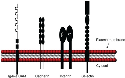
Table 1 Representative CAMs and expression distribution
The selectin family consists of three known members that mediate the initial steps of the leukocyte adhesion cascade,Citation1 the lymphocyte homing receptor (L-selectin), the endothelial leukocyte adhesion molecule (E-selectin), and the platelet-activation-dependent granule-external protein (P-selectin). All selectins interact with sialylated glycans in a Ca2+-dependent manner. As implicated by their names, L-selectin is primarily expressed by leukocytes, E-selectin by activated endothelial cells, and P-selectin by platelets and endothelial cells.Citation2 Soluble forms of all three selectins can be found in both plasma and in cerebrospinal fluid (CSF)Citation3–Citation6 and appear to reflect the activation state of the originating cells.
Integrins belong to a family that includes 18 alpha and 9 beta subunits.Citation7 The subunits form at least 24 different heterodimeric receptors, and through these receptors, cells communicate with the ECM and surrounding cells, both outside-in and inside-out.Citation8 When the ECM or CAMs expressed on neighboring cells adhere to integrins, recruitment of cytosolic non-receptor tyrosine kinases (NRTK) and cytoskeleton proteins (CSK) occur and form focal adhesion complexes (FAC). These complexes associate with the actin cytoskeleton and multiple signaling pathways that involve processes such as cell survival, inflammation, and angiogenesis, while synaptic transmission becomes efficiently regulated.Citation9 Integrins are found on most cells throughout the brain and are region-specific as well as cell-type-specific.Citation9 In addition, some integrins are known to actively shed from the cell surface in response to environmental changes such as inflammation.Citation10
The Ig-CAMs are defined by one or more Ig repeats of 60 to 100 amino acids forming the active adhesion site. CAMs in this family can be found both in the periphery, where Ig-CAM mediated interactions are mostly heterophilic, and in the nervous system, where interactions are predominantly homophilic.Citation11 Several Ig-CAM members appear to be specific to nervous tissueCitation12,Citation13 where they play vital roles in, for example, neurogenesis, neurite elongation, and brain plasticity. Furthermore, Ig-CAMs can be shed from cell membranesCitation14 and the soluble versions may have separate effects from the cell-bound forms.Citation15
Cadherins, particularly the classical cadherin and protocadherin subfamilies, are found throughout the nervous system. The two cadherin subfamilies are found mostly on neuronal synapsesCitation16,Citation17 and extensive studies have shown that classical cadherins play a crucial role in neuronal plasticity and synaptogenesis.Citation17 The functions of classical cadherins are mediated by a complex that is formed between the cytoplasmic tail of the cadherin and the cytosolic catenins, which are linked to the actin cytoskeleton.Citation17 Like the other CAMs, cadherins are sensitive to proteolytic shedding.Citation18
Collectively, CAMs orchestrate important functions in many vital physiological processes. However, increasing evidence suggests that CAMs are involved in the pathophysiol-ogy of several neurological diseases. This review summarizes current knowledge on the role of CAMs in events considered central to the pathogenesis and progression of Alzheimer’s disease (AD) such as amyloid-β (Aβ) metabolism, neuronal plasticity, inflammation, and vascular changes (data summarized in ). We also discuss the potential impact of the involvement of CAMs in these processes in terms of cognitive symptoms in AD patients. Finally, this paper reviews current AD treatment strategies and the possible use of CAMs as targets in current and future AD treatments.
Table 2 CAMs linked to AD pathological events
Alzheimer’s disease pathogenesis
Neuropathologically, AD is primarily characterized by intraneuronal neurofibrillary tangles (NFTs) of hyperphosphorylated tau and extracellular deposits of mainly aggregated Aβ peptide, known as senile or neuritic plaques.Citation19 According to the amyloid cascade hypothesis, the aggregation and deposition of Aβ in brain tissue is key to AD pathogenesis with the formation of tau pathology, inflammatory processes, and neurodegeneration as downstream events.Citation20 The Aβ peptide is generated by proteolytic processing of the amyloid precursor protein (APP) via sequential cleavage by the enzymes β-secretase 1 (BACE 1) and γ-secretase of which the latter is a complex consisting of presenilin 1 (PSEN1), presenilin 2 (PSEN2), and nicastrin.Citation21 Genetic evidence links mutations of the genes encoding PSEN1 and PSEN2 to the familial early onset form of AD (EOAD).Citation22 Furthermore, the presence of the ε4 allele of apolipoprotein E (APOE) – the most well described genetic risk factor for sporadic AD, known as late onset AD (LOAD)Citation23 – was shown to promote Aβ fibrillogenesis, deposition, and plaque formation.Citation24 Therefore, imbalanced Aβ metabolism with increased Aβ production and insufficiently augmented Aβ clearance in EOAD, as well as normal Aβ production, but defective Aβ clearance in LOAD, are believed to be central events in the development of AD.
Next to the core pathological events of Aβ deposition and intraneuronal tau accumulation, the presence of inflammatory processes, mainly driven by glial cells, are well described findings in the brains of patients with AD.Citation25 Numerous studies have demonstrated increased levels of inflammatory markers in the brain tissue as well as in the CSF and plasma of AD patients,Citation26,Citation27 and epidemiological studies have suggested that non-steroidal anti-inflammatory treatment may reduce the risk and slow down the progression of AD.Citation25 Also, neurovascular alterations and white matter lesions reflecting cerebrovascular pathology as well as blood brain barrier (BBB) alterations have frequently been described in dementia disorders such as AD.Citation28 Vascular pathology has even been proposed as a causal or contributing factor in at least 50% of all dementia cases.Citation29 Importantly, most AD patients are affected by the vascular deposition of Aβ, cerebral amyloid angiopathy (CAA).Citation30
The alteration of the glutamatergic and cholinergic systems are other well-described AD features. The glutamateric aberration, including glutamate excitotoxicity mediated through glutamate receptors such as N-methyl-D-aspartate receptors (NMDARs), can either be caused by an Aβ-induced increase in glutamate release or a decrease in glutamate uptake. Furthermore, the loss of cholinergic neurons and various subtypes of acetylcholine receptors are hallmarks of AD, and may interfere with the cholinergic mechanisms that contribute to glutamatergic transmission and synapse plasticity. Together, these alterations may contribute to a decline in cognitive functions.Citation31–Citation33
Not all of the described pathological events can be directly linked to the cognitive deterioration associated with AD, which causes uncertainty surrounding the potential causal roles of these events. Approximately one-third of cognitively normal elderly subjects display some degree of AD pathology and many of these individuals would fulfill the criteria for postmortem AD diagnoses despite the absence of cognitive symptoms. Attempts to elucidate the correlation between postmortem findings and clinical symptoms displayed a better correlation between neurofibrillary tau pathology, rather than amyloid pathology, and cognitive impairment.Citation34,Citation35 However, the best morphological correlate of cognitive dysfunction in clinical AD appears to be loss of synapsesCitation36,Citation37 with synaptic injury developing early in AD pathogenesis.Citation38,Citation39
Taken together, the current understanding supports the notion that AD results from a series of slowly developing neuropathological changes with a long asymptomatic phase preceding cognitive impairment. Therefore, early treatment strategies may prove to be the most efficient mode of prevention.
CAMs in altered synaptic and neuronal plasticity
The neurodegeneration and subsequent massive neuron loss associated with AD is considered to be paralleled by altered neurogenesis, which may be induced by Aβ-related mechanisms.Citation40 In the adult brain, neurogenesis has been identified in the hippocampus and the subventricular zone (SVZ).Citation41,Citation42 Expression of the neural cell adhesion molecule (NCAM), especially the highly polysialylated NCAM (PSA-NCAM) is considered an indicator of neurogenesis, neuronal remodeling, and plasticity, and has therefore been evaluated in AD brains. Results obtained by the use of immunohistochemistry and enzyme-linked immunosorbent assay (ELISA) suggest that alteration in NCAM expression in AD patients is brain-area dependent. Expression of hippocampal PSA-NCAM in AD patients was shown to increase with disease severity,Citation43,Citation44 whereas fewer NCAM positive neurons as well as lower NCAM expression were found in the frontal and temporal cortex of AD patients versus normal aging controls.Citation45,Citation46 The physiological roles of the soluble forms of NCAM are not completely elucidated but may implicate long-term potentiation (LTP), which is a cellular model of learning and memory.Citation47 In a small study on Parkinson’s disease and AD patients, the latter displayed increased CSF levels of the soluble NCAM-120 splice variant compared to the controls.Citation48 Similarly, another study demonstrated a tendency towards elevated levels of soluble NCAM in the CSF of AD patients versus the controls.Citation49 The latter study also showed that CSF concentrations of the L1 cell adhesion molecule (L1CAM), postulated to be involved in neurite elongation, fasciculation, and migrationCitation50 increased by 48% in AD patients versus the controls.Citation49 The significance of these findings and the impact of altered NCAM and L1 expression on cognitive performance require further investigation.
As mentioned previously, synapse loss is strongly correlated with cognitive decline in AD patients.Citation37 Since cadherins, particularly neuronal cadherins (N-cadherins), are important for synaptic formation and stability,Citation51 they have become interesting research targets for their possible role in AD pathology and clinical disease manifestations. Studies of mice cortically injected with Aβ showed that the pedptide inhibited cleavage,Citation52 but increased the expression, of N-cadherin.Citation53 In contrast, a study on murine primary neurons showed that Aβ42 exposure decreased N-cadherin expression through the glutamate N-Methyl-D-aspartate (NMDA) receptors and that the N-cadherin downregulation was followed by phosphorylation of the p38 mitogen activation protein kinase (p38 MAPK) and tau. Therefore, the decreased N-cadherin expression may be linked to tau pathology and, ultimately, to cognitive malperformance.Citation54 The activation of p38 MAPK is noteworthy since it is an intracellular transduction factor that becomes activated by oxidative stress and cytokine secretion,Citation55 both of which are processes linked to AD pathology.
As previously mentioned, mutations in the genes encoding for the presenilins, foremost known as the catalytic component of the Aβ generating γ-secretase, are linked to EOAD.Citation21 However, presenilin is also recruited to the synaptic adhesion site, where it binds to the cadherin-catenin complex and, in response to membrane depolarization or NMDAR stimulation, cleaves N-cadherin.Citation18 Support for its proteolytic effect on N-cadherin is found in functional studies showing a complete loss of N-cadherin cleavage caused by mutations in the PSEN1 gene.Citation56 The cleavage of N-cadherin was also proven to generate a cytosolic protein fragment, termed the carboxy-terminal fragment of N-cadherin (N-Cad/CTF2). This fragment was also shown to disturb the interaction between cyclic adenosine monophosphate (cAMP), response element-binding (CREB), and CREB binding protein (CBP),Citation57 a complex that is critical to several processes affected by AD, such as synaptic plasticity and memory.Citation58 Interestingly, recruitment of presenilin to the cadherin-catenin complex occurs at the expense of the presenilin/γ-secretase cleavage of APP,Citation59 which inhibits the amyloidogenic pathway. Finally, presenilin may also play a role in the trafficking of N-cadherin since cells that express mutant presenilin express less N-cadherinCitation60 which, as previously mentioned, may lead to tau phosphorylation.Citation54
Numerous studies have shown that integrins are also involved in synaptic transmission, synaptic plasticity, and LTP.Citation9 Therefore, it is interesting that a preclinical study demonstrated the preventative role of integrin αv on Aβ-induced LTP inhibition,Citation61 which when extrapolated, may counteract the mild cognitive impairment and synaptic loss observed in the early stages of AD.
Taken together, increasing evidence suggests that molecules belonging to several CAM families are engaged in neurogenesis, synaptic, and neuronal plasticity, all of which are affected by AD. Therefore, specific CAMs may constitute novel targets in strategies aimed at restoring these processes in AD, which may ultimately lead to preserved cognitive functions.
Evidence linking CAMs to Aβ metabolism
The significant association between Aβ- and AD-related synaptic alterations highlights the importance of elucidating the potential links between CAMs and the production, degradation, and biological functions of Aβ.
Immunohistological studies of brain tissue suggest that integrins may play a specific role in Aβ pathology. Expression of the α4 integrin unit in hippocampal pyramidal neurons and some neocortical neurons is increased in aged individuals.Citation62 Furthermore, increased expression of α4 and β3, their ligands fibronectin, and vintronectin, as well as the CSK proteins, paxillin and hydrogen peroxide-inducible clone 5 (Hic-5), were found in plaques and/or tangles in AD brain tissue.Citation62–Citation64 Moreover, integrins may also mediate Aβ neurotoxicity. For instance, when bound to integrins, Aβ induced rapid phosphorylation of the NRTK focal adhesion kinase (FAK) and paxillin, which in turn activated an incomplete anti-apoptotic signaling pathway and tau phosphorylation.Citation65 It is worth noting that neuronal cells can be rescued from Aβ-induced downregulation of α1β1 and cell cycle arrest in the presence of 17β-estradiol, which suggesed an interesting mechanism underlying the previously demonstrated increase of AD in menopausal women.Citation66 Interestingly, an in vitro study on human neuroblastoma cells suggested that the integrin receptor, α5β1 (cluster of differentiation [CD] 49e), may play a role in the internalization and degradation of exogenous Aβ.Citation67 Furthermore, the β1 integrin could mediate the microglial internalization of fibrillar,Citation68 but not soluble Aβ,Citation69 suggesting that this integrin may be important at different stages of AD, where for instance, Aβ becomes more fibrillar at more advanced disease stages.
Additional evidence supporting links between CAMs and AD pathology shows that adhesion based on N-cadherin increased APP dimerization and Aβ release, whereas the ratio of Aβ42/40 was decreased.Citation70,Citation71 Another classical cadherin, epithelial cadherin (E-cadherin), also affected Aβ levels. This cadherin subtype is primarily expressed on epithelial cells,Citation72 but is also found on hippocampal synapses.Citation73,Citation74 E-cadherin-catenin complexes recruit and can be cleaved by presenilin, similar to N-cadherin-catenin complexes. The cleavage product in this pathway, called carboxy-terminal fragment of E-cadherin (E-cad/CTF2), has been shown to promote the lysosomal degradation of APP derivates and to inhibit Aβ production.Citation75
CAMs in AD neuroinflammatory events
Transendothelial migration of leukocytes to sites of inflammation is central to immunological processes that respond to tissue damage in the periphery. However, under certain conditions such as in multiple sclerosis (MS),Citation76 brain malignancies,Citation77,Citation78 and brain infections,Citation79–Citation82 leukocytes can also infiltrate the central nervous system (CNS) by crossing the BBB. The migration of leukocytes depends on integrin expression on the surface of the leukocytes and Ig-CAMs, including the intercellular cell adhesion molecule 1 (ICAM-1), vascular cell adhesion molecule 1 (VCAM-1), and the platelet endothelial cell adhesion molecule (PECAM-1) present on endothelial cells.Citation1 Expression of ICAM-1 can be upregulated in vitro on primary cultures of human brain microvessel endothelial cells by cytokines such as γ-interferon, interleukin (IL)-1, tumor necrosis factor-α (TNF-α), and endotoxin.Citation83 Experimental studies performed on human brain endothelial cells demonstrated an Aβ-induced time-dependent increase in ICAM-1 and VCAM-1 expression. The same study showed the augmented adhesion and transendothelial migration of monocytic cells involving PECAM-1 upon Aβ interaction.Citation84 Interestingly, soluble ICAM-1 could block lymphocyte adhesion to cerebral endothelial cells, which stands in direct opposition to the role of cell-bound ICAM-1.Citation15 Furthermore, it has been demonstrated that ICAM-1, VCAM-1, and PECAM-1 not only engage in transendothelial migration of leukocytes but also serve as signal transducers that initiate endothelial signaling and influence the progression of neuroinflammation.Citation85 Increased levels of both ICAM-1 and PECAM-1 have also been proposed as indicators of inflammatory conditions in the CNSCitation86,Citation87 and could originate from activated glial cells.Citation88 Similarly, Rentzos et al found increased ICAM-1 levels in AD in the absence of endothelial activation and, therefore, proposed a neural, rather than endothelial, origin of ICAM-1.Citation89,Citation90 The authors also displayed a positive correlation between increased levels of CSF ICAM-1 and disease severity, as assessed by mini–mental state examination (MMSE) scores.
The concentrations of the soluble forms of ICAM-1, VCAM-1, and PECAM-1 in patients with AD as well as other neurodegenerative diseases such as dementia with Lewy bodies (DLB) have been evaluated in several studies.Citation89–Citation92 In patients with LOAD, Zuliani et al found increased levels of plasma VCAM-1,Citation93 while this study found that AD and DLB patients exhibited higher plasma levels of soluble ICAM-1 and PECAM-1, but not VCAM-1.Citation92 Discrepancies between the VCAM-1 plasma levels in these studies might be due to dementia severity – AD patients with moderate dementia may exhibit normal plasma VCAM-1 levels while patients with more severe dementia may exhibit different levels.Citation92,Citation93 Altered plasma levels of ICAM-1 were shown to be unrelated to the risk of dementia in non-demented individuals; therefore, increased levels might be secondary to the processes that lead to the onset of dementia.Citation91 In direct opposition to the results on plasma, this review found that the CSF concentrations of VCAM-1 were significantly lower in patients with AD and DLB. In patients with AD, brain tissue ICAM-1 was repeatedly identified in plaques;Citation94–Citation96 however, this study only found increased CSF concentrations of soluble ICAM-1 in patients with DLB.Citation92 Therefore, the soluble fraction of these CAMs may not necessarily reflect the amount of the cell-membrane-bound forms. In a recent study we also showed that CSF levels of ICAM-1 in female AD patients correlated with levels of hyaluronic acid, an adhesion molecule implicated in both inflammatory events and vascular changes.Citation97 The main significance of the described altered levels of Ig-CAMs in AD requires further investigation as neuroinflammation in AD appears to take place without any apparent influx of leukocytes from the blood.Citation98 Thus, the altered levels of the soluble forms of Ig-CAMs may reflect inflammatory processes unrelated to cognitive performance (as assessed with MMSE). Alternatively, they may mediate functions that are currently unknown in relation to AD pathology.
In addition to Ig-CAMs, integrins can also be linked to AD neuroinflammation. The integrins on the surface of leukocytes are considered important for the presentation of antigen in the CNS.Citation99 Similarly, microglia express several integrins that can be upregulated when the cells are activated in response to inflammation and injury. A recent study showed that microglia could interact with fibrillary Aβ through a complex including α6β1 and B-class scavenger receptor, CD36. This interaction induced proinflammatory activation of the microglia, which led to increased production of cytokines and chemokines.Citation100 Moreover, results obtained from mixed neuron-glia cell cultures from cerebellum showed that neurons, when exposed to Aβ, signal a phagocytic request to microglia that is mediated through binding between phosphatidylserine (PS) on the neuron surface and the αvβ3/5 integrin receptors on microglia.Citation101 Other integrins such as αLβ2 (CD11a), αMβ2 (CD11b), and αXβ2 (CD11c) found in immunohistological studies on the temporal cortexes of AD patients, were found to be strongly upregulated on the surface of activated microglia in presence of Aβ.Citation102 Antibodies blocking the microglial αMβ2 (CD11b) have also been shown to attenuate inflammatory reactions, vascular responses, and neurodegeneration induced by the injection of Aβ into rat hippocampuses.Citation103
The proinflammatory activation of microglia is also associated with changes in β-catenin, a protein that links cadherins to the intracellular actin. A study performed on brain tissue from AD patients and transgenic AD mice demonstrated the increased expression of β-catenin in activated morphologically transformed microglia. The authors of this study also used in vitro studies to show that Wnt signaling, a pathway controlling β-catenin metabolism, regulates the gene expression of microglial proinflammatory cytokines.Citation104
Collectively, these data indicate that CAMs of the integrin and Ig-CAM families are involved in AD neuroinflammatory events, and some of these molecules may function as inflammation markers whereas others may play active roles in mediating inflammation.
Role of CAMs in vascular changes linked to AD
Several CAMs that are specifically involved in endothelial alterations, as imposed by vascular alterations and CAA in AD, have been the focus of attention in studies aimed at characterizing the vascular component frequently found in AD. Based on the clinical and epidemiological evidence supporting the links between vascular risk factors and AD, Borroni et al investigated the peripheral blood concentrations of E-selectin in age-matched controls and AD patients with mild dementia. In line with the notion of endothelial dysfunction in AD, the study demonstrated significantly increased E-selectin levels in the AD group, which indicates that endothelial dysfunction in AD could potentially be monitored in peripheral blood.Citation105 However, two recent studies could not confirm altered E-selectin levels in AD patients. The authors of the latter studies investigated soluble CAMs in patients with various neurological disorders including AD patients without the vascular component.Citation90,Citation106 However, when the authors divided the subjects into groups with small or large vessel disease confirmed by computer tomography (CT) scans, the patients with vessel disease exhibited significantly increased E-selectin compared to the group with no vascular lesions.Citation93 Therefore, altered E-selectin levels in AD appear to be related to cerebrovascular lesions.
In the periphery, platelets contain and metabolize the Aβ precursor protein APP, and altered APP processing in platelets of AD patients has been suggested to reflect chronic platelet activation in these patients.Citation107 Furthermore, treatment with acetylcholinesterase inhibitors (AChEI) influences APP metabolism in platelets, which may be modulated by the APOE genotype.Citation108 Interestingly, platelet activation was recently reinvestigated and suggested as a prognostic biomarker for cognitive decline in AD patients. Patients with faster cognitive decline demonstrated significantly higher baseline expression of platelet activation markers including P-selectin.Citation109 The authors of the study proposed several mechanisms for how platelet activity could contribute to dementia progression including triggering perivascular inflammation, induction of vasoconstriction, and consecutive brain hypoperfusion in addition to contributing to the peripheral Aβ pool. In a recent study of platelet activation in sporadic AD patients, Järemo and colleagues found that circulating a lower density platelet population exhibits a reduced activation state, as assessed by the amount of platelet surface bound fibrinogen.Citation110 Therefore, the activation of different platelet populations appears to be related to AD. The role of P-selectins in these processes is yet to be determined, but P-selectin antagonists, as modulators of inflammation, have been proposed in vascular pathologies such as atherosclerosis.Citation111
Next to CAMs of the selectin family, integrins are also related to vascular processes. For instance, the increased expression of β3 integrin on endothelial cells is a well-described marker for ongoing angiogenesis.Citation112 Studies on brain tissue demonstrated upregulated αvβ3 (CD51) expression in AD patients,Citation113 a finding that supports previous results proposing increased angiogenesis in AD patients.Citation114 The upregulation of this integrin was also shown to correlate with Aβ load in the hippocampus and neurofibrillary tangles in the midfrontal cortex.Citation113 Further investigation is required to determine whether there is a direct or an indirect link between αvβ3, AD pathology, and cognitive deterioration. In sum, the increasing evidence regarding the potential causal role of vascular factors in AD pathogenesis warrants the further investigation of CAMs.
Currently available AD treatment options
To date, there is no approved pharmaceutical drug for the prevention or cure of AD. Instead, the available therapy consists of symptomatic treatment based on glutamatergic and cholinergic alterations, which at certain stages of AD can slow the disease’s progression by boosting neuronal activity. Acetylcholinesterase inhibitors were the first AD pharmaceuticals to be approved, and today, the second generation of AChEIs (donezepil, rivastigmine, and galantamine) is the first line of pharmacotherapy for mild to moderate AD. By blocking the enzyme, acetylcholinesterase, AChEIs prevent the breakdown of acetylcholine in the synaptic cleft and partly replace the cholinergic transmission that is lost in AD patients.Citation115 AChEIs have also shown favorable effects on the documented impaired vasodilationCitation116 and neurovascular coupling (regulation of blood flow demand in brain areas)Citation117,Citation118 found in AD patients. Moreover, a number of studies have presented evidence of the antiinflammatory and antiamyloidogenic properties associated with AChEI.Citation119,Citation120 These findings can be related to the previously mentioned effects of AChEI on platelet APP metabolism.Citation108
Memantine, a noncompetitive antagonist of the glutamate NMDA receptor, is the second type of medication approved for AD treatment and has documented effects alone or in combination with AChEI on moderate to severe AD. Its primary effect is the inhibition of pathological activation of NMDARs, which prevent excessive glutamatergically-induced Ca2+ influx and cell death.Citation121 In addition, results from preclinical studies suggest that memantine and NMDA antagonists could reduce the secretion of proinflammatory cytokines by blocking glutamate receptors on glial cells and thereby slowing neuroinflammation,Citation122 protecting against BBB breakdown,Citation123 reducing Aβ production,Citation124,Citation125 and Aβ neurotoxicity.Citation126 However, AD is without a remedy despite the various beneficial effects of AChEIs and memantine and the laborious efforts of trying to find a cure. Therefore, the importance of research aimed at identifying the underlying AD mechanisms, which could yield new pharmaceutical targets, cannot be exaggerated.
Significance of CAMs in potential treatment strategies
In light of the reported involvement of CAMs in the various aspects of AD, these molecules may offer new unexplored treatment opportunities and when used as biomarkers that can provide potential tools to enable the clinical monitoring of AD related inflammation and cerebrovascular complications, disease progression, and treatment response. Depending on the overall treatment goal – prevention, disease-modification, or cure – different aspects of AD may need to be targeted.
Strategies to counteract neuroinflammation
In investigations of long-term use of anti-inflammatory agents, nonsteroidal anti-inflammatory drugs (NSAIDs) have shown beneficial effects in terms of reducing the risk of developing AD.Citation127 Moreover, long-term treatment responses to cholinesterase inhibitors were better in individuals treated with NSAIDs.Citation128 However, recently published extended results of the AD anti-inflammatory prevention trial (ADAPT) demonstrated that treatment with NSAIDs might have an adverse effect in the later stages of AD,Citation129 which indicates that inflammatory processes might shift from acute to chronic, meaning different pathways may be engaged. Anti-selectin substances have been suggested as another potential treatment to control inflammatory disease. However, similar to NSAID treatment, the timing of treatment may be of crucial importance.Citation130 Finally, due to the significant association between inflammatory events, AD pathology,Citation131,Citation132 and increased levels of ICAM-1, it is necessary to emphasize the importance of evaluating the various roles of the soluble and membrane-bound forms of ICAM-1. Targeting ICAM-1 in animal models of reperfusion injuries demonstrated a neuroprotective effect of anti-ICAM administration.Citation133,Citation134 In addition, ICAM-1 levels in cognitively conserved elderly patients have been linked to decreased parietal blood flow.Citation135 Therefore, the elucidation of altered ICAM-1 expression as a consequence or disease modifying component may identify this molecule as a potential target for future anti-inflammatory AD treatment options.
Strategies aimed at altering Aβ metabolism
One of the reported effects of the AChEI rivastigmine is the modulation of secretase activity, which results in decreased Aβ secretion.Citation136 Therefore, interference with the proteolytic generation of the Aβ peptide by inhibition of β-secretase and γ-secretase activity could prove beneficial. However, β and γ-secretase inhibitors, as well as γ-secretase modulators, are yet to prove successful in clinical trials.Citation137 An alternative approach could be to target competition for secretase activity by taking advantage of certain mechanisms such as the recruitment of presenilin to the cadherin-catenin complex at the expense of presenilin/γ-secretase cleavage of APP.Citation59
Elimination of already deposited Aβ is another strategy for eradicating Aβ toxicity. For this purpose, both active and passive immunization against Aβ have been evaluated. The administration of pre-aggregated or soluble Aβ generated an antibody response and Aβ plaque reduction in aged animals. However, due to serious adverse effects with aseptic meningoencephalitis caused by T-cell activation in a subset of patients,Citation138 the initial clinical trial was interrupted. A new generation of active Aβ immunization using Aβ fragments, without the ability to activate T-cells, has currently entered Phase I trials.Citation137 An anti-CAM treatment could be useful to circumvent adverse immunization effects such as the influx of leukocytes into the CNS. Lessons learned from the field of MS research, including its animal model experimental autoimmune encephalomyelitis (EAE), suggest anti-integrin (α4) treatment as a measure of preventing leukocyte influx.Citation139 Passive immunization strategies using humanized monoclonal antibodies directed against different sites of Aβ have shown improved cognitive functions in APOE carriers as well as reduced brain atrophy in APOE non-carrier in Phase II trials. Moreover, intravenously delivered immunoglobulins isolated from pooled human blood (IVIgs) were found to significantly reduce both Aβ plaques and inflammatory events in AD patients.Citation137
Enhanced Aβ clearance can also be achieved via targeted gene delivery by use of CAM-dependent transendothelial migration. For instance, a recent study convincingly showed that endogenous CD11b+ bone marrow cells home to Aβ plaques in the brain of AD transgenic mice. The study further demonstrated that subcutaneous infusion of CD11b+ bone marrow cells transfected with the gene coding for neprilysin, an Aβ-degrading enzyme, and completely arrested Aβ deposition.Citation140 Therefore, using CAM-dependent mechanisms to introduce cells harboring the appropriate genes into the CNS has large-scale potential.
Strategies targeting vascular alterations
Given the reported association between vascular risk factors and AD,Citation29 it is worth noting that activation of different platelet populations, which is also linked to vascular alterations and stroke,Citation141 appears also related to ADCitation110 and altered APP metabolism.Citation107 Since the activation of platelets involves alterations in P-selectin expressionCitation142 as well as integrin αIIbβ complex activation,Citation142 and since these molecules possibly regulate inflammatory processes,Citation112 which are known to affect the vasculature, it is necessary to perform intensified studies that elucidate the potential of targeting platelet adhesion molecules in AD.
Conclusion
This review summarized current knowledge of CAMs associated with AD, as well as the potential clinical utility of CAMs as disease biomarkers or pharmaceutical targets in novel AD treatment strategies. It has also identified a major need for both experimental and clinical studies to characterize the specific mechanisms by which CAMs relate to AD. Numerous observational studies have reported altered levels of CAMs in AD, but the specific roles of CAMs as markers or mediators of biological events leading to cognitive deterioration and dementia remain undetermined.
Acknowledgments
The authors wish to acknowledge the Swedish Brain Power initiative and ALF (the regional agreement on medical training and clinical research between the Skåne County Council and Lund University) for financial support.
Disclosure
The authors report no conflicts of interest in this work.
References
- LeyKLaudannaCCybulskyMINoursharghSGetting to the site of inflammation: the leukocyte adhesion cascade updatedNat Rev Immunol20077967868917717539
- BevilacquaMPNelsonRMSelectinsJ Clin Invest19939123793877679406
- BuhrerCHeroldRStibenzDHenzeGObladenMCerebrospinal fluid soluble L-selectin (sCD62L) in meningoencephalitisArch Dis Child19967442882928669926
- Dore-DuffyPNewmanWBalabanovRCirculating, soluble adhesion proteins in cerebrospinal fluid and serum of patients with multiple sclerosis: correlation with clinical activityAnn Neurol199537155627529475
- WhalenMJCarlosTMKochanekPMSoluble adhesion molecules in CSF are increased in children with severe head injuryJ Neurotrauma199815107777879814634
- GearingAJNewmanWCirculating adhesion molecules in diseaseImmunol Today199314105065127506035
- HynesROIntegrins: bidirectional, allosteric signaling machines. CellSeptember 2020021106673687
- ReichardtLFProkopAIntroduction: The role of extracellular matrix in nervous system development and maintenance. Developmental neurobiologyNov20117111883888
- WuXReddyDSIntegrins as receptor targets for neurological disordersJ Pharmacol Pharmacother201213416881
- EvansBJMcDowallATaylorPCHoggNHaskardDOLandis RC Shedding of lymphocyte function-associated antigen-1 (LFA-1) in a human inflammatory responseBlood5 1, 200610793593359916418329
- MarchettiPJO’ConnorPCellular adhesion molecules in neurologyCan J Neurol Sci19972432002099276104
- WilliamsAFBarclayANThe immunoglobulin superfamily – domains for cell surface recognitionAnnu Rev Immunol198863814053289571
- LaiCWatsonJBBloomFESutcliffeJGMilnerRJNeural protein 1B236/myelin-associated glycoprotein (MAG) defines a subgroup of the immunoglobulin superfamilyImmunol Rev19871001291512450062
- GoliasCTsoutsiEMatziridisAMakridisPBatistatouACharalabopoulosKReview. Leukocyte and endothelial cell adhesion molecules in inflammation focusing on inflammatory heart diseaseIn Vivo200721575776918019409
- RieckmannPMichelUAlbrechtMBruckWWockelLFelgenhauerKSoluble forms of intercellular adhesion molecule-1 (ICAM-1) block lymphocyte attachment to cerebral endothelial cellsJ Neuroimmunol1995601–29157642752
- FrankMKemlerRProtocadherinsCurr Opin Cell Biol200214555756212231349
- ArikkathJReichardtLFCadherins and catenins at synapses: roles in synaptogenesis and synaptic plasticityTrends Neurosci200831948749418684518
- KopanRIlaganMXγ-Secretase: proteasome of the membrane?Nat Rev Mol Cell Biol20045649950415173829
- HymanBTTrojanowskiJQConsensus recommendations for the postmortem diagnosis of Alzheimer disease from the National Institute on Aging and the Reagan Institute Working Group on diagnostic criteria for the neuropathological assessment of Alzheimer diseaseJ Neuropathol Exp Neurol19975610109510979329452
- HardyJAHigginsGAAlzheimer’s disease: the amyloid cascade hypothesisScience199225650541841851566067
- CitronMAlzheimer’s disease: strategies for disease modificationNat Rev Drug Discov20109538739820431570
- BertramLLillCMTanziREThe genetics of Alzheimer disease: back to the futureNeuron201068227028120955934
- CorderEHSaundersAMStrittmatterWJGene dose of apolipoprotein E type 4 allele and the risk of Alzheimer’s disease in late onset familiesScience199326151239219238346443
- HoltzmanDMBalesKRTenkovaTApolipoprotein E isoform-dependent amyloid deposition and neuritic degeneration in a mouse model of Alzheimer’s diseaseProc Natl Acad Sci U S A20009762892289710694577
- LeeYJHanSBNamSYOhKWHongJTInflammation and Alzheimer’s diseaseArch Pharm Res201033101539155621052932
- EikelenboomPvan ExelEHoozemansJJVeerhuisRRozemullerAJvan GoolWANeuroinflammation – an early event in both the history and pathogenesis of Alzheimer’s diseaseNeurodegener Dis201071–3384120160456
- RojoLEFernandezJAMaccioniAAJimenezJMMaccioniRBNeuroinflammation: implications for the pathogenesis and molecular diagnosis of Alzheimer’s diseaseArch Med Res200839111618067990
- de la TorreJCVascular basis of Alzheimer’s pathogenesisAnn N Y Acad Sci200297719621512480752
- BretelerMMVascular risk factors for Alzheimer’s disease: an epidemiologic perspectiveNeurobiol Aging200021215316010867200
- LoveSMinersSPalmerJChalmersKKehoePInsights into the pathogenesis and pathogenicity of cerebral amyloid angiopathyFront Biosci20091447784792
- OndrejcakTKlyubinIHuNWBarryAECullenWKRowanMJAlzheimer’s disease amyloid beta-protein and synaptic functionNeuromolecular Med2010121132619757208
- RupsinghRBorrieMSmithMWellsJLBarthaRReduced hippocampal glutamate in Alzheimer diseaseNeurobiol Aging201132580281019501936
- SimsNRBowenDMAllenSJPresynaptic cholinergic dysfunction in patients with dementiaJ Neurochem19834025035096822833
- BennettDASchneiderJAWilsonRSBieniasJLArnoldSENeurofibrillary tangles mediate the association of amyloid load with clinical Alzheimer disease and level of cognitive functionArch Neurol200461337838415023815
- McKhannGMKnopmanDSChertkowHThe diagnosis of dementia due to Alzheimer’s disease: recommendations from the National Institute on Aging-Alzheimer’s Association workgroups on diagnostic guidelines for Alzheimer’s diseaseAlzheimers Dement20117326326921514250
- DeKoskySTScheffSWSynapse loss in frontal cortex biopsies in Alzheimer’s disease: correlation with cognitive severityAnn Neurol19902754574642360787
- TerryRDMasliahESalmonDPPhysical basis of cognitive alterations in Alzheimer’s disease: synapse loss is the major correlate of cognitive impairmentAnn Neurol19913045725801789684
- MasliahEMalloryMAlfordMAltered expression of synaptic proteins occurs early during progression of Alzheimer’s diseaseNeurology200156112712911148253
- ScheffSWPriceDASchmittFADeKoskySTMufsonEJSynaptic alterations in CA1 in mild Alzheimer disease and mild cognitive impairmentNeurology200768181501150817470753
- RodriguezJJVerkhratskyANeurogenesis in Alzheimer’s diseaseJ Anat20112191788921323664
- ErikssonPSPerfilievaEBjork-ErikssonTNeurogenesis in the adult human hippocampusNat Med1998411131313179809557
- Alvarez-BuyllaAGarcia-VerdugoJMNeurogenesis in adult subventricular zoneJ Neurosci200222362963411826091
- MikkonenMSoininenHTapiolaTAlafuzoffIMiettinenRHippocampal plasticity in Alzheimer’s disease: changes in highly polysialylated NCAM immunoreactivity in the hippocampal formationEur J Neurosci19991151754176410215928
- JinKPeelALMaoXOIncreased hippocampal neurogenesis in Alzheimer’s disease. Proc Natl Acad Sci U S A620041011343347
- AisaBGil-BeaFJSolasMAltered NCAM expression associated with the cholinergic system in Alzheimer’s diseaseJ Alzheimers Dis201020265966820164549
- YewDTLiWPWebbSELaiHWZhangLNeurotransmitters, peptides, and neural cell adhesion molecules in the cortices of normal elderly humans and Alzheimer patients: a comparisonExp Gerontol199934111713310197733
- FazeliMSBreenKErringtonMLBlissTVIncrease in extracellular NCAM and amyloid precursor protein following induction of long-term potentiation in the dentate gyrus of anaesthetized ratsNeurosci Lett19941691–277808047297
- YinGNLeeHWChoJYSukKNeuronal pentraxin receptor in cerebrospinal fluid as a potential biomarker for neurodegenerative diseasesBrain Res2009126515817019368810
- StrekalovaHBuhmannCKleeneRElevated levels of neural recognition molecule L1 in the cerebrospinal fluid of patients with Alzheimer disease and other dementia syndromesNeurobiol Aging20062711916298234
- MiuraMAsouHKobayashiMUyemuraKFunctional expression of a full-length cDNA coding for rat neural cell adhesion molecule L1 mediates homophilic intercellular adhesion and migration of cerebellar neuronsJ Biol Chem19922671510752107581587850
- GiagtzoglouNLyCVBellenHJCell adhesion, the backbone of the synapse: “vertebrate” and “invertebrate” perspectivesCold Spring Harb Perspect Biol200914a00307920066100
- UemuraKKuzuyaAAoyagiNAmyloid beta inhibits ectodomain shedding of N-cadherin via down-regulation of cell-surface NMDA receptorNeuroscience2007145151017257767
- KongLNZuoPPMuLLiuYYYangNGene expression profile of amyloid beta protein-injected mouse model for Alzheimer diseaseActa Pharmacol Sin200526666667215916731
- AndoKUemuraKKuzuyaAN-cadherin regulates p38 MAPK signaling via association with JNK-associated leucine zipper protein: implications for neurodegeneration in Alzheimer diseaseJ Biol Chem201128697619762821177868
- HerlaarEBrownZp38 MAPK signalling cascades in inflammatory diseaseMol Med Today199951043944710498912
- HeiligEAXiaWShenJKelleherRJ3rdA presenilin-1 mutation identified in familial Alzheimer disease with cotton wool plaques causes a nearly complete loss of gamma-secretase activityJ Biol Chem201028529223502235920460383
- MarambaudPWenPHDuttAA CBP binding transcriptional repressor produced by the PS1/epsilon-cleavage of N-cadherin is inhibited by PS1 FAD mutationsCell2003114563564513678586
- SauraCAValeroJThe role of CREB signaling in Alzheimer’s disease and other cognitive disordersRev Neurosci201122215316921476939
- KouchiZBarthetGSerbanGGeorgakopoulosAShioiJRobakisNKp120 catenin recruits cadherins to gamma-secretase and inhibits production of Abeta peptideJ Biol Chem200928441954196119008223
- UemuraKKuzuyaAShimohamaSProtein trafficking and Alzheimer’s diseaseCurr Alzheimer Res20041111015975080
- RowanMJKlyubinIWangQHuNWAnwylRSynaptic memory mechanisms: Alzheimer’s disease amyloid beta-peptide-induced dysfunctionBiochem Soc Trans200735Pt 51219122317956317
- Van GoolDCarmelietGTriauECassimanJJDomRAppearance of localized immunoreactivity for the alpha 4 integrin subunit and for fibronectin in brains from Alzheimer’s, Lewy body dementia patients and aged controlsNeurosci Lett1994170171738041517
- AkiyamaHKawamataTDedharSMcGeerPLImmunohistochemical localization of vitronectin, its receptor and beta-3 integrin in Alzheimer brain tissueJ Neuroimmunol199132119281705945
- CaltagaroneJHamiltonRLMurdochGJingZDeFrancoDBBowserRPaxillin and hydrogen peroxide-inducible clone 5 expression and distribution in control and Alzheimer disease hippocampiJ Neuropathol Exp Neurol201069435637120448481
- CaltagaroneJJingZBowserRFocal adhesions regulate Abeta signaling and cell death in Alzheimer’s diseaseBiochim Biophys Acta20071772443844517215111
- BozzoCGraziolaFChiocchettiACanonicoPLEstrogen and beta-amyloid toxicity: role of integrin and PI3-KMol Cell Neurosci2010452859120538057
- MatterMLZhangZNordstedtCRuoslahtiEThe alpha5beta1 integrin mediates elimination of amyloid-beta peptide and protects against apoptosisJ Cell Biol19981414101910309585419
- KoenigsknechtJLandrethGMicroglial phagocytosis of fibrillar beta-amyloid through a beta1 integrin-dependent mechanismJ Neurosci200424449838984615525768
- MandrekarSJiangQLeeCYKoenigsknecht-TalbooJHoltzmanDMLandrethGEMicroglia mediate the clearance of soluble Abeta through fluid phase macropinocytosisJ Neurosci200929134252426219339619
- Asada-UtsugiMUemuraKNodaYN-cadherin enhances APP dimerization at the extracellular domain and modulates Aβ productionJ Neurochem2011119235436321699541
- UemuraKLillCMBanksMN-cadherin-based adhesion enhances Abeta release and decreases Abeta42/40 ratioJ Neurochem2009108235036019046403
- ShapiroLLoveJColmanDRAdhesion molecules in the nervous system: structural insights into function and diversityAnnu Rev Neurosci20073045147417600523
- FiederlingAEwertRAndreyevaAJunglingKGottmannKE-cadherin is required at GABAergic synapses in cultured cortical neuronsNeurosci Lett2011501316717221782891
- TangLHungCPSchumanEMA role for the cadherin family of cell adhesion molecules in hippocampal long-term potentiationNeuron1998206116511759655504
- AgiostratidouGMurosRMShioiJMarambaudPRobakisNKThe cytoplasmic sequence of E-cadherin promotes non-amyloidogenic degradation of A beta precursorsJ Neurochem20069641182118816417575
- EngelhardtBImmune cell entry into the central nervous system: involvement of adhesion molecules and chemokines. J Neurol Sci1520082741–22326
- D’AbacoGMKayeAHIntegrins: molecular determinants of glioma invasionJ Clin Neurosci200714111041104817954373
- BaramiKLewis-TuffinLAnastasiadisPZThe role of cadherins and catenins in gliomagenesisNeurosurg Focus2006214E13
- BrankinBHartMNCosbySLFabryZAllenIVAdhesion molecule expression and lymphocyte adhesion to cerebral endothelium: effects of measles virus and herpes simplex 1 virusJ Neuroimmunol1995561187822475
- OuRZhangMHuangLFlavellRAKoniPAMoskophidisDRegulation of immune response and inflammatory reactions against viral infection by VCAM-1J Virol20088262952296518216105
- SassevilleVGLaneJHWalshDRinglerDJLacknerAAVCAM-1 expression and leukocyte trafficking to the CNS occur early in infection with pathogenic isolates of SIVJ Med Primatol19952431231318751051
- SassevilleVGNewmanWBrodieSJHesterbergPPauleyDRinglerDJMonocyte adhesion to endothelium in simian immunodeficiency virus-induced AIDS encephalitis is mediated by vascular cell adhesion molecule-1/alpha 4 beta 1 integrin interactionsAm J Pathol1994144127407507300
- WongDDorovini-ZisKUpregulation of intercellular adhesion molecule-1 (ICAM-1) expression in primary cultures of human brain microvessel endothelial cells by cytokines and lipopolysaccharideJ Neuroimmunol1992391–211211352310
- GiriRShenYStinsMbeta-amyloid-induced migration of monocytes across human brain endothelial cells involves RAGE and PECAM-1Am J Physiol Cell Physiol20002796C1772C178111078691
- GreenwoodJEtienne-MannevilleSAdamsonPCouraudPOLymphocyte migration into the central nervous system: implication of ICAM-1 signalling at the blood-brain barrierVascul Pharmacol200238631532212529926
- KalinowskaALosyJPECAM-1, a key player in neuroinflammationEur J Neurol200613121284129017116209
- RieckmannPNunkeKBurchhardtMSoluble intercellular adhesion molecule-1 in cerebrospinal fluid: an indicator for the inflammatory impairment of the blood-cerebrospinal fluid barrierJ Neuroimmunol19934721331408103775
- LeeSJBenvenisteENAdhesion molecule expression and regulation on cells of the central nervous system. J Neuroimmunol319999827788
- RentzosMMichalopoulouMNikolaouCSerum levels of soluble intercellular adhesion molecule-1 and soluble endothelial leukocyte adhesion molecule-1 in Alzheimer’s diseaseJ Geriatr Psychiatry Neurol200417422523115533994
- RentzosMMichalopoulouMNikolaouCThe role of soluble intercellular adhesion molecules in neurodegenerative disorders. J Neurol Sci1520052282129135
- EngelhartMJGeerlingsMIMeijerJInflammatory proteins in plasma and the risk of dementia: the Rotterdam studyArch Neurol200461566867215148142
- NielsenHMLondosEMinthonLJanciauskieneSMSoluble adhesion molecules and angiotensin-converting enzyme in dementiaNeurobiol Dis2007261273517270454
- ZulianiGCavalieriMGalvaniMMarkers of endothelial dysfunction in older subjects with late onset Alzheimer’s disease or vascular dementiaJ Neurol Sci20082721–216417018597785
- EikelenboomPZhanSSKamphorstWvan der ValkPRozemullerJMCellular and substrate adhesion molecules (integrins) and their ligands in cerebral amyloid plaques in Alzheimer’s diseaseVirchows Arch199442444214277515758
- FrohmanEMFrohmanTCGuptaSde FougerollesAvan den NoortSExpression of intercellular adhesion molecule 1 (ICAM-1) in Alzheimer’s diseaseJ Neurol Sci199110611051111685745
- RozemullerJMEikelenboomPPalsSTStamFCMicroglial cells around amyloid plaques in Alzheimer’s disease express leucocyte adhesion molecules of the LFA-1 familyNeurosci Lett198910132882922549464
- NielsenHMPalmqvistSMinthonLLondosEWennstromMGender-dependent levels of hyaluronic acid in cerebrospinal fluid of patients with neurodegenerative dementiaCurr Alzheimer Res20129325726622191565
- EikelenboomPRozemullerAJHoozemansJJVeerhuisRvan GoolWANeuroinflammation and Alzheimer disease: clinical and therapeutic implicationsAlzheimer Dis Assoc Disord200014Suppl 1S54S6110850731
- HoggNPatzakIWillenbrockFThe insider’s guide to leukocyte integrin signalling and functionNat Rev Immunol201111641642621597477
- BambergerMEHarrisMEMcDonaldDRHusemannJLandrethGEA cell surface receptor complex for fibrillar beta-amyloid mediates microglial activationJ Neurosci20032372665267412684452
- NeniskyteUNeherJJBrownGCNeuronal death induced by nanomolar amyloid beta is mediated by primary phagocytosis of neurons by microgliaJ Biol Chem201128646399043991321903584
- AkiyamaHMcGeerPLBrain microglia constitutively express beta-2 integrinsJ Neuroimmunol199030181931977769
- RyuJKMcLarnonJGA leaky blood-brain barrier, fibrinogen infiltration and microglial reactivity in inflamed Alzheimer’s disease brainJ Cell Mol Med2009139A2911292518657226
- HalleskogCMulderJDahlstromJWNT signaling in activated microglia is proinflammatoryGlia201159111913120967887
- BorroniBVolpiRMartiniGPeripheral blood abnormalities in Alzheimer disease: evidence for early endothelial dysfunctionAlzheimer Dis Assoc Disord200216315015512218645
- RentzosMMichalopoulouMNikolaouCSerum levels of soluble intercellular adhesion molecule-1 and soluble endothelial leukocyte adhesion molecule-1 in Alzheimer’s diseaseJ Geriatr Psychiatry Neurol200417422523115533994
- RosenbergRNBaskinFFosmireJAAltered amyloid protein processing in platelets of patients with Alzheimer diseaseArch Neurol19975421391449041854
- BorroniBColciaghiFPastorinoLApoE genotype influences the biological effect of donepezil on APP metabolism in Alzheimer disease: evidence from a peripheral modelEur Neuropsychopharmacol200212319520012007670
- StellosKPanagiotaVKogelALeyheTGawazMLaskeCPredictive value of platelet activation for the rate of cognitive decline in Alzheimer’s disease patientsJ Cereb Blood Flow Metab201030111817182020717123
- JaremoPMilovanovicMBullerCNilssonSWinbladBLow-density platelet populations demonstrate low in vivo activity in sporadic Alzheimer diseasePlatelets201223211612022150375
- WoollardKJChin-DustingJP-selectin antagonism in inflammatory diseaseCurr Pharm Des201016374113411821247396
- BeerAJSchwaigerMImaging of integrin alphavbeta3 expressionCancer Metastasis Rev200827463164418523730
- DesaiBSSchneiderJALiJLCarveyPMHendeyBEvidence of angiogenic vessels in Alzheimer’s diseaseJ Neural Transm2009116558759719370387
- GrammasPSanchezATripathyDLuoEMartinezJVascular signaling abnormalities in Alzheimer diseaseCleve Clin J Med201178Suppl 1S50S5321972332
- FrancisPTPalmerAMSnapeMWilcockGKThe cholinergic hypothesis of Alzheimer’s disease: a review of progressJ Neurol Neurosurg Psychiatry199966213714710071091
- BarKJBoettgerMKSeidlerNMentzelHJTerborgCSauerHInfluence of galantamine on vasomotor reactivity in Alzheimer’s disease and vascular dementia due to cerebral microangiopathyStroke200738123186319217962592
- RosengartenBLutzHKapsMThe neurovascular coupling bears properties of a feedforward and feedback regulative mechanismUltrasound Med Biol20083411617720302
- RosengartenBPaulsenSMolnarSKaschelRGallhoferBKapsMAcetylcholine esterase inhibitor donepezil improves dynamic cerebrovascular regulation in Alzheimer patientsJ Neurol20062531586416096820
- HwangJHwangHLeeHWSukKMicroglia signaling as a target of donepezilNeuropharmacology20105871122112920153342
- YoshiyamaYKojimaAIshikawaCAraiKAnti-inflammatory action of donepezil ameliorates tau pathology, synaptic loss, and neurodegeneration in a tauopathy mouse modelJ Alzheimers Dis201022129530620847440
- OsbornGGSaundersAVCurrent treatments for patients with Alzheimer diseaseJ Am Osteopath Assoc20101109 Suppl 8S16S2620926739
- RosiSRamirez-AmayaVVazdarjanovaAAccuracy of hippocampal network activity is disrupted by neuroinflammation: rescue by memantine. BrainSep2009132Pt 924642477
- PaulCBoltonCModulation of blood-brain barrier dysfunction and neurological deficits during acute experimental allergic encephalomyelitis by the N-methyl-D-aspartate receptor antagonist memantineJ Pharmacol Exp Ther20023021505712065699
- BordjiKBecerril-OrtegaJBuissonASynapses, NMDA receptor activity and neuronal Abeta production in Alzheimer’s diseaseRev Neurosci201122328529421568789
- RayBBanerjeePKGreigNHLahiriDKMemantine treatment decreases levels of secreted Alzheimer’s amyloid precursor protein (APP) and amyloid beta (A beta) peptide in the human neuroblastoma cellsNeurosci Lett201047011519948208
- HarkanyTHortobagyiTSasvariMNeuroprotective approaches in experimental models of beta-amyloid neurotoxicity: relevance to Alzheimer’s diseaseProg Neuropsychopharmacol Biol Psychiatry1999236963100810621945
- in’t VeldBARuitenbergAHofmanANonsteroidal antiinflammatory drugs and the risk of Alzheimer’s diseaseN Engl J Med2001345211515152111794217
- WattmoCWallinAKLondosEMinthonLPredictors of long-term cognitive outcome in Alzheimer’s diseaseAlzheimers Res Ther2011342321774798
- BreitnerJCBakerLDMontineTJExtended results of the Alzheimer’s disease anti-inflammatory prevention trialAlzheimers Dement20117440241121784351
- RossiBConstantinGAnti-selectin therapy for the treatment of inflammatory diseasesInflamm Allergy Drug Targets200872859318691137
- VerbeekMMOtte-HollerIWesselingPRuiterDJde WaalRMDifferential expression of intercellular adhesion molecule-1 (ICAM-1) in the A beta-containing lesions in brains of patients with dementia of the Alzheimer typeActa Neuropathol19969166086158781660
- VerbeekMMOtte-HollerIWestphalJRWesselingPRuiterDJde WaalRMAccumulation of intercellular adhesion molecule-1 in senile plaques in brain tissue of patients with Alzheimer’s diseaseAm J Pathol199414411041167904796
- CaoJShiXLiWLiuJMiaoXXuJProtective effect of anti-intercellular adhesion molecule-1 antibody on global cerebral ischemia/reperfusion injury in the ratBiosci Trends200932485220103946
- KanemotoYNakaseHAkitaNSakakiTEffects of anti-intercellular adhesion molecule-1 antibody on reperfusion injury induced by late reperfusion in the rat middle cerebral artery occlusion modelNeurosurgery200251410341041 discussion 1041–104212234414
- JanciauskieneSMEriksonCWarkentinSA link between sICAM-1, ACE and parietal blood flow in the aging brainNeurobiol Aging20093091504151118243419
- BaileyJARayBGreigNHLahiriDKRivastigmine lowers Abeta and increases sAPPalpha levels, which parallel elevated synaptic markers and metabolic activity in degenerating primary rat neuronsPLoS One201167e2195421799757
- HaasCStrategies, development, and pitfalls of therapeutic options for Alzheimer’s diseaseJ Alzheimers Dis201228224128121987594
- BocheDZotovaEWellerROConsequence of Abeta immunization on the vasculature of human Alzheimer’s disease brainBrain2008131Pt 123299331018953056
- EngelhardtBThe blood-central nervous system barriers actively control immune cell entry into the central nervous systemCurr Pharm Des200814161555156518673197
- LebsonLNashKKamathSTrafficking CD11b-positive blood cells deliver therapeutic genes to the brain of amyloid-depositing transgenic miceJ Neurosci201030299651965820660248
- Fateh-MoghadamSHtunPTomandlBHyperresponsiveness of platelets in ischemic strokeThromb Haemost200797697497817549300
- StellosKBigalkeBStakosDHenkelmannNGawazMPlatelet-bound P-selectin expression in patients with coronary artery disease: impact on clinical presentation and myocardial necrosis, and effect of diabetes mellitus and anti-platelet medicationJ Thromb Haemost20108120520719874461
- AkiyamaHMcGeerPLBrain microglia constitutively express beta-2 integrinsJ Neuroimmunol199030181931977769