Abstract
Considering there are no treatments for progressive forms of multiple sclerosis (MS), a comprehensive understanding of the role of neurodegeneration in the pathogenesis of MS should lead to novel therapeutic strategies to treat it. Many studies have implicated viral triggers as a cause of MS, yet no single virus has been exclusively shown to cause MS. Given this, human and animal viral models of MS are used to study its pathogenesis. One example is human T-lymphotropic virus type 1-associated myelopathy/tropical spastic paraparesis (HAM/TSP). Importantly, HAM/TSP is similar clinically, pathologically, and immunologically to progressive MS. Interestingly, both MS and HAM/TSP patients were found to make antibodies to heterogeneous nuclear ribonucleoprotein (hnRNP) A1, an RNA-binding protein overexpressed in neurons. Anti-hnRNP A1 antibodies reduced neuronal firing and caused neurodegeneration in neuronal cell lines, suggesting the autoantibodies are pathogenic. Further, microarray analyses of neurons exposed to anti-hnRNP A1 antibodies revealed novel pathways of neurodegeneration related to alterations of RNA levels of the spinal paraplegia genes (SPGs). Mutations in SPGs cause hereditary spastic paraparesis, genetic disorders clinically indistinguishable from progressive MS and HAM/TSP. Thus, there is a strong association between involvement of SPGs in neurodegeneration and the clinical phenotype of progressive MS and HAM/TSP patients, who commonly develop spastic paraparesis. Taken together, these data begin to clarify mechanisms of neurodegeneration related to the clinical presentation of patients with chronic immune-mediated neurological disease of the central nervous system, which will give insights into the design of novel therapies to treat these neurological diseases.
Clinical phenotype of progressive neurodegenerative diseases
Multiple sclerosis (MS) is the most common human demyelinating disease of the central nervous system (CNS), affecting as much as 0.2% of the population in high-prevalence areas.Citation1 MS most frequently affects middle-aged people, and there are an estimated 2 million cases worldwide,Citation1 of which 400,000 are in the United States.Citation2 Initially, two-thirds of patients develop relapsing-remitting MS (RRMS), in which neurological symptoms occur followed by complete or incomplete recovery.Citation1,Citation3,Citation4 Over time, a significant proportion (up to 90% within 25 yearsCitation2) of these patients may develop neurological deterioration independent of relapses and thus develop “secondary progressive MS” (SPMS).Citation1,Citation3,Citation4 Approximately 15% of people develop primary progressive MS (PPMS), in which neurological symptoms progress over time without relapses.Citation1,Citation3,Citation4 Thus, the majority of patients develop progressive forms of MS during their lifetime.Citation1,Citation2,Citation4 Progression in individual patients is highly variable, and may be related to whether patients have plaques in the brain, spinal cord, or both.Citation5,Citation6 Common symptoms of progressive forms of MS include spastic paraparesis, sensory dysfunction including neuropathic pain, and urinary disturbance.Citation7
Despite decades of research, the etiology of MS remains elusive. Evidence indicates that exposure to an environmental agent (such as a virus) in a genetically susceptible person results in a series of immunological events that lead to neurological damage. Importantly, a number of hypotheses have attempted to link exposure to environmental agents with stimulation of the immune response, which in turn leads to CNS damage. One example is molecular mimicry. The challenge in studying molecular mimicry in MS is that an infectious agent has not unequivocally been shown to cause it, although data suggest Chlamydia pneumoniae, human herpes virus 6, or Epstein–Barr virus may play a role.Citation8–Citation10 To address this, we use human T-lymphotropic virus type 1 (HTLV-1)-associated myelopathy/tropical spastic paraparesis (HAM/TSP) as a model to study molecular mimicry in autoimmune diseases of the CNS.Citation11–Citation15 HAM/TSP is caused by HTLV-1, which allows for the direct comparison of the infecting agent with host antigens.Citation16–Citation24 Importantly, HAM/TSP patients are similar clinically, pathologically, and immunologically to people with progressive MS. In fact, many HAM/TSP patients were initially diagnosed with PPMS.Citation11–Citation13,Citation19,Citation20,Citation25,Citation26
In addition to HAM/TSP, progressive forms of MS are clinically and pathologically similar to the hereditary spastic paraplegias (HSPs) ().Citation7,Citation27,Citation28 The HSPs are a clinically and genetically diverse group of diseases, which like progressive MS and HAM/TSP are characterized by spastic paraparesis, urinary symptoms, and posterior column dysfunction. HSPs are caused by mutations in the spinal paraplegia genes (SPGs). The predominant manifestation of “pure or uncomplicated” HSP is spastic paraparesis. Patients with “complicated” HSPs develop spastic paraparesis concurrent with other neurologic abnormalities, such as ataxia, optic atrophy, peripheral neuropathy, retinopathy, and dementia.Citation27,Citation28 MS and HAM/TSP patients also exhibit some of these symptoms.Citation21–Citation24 Pathologically, progressive MS, HAM/TSP, and HSP are characterized by damage to and neurodegeneration of the long tracts of the CNS, including the corticospinal tracts and posterior column/medial lemniscal sensory system ().Citation7,Citation11,Citation27–Citation33
Table 1 Clinical and pathological features of progressive MS, HAM/TSP, and HSP
Contribution of neurodegeneration to the pathogenesis of immune-mediated neurological diseases
Over the past several years, there has been a major shift in thinking about the pathogenesis of progressive forms of MS.Citation1,Citation34–Citation47 Prevailing hypotheses suggested that progressive disease and disability were related to the location and volume of MS white-matter plaques (the “plaque-centric” viewpoint), and that neurodegeneration was present only in the latter stages of progressive MS. There is now evidence that neurodegeneration is present in all stages of the disease.Citation34,Citation42,Citation48,Citation49 In addition, neurodegeneration in progressive forms of MS is not related to white-matter plaques or their location, but rather correlates with gray matter and cortical pathology.Citation2,Citation34,Citation35,Citation48,Citation50–Citation55 Brain magnetic resonance imaging data support the concept that gray-matter disease and neurodegeneration is a more sensitive marker of progression than white-matter disease.Citation53–Citation55 For example, gray-matter atrophy is increased when comparing SPMS patients to controls.Citation53 In addition, there was significantly greater gray-matter than white-matter atrophy when comparing SPMS to other forms of MS, and it was gray-matter atrophy that correlated with disability.Citation54 Pathologically, cortical demyelination has been shown to contribute significantly to the pathogenesis of MS.Citation2,Citation34,Citation35,Citation48,Citation50–Citation55 Inflammatory cells and neurodegeneration are present in all stages of MS. However, the type and quantity of inflammatory cells and the extent of neurodegeneration are related to MS subtype. For example, in early forms of MS (acute and RRMS), T cells and B cells predominate and correlate with demyelination (plaque location) and neuronal injury, as detected by amyloid precursor protein (APP) staining.Citation36,Citation48 In progressive forms of the disease, the inflammatory response is more diffuse (involving CNS parenchyma and meninges) and immunoglobulin G (IgG)-positive plasma cells predominate.Citation48 Importantly, B-cell follicle-like structures present in the meninges in SPMS are associated with cortical pathology such as subpial cortical demyelination and atrophy.Citation56,Citation57 Also, there is neuronal loss and apoptosis within the motor cortex, particularly in motor pyramidal neurons in layers III and V, which contain the cells of origin of the corticospinal tract.Citation57 This is particularly relevant, because in progressive MS patients spastic paraparesis predominates and there is a strong clinical correlation between axonal damage and neurological disability related to the long tracts of the CNS.Citation1 For example, analysis of the corticospinal tract and posterior columns in the spinal cord of MS patients showed a significant reduction of nerve-fiber density independent of cerebral plaque location, which correlated strongly with progression.Citation32,Citation58–Citation61
The neuropathology of the spinal cord in HAM/TSP closely resembles that seen in progressive forms of MS, and forms the basis for hypotheses related to the pathogenesis of viral-induced, immune-mediated neurological disease. Like MS, the corticospinal tract and posterior columns are preferentially damaged and demonstrate axonal dystrophy and demyelination.Citation20,Citation30,Citation33,Citation62,Citation63 In addition, there is increased APP expression indicative of neurodegeneration within the corticospinal tract.Citation29 In both HAM/TSP and MS, magnetic resonance images show spinal cord atrophy.Citation64,Citation65 There is blood–brain barrier breakdown and infiltration of the CNS with inflammatory cells, which localize predominantly to the thoracic spinal cord, where a majority of cells concentrate in the lateral (inclusive of the corticospinal tract) and posterior columns.Citation20,Citation30,Citation33,Citation62,Citation63 These areas also correlate with relatively low blood flow within the spinal cord parenchyma.Citation33 CD4+ and CD8+ T lymphocytes as well as B cells and natural killer cells have been localized to the spinal cord, and their ratios are related to disease chronicity.Citation19,Citation66 Some CD8+ cells costained with T cells restricted intracellular antigen 1, a marker of cytotoxic T cells.Citation33,Citation66 Importantly, both HTLV-1 tax provirus and RNA were localized to infiltrating T cells in the spinal cord.Citation67,Citation68 There is little evidence of direct infection of neural elements with HTLV-1. Thus, data indicate that immune-mediated mechanisms contribute to the pathogenesis of HAM/TSP.Citation12,Citation13,Citation20,Citation22,Citation24,Citation69–Citation73 For example, HAM/TSP patients make immune responses to a number of viral targets including HTLV-1 tax, env, and gag that separates them from control populations.Citation74–Citation77 Immunologically, HAM/TSP patients develop CD8+, human leukocyte antigen (HLA)-2-restricted cytotoxic T lymphocytes (CTLs) specific for tax and CD4+ responses to both tax and env that are thought to contribute to disease.Citation74,Citation75,Citation78 In contrast, other studies suggest that tax-specific CTLs are protective rather than pathogenic.Citation74,Citation79 In addition, CTLs to the newly discovered HTLV-1 basic leucine zipper factor appear to have a protective influence for the development of HAM/TSP.Citation80 Interestingly, CD8+ lymphocytes also play an important role in the pathogenesis of MS.Citation81 For example, MS patients have elevated cytotoxic responses by interleukin-15-exposed CD8+ lymphocytes and cytotoxic C8+ lymphocytes are present in MS plaques.Citation81,Citation82 In HAM/TSP, other inflammatory cells might also contribute to its pathogenesis including natural killer cells and interferon-γ-secreting Foxp3-CD4+CD25+CCR4+ T cells.Citation23,Citation83,Citation84 Interestingly, the HLA class 2 gene HLA-DRB1*0101 increased HTLV-1 viral load (blood and spinal fluid) and elevated HTLV-1 antibody titers were found to increase the risk of HAM/TSP.Citation20,Citation74,Citation79,Citation85–Citation87 Elevated levels of proinflammatory cytokines such as tumor necrosis factor (TNF)-α and interferon-γ are also associated with HAM/TSP, as are interferon-stimulated genes such as STAT1, STAT2, TAP1, and CXCL10.Citation22,Citation88,Citation89 Taken together, these data are closely related pathologically and immunologically to MS, and suggest that immune-mediated mechanisms contribute to the pathogenesis of both MS and HAM/TSP.
Mechanisms of neurodegeneration in MS are similar to other neurodegenerative diseases
The mechanisms underlying neurodegeneration in MS are an area of intense investigation and remain an open question. There is evidence that neurodegeneration might result from lack of trophic support from myelin,Citation41 abnormal ion- and sodium-channel expression on axons,Citation1,Citation34,Citation90 calcium accumulation in damaged axons, or Wallerian degeneration.Citation34,Citation91 Inflammatory cytokines may also play a role.Citation34 Several studies have shown that mitochondrial injury and associated oxidative damage, production of reactive oxygen species, and induction of apoptosis contribute to axonal injury.Citation42,Citation92–Citation94 Other studies implicate increased energy demands of demyelinating axons and the relative reduction of axonal adenosine triphosphate (ATP) production, which results in a state of “virtual hypoxia.”Citation91,Citation95
Several studies have demonstrated axonal changes in association with increased APP levels.Citation42,Citation96 Importantly, APP accumulation develops in the setting of abnormal fast axonal transport and has been shown not only to accumulate in damaged axons in MS brainsCitation48,Citation97 but is also an early marker of axonopathy related to impaired axonal transport in Alzheimer’s disease.Citation98 Notably, increased APP expression is also present within the corticospinal tracts of HAM/TSP patients.Citation29,Citation33 Abnormal axonal transport results in neurodegeneration in a number of neurodegenerative diseases.Citation99 This is particularly evident in the HSPs. For example, some HSP genes, such as spinal paraplegia gene 4 (SPG4 – spastin), SPG7 (paraplegin), and SPG20 (spartin), have all been shown to contribute to axonal transport.Citation27,Citation28,Citation99–Citation102
In addition to these mechanisms, recent studies have implicated RNA-binding proteins (RBPs) and their function in RNA metabolism (ie, transport, stability, translation) as major contributors to abnormal neuronal function and neurodegeneration in amyotrophic lateral sclerosis, spinal muscle atrophy, and dementia.Citation103,Citation104 For example, the RBP TAR DNA-binding protein 43 has been identified as a major contributor to amyotrophic lateral sclerosis and frontotemporal dementia.Citation105,Citation106 Also, RNA metabolism related to fragile X mental retardation protein appears to contribute to the pathogenesis of fragile X syndrome and fragile X syndrome-associated tremor/ataxia.Citation103 Further, patients with paraneoplastic neurologic syndromes develop antibodies to RBPs, such as Hu and Nova.Citation103,Citation107,Citation108 Taken together, these data show that neurodegeneration in MS involves mechanisms that are also present in other neurodegenerative diseases.
Antibodies as contributors to neurodegeneration and the pathogenesis of MS
In addition to T cells, recent studies have emphasized the role of humoral autoimmunity in the pathogenesis of MS.Citation47,Citation109,Citation110 Clinically, intrathecal IgG and oligoclonal bands are a hallmark of MS.Citation109,Citation110 Some types of MS show Ig and complement deposition in MS lesions, and some MS patients exhibit a therapeutic response to plasma exchange or B-cell depletion with anti-CD20 antibodies.Citation111–Citation117
The humoral immune response may be particularly important when studying neurodegeneration.Citation1,Citation4,Citation37–Citation41,Citation118 For example, patients with progressive disease are more likely to have B-cell follicle-like structures in the cerebral meninges.Citation56,Citation57,Citation110,Citation117 IgG-containing plasma cells are found in the meninges and throughout the brain in MS patients, and persist in progressive forms of MS when T cells and B cells diminish to control levels.Citation48,Citation117 Importantly, studies also show that antibodies to neurons and axons contribute to the pathogenesis of neurodegeneration in MS.Citation119,Citation120 In one study, MS patients were found to make antibodies to neurofascin.Citation119,Citation120 The autoantibodies reacted to both neurofascin 186 (a neuronal protein located on axons at the nodes of Ranvier) and neurofascin 155, an oligodendrocyte protein.Citation119,Citation120 Application of these antibodies to hippocampal slice cultures inhibited axonal conduction.Citation120 Following induction of experimental allergic encephalomyelitis with myelin oligodendrocyte glycoprotein-specific T cells, the addition of antineurofascin antibodies augmented disease.Citation120 The antibodies bound to the nodes of Ranvier, resulting in complement deposition and axonal injury. Taken together, these data indicate antibodies that target neuronal antigens are pathogenic.Citation119,Citation120
These data suggest that antibodies to CNS targets other than myelin, such as neurons and axons, may make major contributions to the pathogenesis of MS. For example, studies in MS patients revealed that there were elevated antibodies to the “axolemma-enriched fraction,” neurofilament, and gangliosides.Citation121–Citation123 Also, Owens et alCitation109 tested recombinant monoclonal antibodies derived from the light-variable region sequences of plasma and B-cell clones isolated from the cerebrospinal fluid (CSF) of MS patients for immunoreactivity to myelin antigens. Remarkably, none of the recombinant antibodies reacted with myelin basic protein, proteolipid protein, or myelin oligodendrocyte glycoprotein.Citation109 These data suggest the myelin compartment may not always be a target for a pathogenic humoral response in MS patients and that other autoantigens might be important. For example, a recent study showed that autoantibodies to the potassium channel KIR4.1, a glial protein, might contribute to the pathogenesis of MS.Citation124,Citation125
A potential role for antibodies to the RBP hnRNP A1 in the pathogenesis of neurodegeneration in MS and HAM/TSP
Data generated in our lab also indicate that antibodies to the nonmyelin compartment, specifically neurons, may contribute to the pathogenesis of MS and HAM/TSP.Citation11–Citation14,Citation126–Citation129 Initially, using neuronal proteins purified from human brain, IgG isolated from HAM/TSP patients was found to immunoreact with a 33 kDa protein by Western blot.Citation126 Subsequently, the protein was identified as heterogeneous nuclear ribonuclear protein A1 (hnRNP A1).Citation14 hnRNP A1 is an RBP that has multiple functions, including mRNA nucleocytoplasmic transport, metabolism, and translation.Citation130,Citation131 Importantly, HAM/TSP IgG reacted specifically with neurons, but not with proteins isolated from systemic organs.Citation14,Citation132 Further, HAM/TSP IgG reacted preferentially with neurons isolated from human precentral gyrus, including Betz cells, the neuronal cells of origin of the corticospinal tract.Citation14 In HAM/TSP brain, in situ IgG localized to the corticospinal tract, pyramidal neurons of the precentral gyrus, and the posterior column/medial lemniscal sensory system, areas that are commonly damaged in HAM/TSP.Citation30 In addition, anti-hnRNP A1 antibodies decreased neuronal firing in ex vivo patch-clamp experiments of rat brain slices.Citation14,Citation133 The human epitope of hnRNP A1 recognized by HAM/TSP IgG was mapped to an amino acid sequence (AA 293-GQYFAKPRNQGG-304) contained within M9 (the nuclear transport sequence of hnRNP A1), which is required for its nucleocytoplasmic transport ().Citation12,Citation127 Importantly, an HTLV-1 tax monoclonal antibody showed cross-reactivity with human neurons and hnRNP A1 indicative of molecular mimicry between the two proteins.Citation12–Citation14,Citation132 The cross-reactivity between HTLV-1 tax monoclonal antibodies with a 33 kDa brain-derived protein was replicated independently.Citation134 Taken together, these data suggest that HAM/TSP patients develop a highly specific antibody response to neurons that is biologically active and potentially contributory to the pathogenesis of this immune-mediated neurologic disease.Citation12,Citation13
Figure 1 Human T-lymphotropic virus type 1-associated myelopathy/tropical spastic paraparesis (HAM/TSP) and multiple sclerosis (MS) immunoglobulin G (IgG) react with the same epitope in M9. Schematic of heterogeneous nuclear ribonucleoprotein (hnRNP) A1 shows the RNA-binding domains (RBDs), the RGG domain, and M9. Overlapping protein-fragment experiments showed HAM/TSP and MS IgG reacted with an epitope (amino acids [AA] 293-GQYFAKPRNQGG-304, red box), which is contained within the M9 sequence (AA 268–305).Citation11,Citation127
![Figure 1 Human T-lymphotropic virus type 1-associated myelopathy/tropical spastic paraparesis (HAM/TSP) and multiple sclerosis (MS) immunoglobulin G (IgG) react with the same epitope in M9. Schematic of heterogeneous nuclear ribonucleoprotein (hnRNP) A1 shows the RNA-binding domains (RBDs), the RGG domain, and M9. Overlapping protein-fragment experiments showed HAM/TSP and MS IgG reacted with an epitope (amino acids [AA] 293-GQYFAKPRNQGG-304, red box), which is contained within the M9 sequence (AA 268–305).Citation11,Citation127](/cms/asset/b116ddd2-38c1-49e9-9938-d8214a8d0ccf/dnnd_a_38353_f0001_c.jpg)
Because of similarities between the chronic, progressive nature and pathology of HAM/TSP and progressive forms of MS, we hypothesized that MS patients would also develop antibodies to hnRNP A1. This was found to be the case.Citation11 Specifically, IgG isolated from the serum of MS patients immunoreacted with neurons but not systemic organs. In addition, MS IgG immunoreacted with the identical M9 epitope as the HAM/TSP patients (). The sera of all 37 MS patients examined were positive for the M9 epitope. In contrast, normal controls and patients with Alzheimer’s disease (a control for neurodegenerative disease) showed no immunoreactivity. IgG from the CSF of an MS patient also immunoreacted with the M9 epitope.Citation11 Of note, the study group included 49% RRMS, 27% SPMS, and 24% PPMS. Greater than 89% of patients had clinical evidence of corticospinal dysfunction.Citation11 Other groups have confirmed that IgG isolated from the CSF of HAM/TSP and MS patients immunoreacts with hnRNP A1.Citation135,Citation136 We then hypothesized that the anti-hnRNP A1-M9 antibodies might contribute to neurodegeneration. To test this hypothesis, we transfected neurons with anti-M9 and control antibodies and examined them for signs of neurodegeneration and changes in gene expression using microarray.Citation11,Citation137 In contrast to control neurons, neurons containing the anti-M9 antibodies showed evidence of neurodegeneration, including positive staining with Fluoro Jade C (a fluorescent marker for degenerating neurons)Citation138 and loss of neuronal processes.Citation11 Microarray analyses of total RNA extracted from the cells showed that 866 transcripts were significantly altered by the M9-specific antibodies compared to controls. We investigated the functional significance of the gene-expression changes associated with anti-hnRNP A1 M9 antibody transfection of neurons using a dual bioinformatics approach.Citation11 First, the 866 genes that were significantly altered by the anti-M9 antibodies were functionally classified by Gene Ontology annotation using the Gene Ontology Tree Machine. Remarkably, the anti-M9 antibodies affected almost all aspects of hnRNP A1’s role in nucleocytoplasmic transport and mRNA processing ().Citation11,Citation139–Citation142 The second bioinformatics approach we utilized relied on a text-mining system that queries Medline abstracts called GeneIndexer (Computable Genomix, Memphis, TN).Citation143 We used GeneIndexer to identify genes that correlate with the neural systems affected and the clinical symptoms present in progressive MS and HAM/TSP patients. By querying the identical 866 gene set with the clinical terms paraplegia, spastic, weakness, motor, and sensory and comparing them to nonspecific search terms, GeneIndexer identified networks of genes that correlated strongly with neurodegeneration of the neural systems damaged, and the clinical phenotype expressed, by patients with progressive MS and HAM/TSP (, column 1). Most notably, the SPGs were involved. Following this output, we manually reviewed the manuscripts (identified by GeneIndexer, which were derived from Medline abstracts) related to each of these genes, and categorized them according to their primary functions (unpublished observation) (). Importantly, these changes in gene expression in neuronal cell lines were confirmed in neurons isolated from the brains of HAM/TSP and progressive MS patients, suggesting the changes found in vitro are clinically and pathologically relevant.Citation11
Figure 2 Genes identified by Gene Ontology are directly related to heterogeneous nuclear ribonucleoprotein (hnRNP) A1 function. The gene categories and some individual genes affected by the anti-M9 antibodies are shown in red type. The complete M9 sequence (amino acids [AA] 268–305) is elongated for emphasis and bordered by the black lines within hnRNP A1. Transportin binds the M9 region at AA 263–289. AA 293–304, which are recognized by the multiple sclerosis and human T-lymphotropic virus type 1-associated myelopathy/tropical spastic paraparesis immunoglobulin G anti-M9 immune response, are not bound to transportin, and thus are available for antibody binding.Citation11
![Figure 2 Genes identified by Gene Ontology are directly related to heterogeneous nuclear ribonucleoprotein (hnRNP) A1 function. The gene categories and some individual genes affected by the anti-M9 antibodies are shown in red type. The complete M9 sequence (amino acids [AA] 268–305) is elongated for emphasis and bordered by the black lines within hnRNP A1. Transportin binds the M9 region at AA 263–289. AA 293–304, which are recognized by the multiple sclerosis and human T-lymphotropic virus type 1-associated myelopathy/tropical spastic paraparesis immunoglobulin G anti-M9 immune response, are not bound to transportin, and thus are available for antibody binding.Citation11](/cms/asset/bdda82f7-9fb0-4632-9055-4e4cdb7761b8/dnnd_a_38353_f0002_c.jpg)
Table 2 Gene clusters (color coded) identified by a text-mining database using clinical search terms following anti-hnRNP A1-M9 antibody transfection into neurons (column 1, color coded) and their primary functions (columns 2–7)
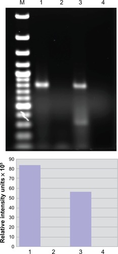
These data suggest a strong association might exist between hnRNP A1 and the SPGs. To prove such an association exists, we tested for molecular interactions between hnRNP A1 and spastin (SPG4). Following immunoprecipitation of neuronal lysates with anti-hnRNP A1 antibodies, we found that spastin protein was bound to hnRNP A1.Citation11 This interaction was confirmed by immunohistochemistry, which revealed that spastin and hnRNP A1 colocalized within the nuclei of neurons.Citation11 In separate experiments (), we performed immunoprecipitation of neuronal lysates using hnRNP A1 antibodies, and examined the resulting complex for the presence of spastin mRNA. Following immunoprecipitation with anti-hnRNP A1 antibodies, there was enrichment of the spastin mRNA signal indicative of the binding of spastin mRNA to hnRNP A1 (unpublished observation) (). In contrast, there was no signal following immunoprecipitation with a nonspecific antibody, confirming the specificity of spastin mRNA binding to hnRNP A1. These experiments confirm a biological interaction between the anti-M9 autoimmune response and neurodegeneration, as suggested by the bioinformatics analyses.Citation11
Figure 3 Spastin mRNA is bound to heterogeneous nuclear ribonucleoprotein (hnRNP) A1 in neuronal cells. Upper panel: agarose gel. Compared to the neuronal lysate without immunoprecipitation or input (lane 3), there is an enriched spastin mRNA signal following immunoprecipitation with anti-hnRNP A1 mouse monoclonal antibodies (lane 1). In contrast, spastin mRNA was not isolated following immunoprecipitation with a nonspecific control antibody – mouse IgG (lane 2). Lane 4 used spastin primers without lysate (control for DNA contamination). Lower panel: the image was analyzed using ImageQuant software, which provided relative fluorescent intensity of the bands.
A link between autoimmunity and mechanisms of neurodegeneration
In summary, HAM/TSP and MS patients make antibodies to an epitope contained within the M9 region of hnRNP A1, an RBP that plays a critical role in RNA metabolism.Citation11,Citation130,Citation131 Alterations in RBP function are associated with neurodegeneration.Citation103 Anti-M9 antibodies caused neurodegeneration, loss of neuronal processes, and altered genes related to hnRNP A1 function and mechanisms of neurodegeneration associated with the clinical phenotype of HAM/TSP and progressive MS patients.Citation11 Thus, a link between autoimmunity, clinical phenotype, and neurodegeneration has been identified. M9 is the nuclear export sequence and nuclear localization sequence of hnRNP A1, which is required for the “nonclassical” (transportin-mediated) pathway of nucleocytoplasmic transport.Citation14,Citation127,Citation141,Citation144 This is of particular interest considering that importins and exportins, which are required for “classical” (β-importin-mediated) nucleocytoplasmic transport, play a crucial role in nerve injury,Citation145–Citation148 and recently were shown to contribute to neurodegeneration in MS.Citation148,Citation149 Specifically, in neurons exposed to TNF-α, the transcription factor histone deacetylase 1 was transported from the nucleus to the cytoplasm utilizing the β-importin-mediated nucleocytoplasmic transport system, which resulted in neurodegeneration in a model of MS.Citation148 Similar findings were observed in experiments involving the transport of hnRNP A1 (unpublished observation) (). Interestingly, spastin contains several nuclear export sequences and nuclear localization sequences, and thus is involved in classical (β-importin-mediated) nucleocytoplasmic transport.Citation150 Our data indicate a molecular interaction exists between spastin and hnRNP A1 (), thus implicating an interaction between classical and nonclassical nucleocytoplasmic transport, which is a novel experimental observation. How might the anti-M9 immune response directed at the nonclassical nucleocytoplasmic transport system alter the classical nucleocytoplasmic transport system? Although this is not yet known, several possibilities exist. Notably, both the nonclassical and classical nucleocytoplasmic transport pathways are tightly regulated and require the binding of RanGTP to a β-karyopherin (transportin for hnRNP A1 and a β-importin for spastin) to function ().Citation11,Citation139–Citation142,Citation151 Thus, both pathways are regulated by the same system and an interruption in one may affect the other. For example, binding of anti-M9 antibodies to hnRNP A1 might interrupt the finely tuned regulation of RanGTP, thus altering the classical nucleocytoplasmic transport of spastin. Alternatively, anti-M9 antibodies might sterically hinder hnRNP A1’s binding to spastin protein. Finally, considering our data showing that spastin RNA binds hnRNP A1, anti-M9 antibodies might interfere with spastin’s transport and subsequent translation or localization to specific neuronal sites. Importantly, in addition to interaction with the nuclear pore and nucleocytoplasmic transport, spastin contributes to the function of other neuronal processes (). Spastin contains an AAA site and thus is a member of the ATPases associated with various cellular activities, which are involved in microtubule regulation, as well as proteosome and endosome function.Citation28,Citation100,Citation102 Spastin contains a microtubule interacting and trafficking protein site,Citation100,Citation152 and has been shown to play a role in microtubule stability and axonal transport in neurons,Citation100 and in turn in normal synaptic growth and transmission.Citation101 In addition, spastin has been shown to play an important role in axonal transport, which when disrupted results in neurodegeneration.Citation28,Citation99,Citation100,Citation152
Figure 4 Heterogeneous nuclear ribonucleoprotein (hnRNP) A1 localization following TNF-α exposure in neurons. Without TNF-α exposure, hnRNP A1 is localized to nuclei (*) in dNT-2 neurons (left panels) (blue nuclear stain is diamidino-2-phenylindole; green stain is immunohistochemistry using an anti-hnRNP A1 antibody). Following exposure to TNF-α (400 ng/mL, 50 mM glutamate, 30 minutes), hnRNP A1 is also found in the cytoplasm (arrowheads) and neuronal processes (arrow) of neurons (lower right panel).
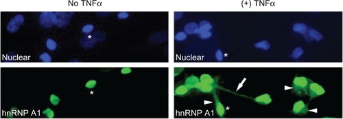
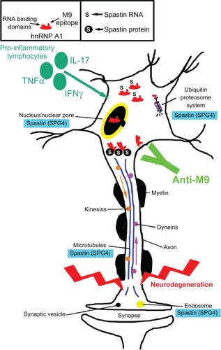
Figure 5 Potential contribution of the anti-heterogeneous nuclear ribonucleoprotein (hnRNP) A1 M9 immune response to neurodegeneration in immune-mediated neurological disease. Multiple sclerosis and human T-lymphotropic virus type 1-associated myelopathy/tropical spastic paraparesis patients develop antibodies to an epitope contained within the M9 region of hnRNP A1 (box). hnRNP A1 has been shown to interact molecularly with spastin RNA and protein (box and figure). The anti-M9 immune response altered spastin RNA levels, which may alter spastin function at multiple sites within neurons (blue boxes). The combination of proinflammatory cytokines and the anti-M9 immune response might contribute to neurodegeneration in immune-mediated neurological disease.
Spastin is just one example of how the anti-hnRNP A1 M9 immune response might contribute to neurodegeneration. As shown in , other genes were altered by the anti-M9 immune response.Citation11 Future studies are needed to tease out the details of how these other groups of genes might contribute to mechanisms of neurodegeneration, including those potentially related to the progressive cognitive decline seen in MS patients,Citation35,Citation153,Citation154 which has not yet been addressed in this model.
Conclusion
Taken together, these data suggest that neurodegeneration in MS involves multiple processes. One of several possible links between autoimmunity and neurodegeneration in neurological disease has been described. The target is hnRNP A1 in neurons and the immune response is antibodies to M9, its shuttling domain required for nucleocytoplasmic transport. Anti-M9 antibodies identified integrated networks of genes that maintain neuronal and axonal function and contribute to mechanisms of neurodegeneration related to the clinical phenotype expressed by patients with immune-mediated neurological diseases, such as progressive MS and HAM/TSP. Comprehensive analyses of this type of phenotype-specific mechanism of neurodegeneration may reveal novel strategies to treat these unremitting progressive neurological diseases.
Acknowledgments
This work is based upon work supported by the Office of Research and Development, Medical Research Service, Department of Veterans Affairs. This study was funded by a VA Merit Review Award (to MCL) and the University of Tennessee Health Science Center Multiple Sclerosis Research Fund.
Disclosure
Drs Michael Levin and Sangmin Lee have a patent pending titled “Biomarker for neurodegeneration in neurological disease.” All other authors report no conflicts of interest in this paper.
References
- DuttaRTrappBDPathogenesis of axonal and neuronal damage in multiple sclerosisNeurology20076822 Suppl 3S22S31 discussion S43–S5417548565
- PetersonJWTrappBDNeuropathobiology of multiple sclerosisNeurol Clin2005231107129vivii15661090
- NoseworthyJLucchinettiCRodgriguezMWeinshenkerBMultiple sclerosisN Engl J Med20003431393895211006371
- LassmannHBruckWLucchinettiCFThe immunopathology of multiple sclerosis: an overviewBrain Pathol200717221021817388952
- FilippiMRovarisMRoccaMAImaging primary progressive multiple sclerosis: the contribution of structural, metabolic, and functional MRI techniques. Mult SclerJun200410Suppl 1S36S44 discussion S44–S45
- BarkhofFCalabresiPAMillerDHReingoldSCImaging outcomes for neuroprotection and repair in multiple sclerosis trialsNat Rev Neurol20095525626619488083
- DeLucaGCRamagopalanSVCaderMZThe role of hereditary spastic paraplegia related genes in multiple sclerosis. A study of disease susceptibility and clinical outcomeJ Neurol200725491221122617420921
- SoldanSBertiRSalemNAssociation of human herpes virus 6 (HHV-6) with multiple sclerosis: increased IgM response to HHV-6 early antigen and detection of serum HHV-6 DNANat Med1997312139413979396611
- SriramSStrattonCYaoSChlamydia pneumonaie infection of the central nervous system in multiple sclerosisAnn Neurol199946161410401775
- ThackerELMirzaeiFAscherioAInfectious mononucleosis and risk for multiple sclerosis: a meta-analysisAnn Neurol200659349950316502434
- LeeSXuLShinYA potential link between autoimmunity and neurodegeneration in immune-mediated neurological diseaseJ Neuroimmunol20112351–2566921570130
- LeeSLevinMCMolecular mimicry in neurological disease: what is the evidence?Cell Mol Life Sci2008657–81161117518193392
- LeeSMMorocosYJangHStuartJMLevinMCHTLV-1 induced molecular mimicry in neurologic diseaseOldstoneMMolecular Mimicry: Infection Inducing Autoimmune DiseaseNew YorkSpringer2005
- LevinMCLeeSMKalumeFAutoimmunity due to molecular mimicry as a cause of neurological disease. Nat MedMay200285509513
- WucherpfennigKWInfectious triggers for inflammatory neurological diseasesNat Med20028545545711984586
- PoeiszBRuscettiFGazdarABunnPMinnaJGalloRDetection and isolation of type C retrovirus particles from fresh and cultured lymphocytes of a patient with cutaneous T cell lymphomaProc Natl Acad Sci19807712741574196261256
- GessainAVernantJMaursLAntibodies to human T-lymphotropic virus I in patients with tropical spastic paraparesisLancet198524074102863442
- OsameMUsukuKIzumoSHTLV-I associated myelopathy, a new clinical entityLancet19861848810311032
- LevinMLehkyTFlerlageNImmunopathogenesis of HTLV-1 associated neurologic disease based on a spinal cord biopsy from a patient with HTLV-1 associated myelopathy/tropical spastic paraparesis (HAM/TSP)N Engl J Med19973368398459062093
- LevinMCJacobsonSHTLV-I associated myelopathy/tropical spastic paraparesis (HAM/TSP): a chronic progressive neurologic disease associated with immunologically mediated damage to the central nervous systemJ Neurovirol1997321261409111175
- AraujoAQSilvaMTThe HTLV-1 neurological complexLancet Neurol20065121068107617110288
- VerdonckKGonzalezEVan DoorenSVandammeAMVanhamGGotuzzoEHuman T-lymphotropic virus 1: recent knowledge about an ancient infectionLancet Infect Dis20077426628117376384
- JacobsonSThe NK cell as a new player in the pathogenesis of HTLV-I associated neurologic diseaseVirulence2010118921178408
- MartinFBanghamCRCiminaleVConference highlights of the 15th International Conference on Human Retrovirology: HTLV and related retroviruses, June 4–8, 2011. Leuven, Gembloux, BelgiumRetrovirology201188622035054
- Puccioni-SohlerMYamanoYRiosMDifferentiation of HAM/TSP from patients with multiple sclerosis infected with HTLV-INeurology200768320621317224575
- KhanRBBertoriniTELevinMCHTLV-1 and its neurological complicationsNeurologist20017527127812803668
- SoderblomCBlackstoneCTraffic accidents: molecular genetic insights into the pathogenesis of the hereditary spastic paraplegiasPharmacol Ther20061091–2425616005518
- SalinasSProukakisCCrosbyAWarnerTTHereditary spastic paraplegia: clinical features and pathogenetic mechanismsLancet Neurol20087121127113819007737
- UmeharaFAbeMKoreedaYIzumoSOsameMAxonal damage revealed by accumulation of beta-amyloid precursor protein in HTLV-I-associated myelopathyJ Neurol Sci200017629510110930590
- JerniganMMorcosYLeeSMDohanFCJrRaineCLevinMCIgG in brain correlates with clinicopathological damage in HTLV-1 associated neurologic diseaseNeurology20036081320132712707436
- DelucaGCEbersGCEsiriMMThe extent of axonal loss in the long tracts in hereditary spastic paraplegiaNeuropathol Appl Neurobiol200430657658415540998
- DeLucaGCEbersGCEsiriMMAxonal loss in multiple sclerosis: a pathological survey of the corticospinal and sensory tractsBrain2004127Pt 51009101815047586
- IzumoSNeuropathology of HTLV-1-associated myelopathy (HAM/TSP)Neuropathology Epub6212010
- TrappBDNaveKAMultiple sclerosis: an immune or neurodegenerative disorder?Annu Rev Neurosci20083124726918558855
- GeurtsJJBarkhofFGrey matter pathology in multiple sclerosisLancet Neurol20087984185118703006
- KutzelniggALucchinettiCFStadelmannCCortical demyelination and diffuse white matter injury in multiple sclerosisBrain2005128Pt 112705271216230320
- KornekBStorchMKWeissertRMultiple sclerosis and chronic autoimmune encephalomyelitis: a comparative quantitative study of axonal injury in active, inactive, and remyelinated lesionsAm J Pathol2000157126727610880396
- GoldRLiningtonCLassmannHUnderstanding pathogenesis and therapy of multiple sclerosis via animal models: 70 years of merits and culprits in experimental autoimmune encephalomyelitis researchBrain2006129Pt 81953197116632554
- Aboul-EneinFWeiserPHoftbergerRLassmannHBradlMTransient axonal injury in the absence of demyelination: a correlate of clinical disease in acute experimental autoimmune encephalomyelitisActa Neuropathol (Berl)2006111653954716718350
- LassmannHMultiple sclerosis: is there neurodegeneration independent from inflammation?J Neurol Sci20072591–23617367814
- BjartmarCWujekJRTrappBDAxonal loss in the pathology of MS: consequences for understanding the progressive phase of the diseaseJ Neurol Sci2003206216517112559505
- LassmannHvan HorssenJThe molecular basis of neurodegeneration in multiple sclerosisFEBS Lett2011585233715372321854776
- BennettJLStuveOUpdate on inflammation, neurodegeneration, and immunoregulation in multiple sclerosis: therapeutic implicationsClin Neuropharmacol200932312113219483479
- SteinmanLMixed results with modulation of TH-17 cells in human autoimmune diseasesNat Immunol2010111414420016509
- McFarlandHFMartinRMultiple sclerosis: a complicated picture of autoimmunityNat Immunol20078991391917712344
- LisakRPNeurodegeneration in multiple sclerosis: defining the problemNeurology20076822 Suppl 3S5S12 discussion S43–S5417548569
- FrohmanEMRackeMKRaineCSMultiple sclerosis – the plaque and its pathogenesisN Engl J Med2006354994295516510748
- FrischerJMBramowSDal-BiancoAThe relation between inflammation and neurodegeneration in multiple sclerosis brains. BrainMay2009132Pt 511751189
- TrappBPetersonJRansohoffRRudickRMorkSBoLAxonal transection in the lesions of multiple sclerosisN Engl J Med199833852782859445407
- LassmannHLucchinettiCFCortical demyelination in CNS inflammatory demyelinating diseasesNeurology200870533233318227414
- LucchinettiCFPopescuBFBunyanRFInflammatory cortical demyelination in early multiple sclerosisN Engl J Med2011365232188219722150037
- PetersonJWBoLMorkSChangATrappBDTransected neurites, apoptotic neurons, and reduced inflammation in cortical multiple sclerosis lesionsAnn Neurol200150338940011558796
- FisherELeeJCNakamuraKRudickRAGray matter atrophy in multiple sclerosis: a longitudinal studyAnn Neurol200864325526518661561
- FisnikuLKChardDTJacksonJSGray matter atrophy is related to long-term disability in multiple sclerosisAnn Neurol200864324725418570297
- GeurtsJJIs progressive multiple sclerosis a gray matter disease?Ann Neurol200864323023218825672
- SerafiniBRosicarelliBMagliozziRStiglianoEAloisiFDetection of ectopic B-cell follicles with germinal centers in the meninges of patients with secondary progressive multiple sclerosisBrain Pathol200414216417415193029
- MagliozziRHowellOWReevesCA gradient of neuronal loss and meningeal inflammation in multiple sclerosisAnn Neurol201068447749320976767
- GanterPPrinceCEsiriMMSpinal cord axonal loss in multiple sclerosis: a post-mortem studyNeuropathol Appl Neurobiol199925645946710632896
- LovasGSzilagyiNMajtenyiKPalkovitsMKomolySAxonal changes in chronic demyelinated cervical spinal cord plaquesBrain2000123Pt 230831710648438
- DeLucaGCWilliamsKEvangelouNEbersGCEsiriMMThe contribution of demyelination to axonal loss in multiple sclerosisBrain2006129Pt 61507151616597651
- EvangelouNDeLucaGCOwensTEsiriMMPathological study of spinal cord atrophy in multiple sclerosis suggests limited role of local lesionsBrain2005128Pt 1293415548559
- IzumoSHiguchiIIjichiTNeuropathological study in two autopsy cases of HTLV-1 associated myelopathy (HAM)IwasakiYNeuropathology of HAM/TSP in JapanProceedings of the First Workshop on Neuropathology of Retrovirus InfectionsAugust 31, 1989Tokyo, JapanSendai, JapanTohuku University Press1989717
- IzumoSUmeharaFOsameMHTLV-I-associated myelopathyNeuropathology200020SupplS65S6811037191
- EvangelouIEOhUMassoudRJacobsonSHTLV-I-associated myelopathy/tropical spastic paraparesis: semiautomatic quantification of spinal cord atrophy from 3-dimensional MR imagesJ Neuroimaging Epub232012
- FurbyJHaytonTAndersonVMagnetic resonance imaging measures of brain and spinal cord atrophy correlate with clinical impairment in secondary progressive multiple sclerosisMult Scler20081481068107518632782
- UmeharaFIzumoSNakagawaMImmunocytochemical analysis of the cellular infiltrate in the spinal cord lesions in HTLV-I-associated myelopathyJ Neuropathol Exp Neurol19935244244308355031
- MatsuokaETakenouchiNHashimotoKPerivascular T cells are infected with HTLV-I in the spinal cord lesions with HTLV-I-associated myelopathy/tropical spastic paraparesis: double staining of immunohistochemistry and polymerase chain reaction in situ hybridizationActa Neuropathol (Berl)19989643403469796997
- MoritoyoTReinhartTMoritoyoHHuman T-lymphotropic virus type I associated myelopathy and tax gene expression in CD4+ T lymphocytesAnn Neurol199640184908687197
- ArayaNSatoTYagishitaNHuman T-lymphotropic virus type 1 (HTLV-1) and regulatory T cells in HTLV-1-associated neuroinflammatory diseaseViruses2011391532154821994794
- MatsuuraEYamanoYJacobsonSNeuroimmunity of HTLV-I InfectionJ Neuroimmune Pharmacol20105331032520437106
- TakenouchiNYaoKJacobsonSImmunopathogensis of HTLV-I associated neurologic disease: molecular, histopathologic, and immunologic approachesFront Biosci200492527253915353305
- BanghamCROsameMCellular immune response to HTLV-1Oncogene200524396035604616155610
- BanghamCRMeekingsKToulzaFThe immune control of HTLV-1 infection: selection forces and dynamicsFront Biosci20091428892903
- BanghamCRThe immune response to HTLV-ICurr Opin Immunol200012439740210899027
- JacobsonSShidaHMcFarlinDEFauciASKoenigSCirculating CD8+ cytotoxic T lymphocytes specific for HTLV-I pX in patients with HTLV-I associated neurological diseaseNature199034862982452482146511
- LalRBGiamC-ZColiganJERudolphDLDifferential immune responsiveness to the immunodominant epitopes of regulatory proteins (tax and rex) in human T cell lymphotropic virus type 1-associated myelopathyJ Infect Dis199416934965038158021
- KubotaRFurukawaYIzumoSUsukuKOsameMDegenerate specificity of HTLV-1-specific CD8+ T cells during viral replication in patients with HTLV-1-associated myelopathy (HAM/TSP)Blood200310183074308112480698
- GoonPKHanonEIgakuraTHigh frequencies of Th1-type CD4(+) T cells specific to HTLV-1 Env and Tax proteins in patients with HTLV-1-associated myelopathy/tropical spastic paraparesisBlood20029993335334111964301
- JefferyKJUsukuKHallSEHLA alleles determine human T-lymphotropic virus-I (HTLV-I) proviral load and the risk of HTLV-I-associated myelopathyProc Natl Acad Sci U S A19999673848385310097126
- MacnamaraARowanAHilburnSHLA class I binding of HBZ determines outcome in HTLV-1 infectionPLoS Pathog201069e100111720886101
- SaxenaAMartin-BlondelGMarsLTLiblauRSRole of CD8 T cell subsets in the pathogenesis of multiple sclerosisFEBS Lett2011585233758376321910991
- SchneiderRMohebianyANIferganIB cell-derived IL-15 enhances CD8 T cell cytotoxicity and is increased in multiple sclerosis patientsJ Immunol201118784119412821911607
- YamanoYArayaNSatoTAbnormally high levels of virus-infected IFN-gamma+ CCR4+ CD4+ CD25+ T cells in a retrovirus-associated neuroinflammatory disorderPloS One200948e651719654865
- NorrisPJHirschkornDFDeVitaDALeeTHMurphyELHuman T cell leukemia virus type 1 infection drives spontaneous proliferation of natural killer cellsVirulence201011192820640055
- NagaiMUsukuKMatsumotoWAnalysis of HTLV-1 proviral load in 202 HAM/TSP patients and 243 asymptomatic HTLV-1 carriers: high proviral load strongly predisposes to HAM/TSPJ Neurovirol19984658659310065900
- OlindoSLezinACabrePHTLV-1 proviral load in peripheral blood mononuclear cells quantified in 100 HAM/TSP patients: a marker of disease progressionJ Neurol Sci20052371–2535915972218
- MontanheiroPAOliveiraACPosada-VergaraMPHuman T-cell lymphotropic virus type I (HTLV-I) proviral DNA viral load among asymptomatic patients and patients with HTLV-I-associated myelopathy/tropical spastic paraparesisBraz J Med Biol Res200538111643164716258633
- TattermuschSSkinnerJAChaussabelDSystems biology approaches reveal a specific interferon-inducible signature in HTLV-1 associated myelopathyPLoS Pathog201281e100248022291590
- GoonPKIgakuraTHanonEHigh circulating frequencies of tumor necrosis factor alpha- and interleukin-2-secreting human T-lymphotropic virus type 1 (HTLV-1)-specific CD4+ T cells in patients with HTLV-1-associated neurological diseaseJ Virol200377179716972212915584
- WaxmanSGAxonal conduction and injury in multiple sclerosis: the role of sodium channelsNat Rev Neurosci200671293294117115075
- DuttaRTrappBMechanisms of neuronal dysfunction and degeneration in multiple sclerosisProg Neurobiol201193111220946934
- DuttaRMcDonoughJYinXMitochondrial dysfunction as a cause of axonal degeneration in multiple sclerosis patientsAnn Neurol200659347848916392116
- CampbellGRZiabrevaIReeveAKMitochondrial DNA deletions and neurodegeneration in multiple sclerosisAnn Neurol201169348149221446022
- HaiderLFischerMTFrischerJMOxidative damage in multiple sclerosis lesionsBrain2011134Pt 71914192421653539
- TrappBDStysPKVirtual hypoxia and chronic necrosis of demyelinated axons in multiple sclerosisLancet Neurol20098328029119233038
- MahadDJZiabrevaICampbellGMitochondrial changes within axons in multiple sclerosisBrain2009132Pt 51161117419293237
- FergusonBMatyszakMKEsiriMMPerryVHAxonal damage in acute multiple sclerosis lesionsBrain1997120Pt 33933999126051
- StokinGBLilloCFalzoneTLAxonopathy and transport deficits early in the pathogenesis of Alzheimer’s diseaseScience200530757131282128815731448
- De VosKJGriersonAJAckerleySMillerCCRole of axonal transport in neurodegenerative diseasesAnnu Rev Neurosci20083115117318558852
- Roll-MecakAValeRDStructural basis of microtubule severing by the hereditary spastic paraplegia protein spastinNature2008451717636336718202664
- TrottaNOrsoGRossettoMGDagaABroadieKThe hereditary spastic paraplegia gene, spastin, regulates microtubule stability to modulate synaptic structure and functionCurr Biol200414131135114715242610
- HazanJFonknechtenNMavelDSpastin, a new AAA protein, is altered in the most frequent form of autosomal dominant spastic paraplegiaNat Genet199923329630310610178
- LukongKEChangKWKhandjianEWRichardSRNA-binding proteins in human genetic diseaseTrends Genet200824841642518597886
- OrrHTFTD and ALS: genetic ties that bindNeuron201172218919022017980
- GaoFBTaylorJPRNA-binding proteins in neurological diseaseBrain Res201214621222682432
- PolymenidouMLagier-TourenneCHuttKRBennettCFClevelandDWYeoGWMisregulated RNA processing in amyotrophic lateral sclerosisBrain Res2012146231522444279
- LicatalosiDDDarnellRBSplicing regulation in neurologic diseaseNeuron20065219310117015229
- DarnellRBPosnerJBParaneoplastic syndromes involving the nervous systemN Engl J Med2003349161543155414561798
- OwensGPBennettJLLassmannHAntibodies produced by clonally expanded plasma cells in multiple sclerosis cerebrospinal fluidAnn Neurol200965663964919557869
- LovatoLWillisSNRodigSJRelated B cell clones populate the meninges and parenchyma of patients with multiple sclerosisBrain2011134Pt 253454121216828
- KeeganMPinedaAAMcClellandRLDarbyCHRodriguezMWeinshenkerBGPlasma exchange for severe attacks of CNS demyelination: predictors of responseNeurology200258114314611781423
- KeeganMKonigFMcClellandRRelation between humoral pathological changes in multiple sclerosis and response to therapeutic plasma exchangeLancet2005366948557958216099294
- ElovaaraIKuusistoHWuXRintaSDastidarPReipertBIntravenous immunoglobulins are a therapeutic option in the treatment of multiple sclerosis relapseClin Neuropharmacol2011342848921301327
- HaasJMaas-EnriquezMHartungHPIntravenous immunoglobulins in the treatment of relapsing remitting multiple sclerosis – results of a retrospective multicenter observational study over five yearsMult Scler200511556256716193894
- HauserSLWaubantEArnoldDLB-cell depletion with rituximab in relapsing-remitting multiple sclerosisN Engl J Med2008358767668818272891
- BosterAAnkenyDPRackeMKThe potential role of B cell-targeted therapies in multiple sclerosisDrugs201070182343235621142258
- MeinlEDerfussTKrumbholzMProbstelAKHohlfeldRHumoral autoimmunity in multiple sclerosisJ Neurol Sci20103061–218018220817206
- BrownDASawchenkoPETime course and distribution of inflammatory and neurodegenerative events suggest structural bases for the pathogenesis of experimental autoimmune encephalomyelitisJ Comp Neurol2007502223626017348011
- DerfussTLiningtonCHohlfeldRMeinlEAxo-glial antigens as targets in multiple sclerosis: implications for axonal and grey matter injuryJ Mol Med (Berl)201088875376120445955
- MatheyEKDerfussTStorchMKNeurofascin as a novel target for autoantibody-mediated axonal injuryJ Exp Med2007204102363237217846150
- RawesJACalabreseVPKhanOADeVriesGHAntibodies to the axolemma-enriched fraction in the cerebrospinal fluid and serum of patients with multiple sclerosis and other neurological diseasesMult Scler1997363633699493635
- NorgrenNEdelstamAStigbrandTCerebrospinal fluid levels of neurofilament light in chronic experimental autoimmune encephalomyelitisBrain Res Bull200567426426816182933
- SadatipourBTGreerJMPenderMPIncreased circulating antiganglioside antibodies in primary and secondary progressive multiple sclerosisAnn Neurol19984469809839851447
- SrivastavaRAslamMKalluriSRPotassium channel KIR4.1 as an immune target in multiple sclerosisN Engl J Med2012367211512322784115
- CrossAHWaubantEAntibodies to potassium channels in multiple sclerosisN Engl J Med2012367217217422784120
- LevinMKrichavskyMBerkJNeuronal molecular mimicry in immune mediated neurologic diseaseAnn Neurol199844187989667596
- LeeSMDunnavantFDJangHZuntJLevinMCAutoantibodies that recognize functional domains of hnRNPA1 implicate molecular mimicry in the pathogenesis of neurological diseaseNeurosci Lett20064011–218819316600502
- LeeSShinYMarlerJLevinMCPost-translational glycosylation of target proteins implicate molecular mimicry in the pathogenesis of HTLV-1 associated neurological diseaseJ Neuroimmunol20082041–214014818793806
- LeeSShinYClarkDGotuzzoELevinMCCross-reactive antibodies to target proteins are dependent upon oligomannose glycosylated epitopes in HTLV-1 associated neurological diseaseJ Clin Immunol201232473674522392044
- DreyfussGKimVNKataokaNMessenger-RNA-binding proteins and the messages they carryNat Rev Mol Cell Biol20023319520511994740
- HanSPTangYHSmithRFunctional diversity of the hnRNPs: past, present and perspectivesBiochem J2010430337939220795951
- LevinMCLeeSMMorcosYBradyJStuartJCross-reactivity between immunodominant human T lymphotropic virus type I tax and neurons: implications for molecular mimicryJ Infect Dis2002186101514151712404172
- KalumeFLeeSMMorcosYCallawayJCLevinMCMolecular mimicry: cross-reactive antibodies from patients with immune-mediated neurologic disease inhibit neuronal firingJ Neurosci Res2004771828915197740
- García-VallejoFDomínguezMCTamayoOAutoimmunity and molecular mimicry in tropical spastic paraparesis/human T-lymphotropic virus-associated myelopathyBraz J Med Biol Res200538224125015785836
- SueokaEYukitakeMIwanagaKSueokaNAiharaTKurodaYAutoantibodies against heterogeneous nuclear ribonucleoprotein B1 in CSF of MS patientsAnn Neurol200456677878615497154
- YukitakeMSueokaESueoka-AraganeNSignificantly increased antibody response to heterogeneous nuclear ribonucleoproteins in cerebrospinal fluid of multiple sclerosis patients but not in patients with human T-lymphotropic virus type I-associated myelopathy/tropical spastic paraparesisJ Neurovirol200814213013518444084
- DouglasJGardnerLLeeSShinYGrooverCLevinMCAntibody transfection into neurons as a tool to study disease pathogenesisJournal of Visualized Experiments201267 pii 4154
- SchmuedLCStowersCCScalletACXuLFluoro-Jade C results in ultra high resolution and contrast labeling of degenerating neuronsBrain Res200510351243115713273
- StewartMMolecular mechanism of the nuclear protein import cycleNat Rev Mol Cell Biol20078319520817287812
- ContiEMüllerCWStewartMKaryopherin flexibility in nucleocytoplasmic transportCurr Opin Struct Biol200616223724416567089
- CookABonoFJinekMContiEStructural biology of nucleocytoplasmic transportAnnu Rev Biochem20077664767117506639
- LeeBJCansizogluAESuelKELouisTHZhangZChookYMRules for nuclear localization sequence recognition by karyopherin beta 2Cell2006126354355816901787
- HomayouniRHeinrichKWeiLBerryMWGene clustering by latent semantic indexing of MEDLINE abstractsBioinformatics200521110411515308538
- MichaelWMChoiMDreyfussGA nuclear export signal in hnRNP A1: a signal-mediated, temperature-dependent nuclear protein export pathwayCell19958334154228521471
- HanzSPerlsonEWillisDAxoplasmic importins enable retrograde injury signaling in lesioned nerveNeuron20034061095110414687545
- ThompsonKROtisKOChenDYZhaoYO’DellTJMartinKCSynapse to nucleus signaling during long-term synaptic plasticity; a role for the classical active nuclear import pathwayNeuron2004446997100915603742
- YudinDHanzSYooSLocalized regulation of axonal RanGTPase controls retrograde injury signaling in peripheral nerveNeuron200859224125218667152
- KimJYShenSDietzKHDAC1 nuclear export induced by pathological conditions is essential for the onset of axonal damageNat Neurosci201013218018920037577
- MillerRHRenegade nuclear enzymes disrupt axonal integrityNat Neurosci201013214314420104203
- BeetzCBrodhunMMoutzourisKIdentification of nuclear localisation sequences in spastin (SPG4) using a novel Tetra-GFP reporter systemBiochem Biophys Res Commun200431841079108415147984
- RebaneAAabASteitzJATransportins 1 and 2 are redundant nuclear import factors for hnRNP A1 and HuRRNA200410459059915037768
- SalinasSCarazo-SalasREProukakisCSchiavoGWarnerTTSpastin and microtubules: functions in health and diseaseJ Neurosci Res200785122778278217348041
- LazeronRHde SonnevilleLMScheltensPPolmanCHBarkhofFCognitive slowing in multiple sclerosis is strongly associated with brain volume reductionMult Scler200612676076817263004
- LazeronRHBoringaJBSchoutenMBrain atrophy and lesion load as explaining parameters for cognitive impairment in multiple sclerosisMult Scler200511552453116193889