Abstract
Background
Ventilator-associated pneumonia is a deadly nosocomial infection caused by contaminated endotracheal tubes. It has been shown that polyvinyl chloride (PVC, the endotracheal tube substrate) coated with elemental selenium nanoparticles reduces bacterial adherence and proliferation on PVC by over 99%. However, it is not known if selenium nanoparticles elicit a cytotoxic effect in vitro. The purpose of this study was to investigate the cytotoxic effects of PVC coated with selenium nanoparticles on fibroblasts, which are mammalian cells central to endotracheal tube intubation.
Methods
Different concentrations of selenium nanoparticles were precipitated onto the PVC surface by reduction of selenium salts using glutathione. Characterization of PVC coated with selenium nanoparticles was done by scanning electron microscopy, energy dispersive x-ray, and contact angle measurements. For the cytotoxicity experiments, fibroblasts were seeded at a density of 5000 cm2 onto PVC coated with three different concentrations of selenium nanoparticles (high, medium, low) and incubated for 4 hours (adhesion) as well as for 24 hours and 72 hours (proliferation). The half-maximal inhibitory concentration (IC50) value was determined after 72 hours using an ultrahigh concentration. MTT assays were used to assess cell viability at the indicated time points.
Results
The three concentrations of selenium nanoparticles did not elicit a cytotoxic effect after 72 hours (P < 0.01, n = 3). It was found that the IC50value was at the ultrahigh concentration of selenium nanoparticles. The nanoparticulate elemental selenium concentration previously shown to decrease the function of bacteria was shown not to cause a cytotoxic effect on fibroblasts in vitro.
Conclusion
These findings demonstrate great selectivity between bacteria and healthy cells, and are a viable option for coating endotracheal tubes in order to prevent ventilator-associated pneumonia.
Introduction
Medical device-related infection is a growing clinical concern. For example, ventilator-associated pneumonia is the second most common nosocomial infection in the US.Citation1 It is also the deadliest hospital-acquired infection in children and adults, affecting 23% of all patients receiving mechanical ventilation for more than 24 hours (approximately 250,000 annually).Citation2 The Staphylococcus aureus bacterium is responsible for 25% of all ventilator-associated pneumonia infections and is of great concern to clinicians due to its multidrug resistant strain, ie, methicillin-resistant S. aureus (MRSA).Citation3 Ventilator-associated pneumonia can also be very costly for both patients and hospitals, adding an estimated $40,000 dollars to each hospital admission due to the extra intensive care required for treatment.Citation4
Inadvertent exposure to bacteria infects up to 54% of all endotracheal tubes.Citation5 These intubated tubes provide a conduit for bacterial access from the extracorporeal environment to the more sterile area of the lungs. Through cell division and biochemical recruitment of extracellular DNA, proteins, and exopolysaccharides, these adherent bacteria form an aggregate of microorganisms called a biofilm. Ventilation through these infected endotracheal tubes, or even condensation of humidified air, breaks off pieces of the biofilm, causing gross and micro aspiration of bacteria into the distal bronchi, followed by bacterial proliferation and parenchymal invasion.Citation6–Citation8 Endotracheal tube intubation also thwarts the cough reflex and compromises mucociliary clearance, making it nearly impossible to expel contaminated secretions.Citation9,Citation10 The current techniques for preventing ventilator-associated pneumonia, ie, sterilization of equipment, better hospital practices, systemic antibiotic administration, and re-engineering of the endotracheal tube, have not proven to be efficacious.
As a result, the most promising and straightforward strategy for decreasing ventilator-associated pneumonia is to fabricate endotracheal tubes with antibacterial agents that resist bacterial attachment. However, currently used materials, such as silver and silver compounds, eg, silver sulfadiazine, have been shown to be ineffective at preventing bacterial attachment in the long term.Citation11–Citation13 Moreover, the use of these silver-coated endotracheal tubes is contraindicated for persons who are allergic to silver;Citation14 silver is also not naturally found in the body. In contrast, selenium is an essential trace element found in the human body. Adults require 55–70 μg of selenium per day, with an upper tolerable limit of 400 μg, to make selenoproteins, which play a vital role in the human antioxidant defense system, redox control of cell reactions, and thyroid hormone metabolism. Elemental selenium also naturally possesses antibacterial properties. Studies of these antibacterial properties have demonstrated that elemental selenium-enriched probioticsCitation15 and organoselenium compoundsCitation16,Citation17 have antibacterial effects in vitro and in vivo against Escherichia coli and S. aureus, respectively. Furthermore, studies have demonstrated that selenium nanoparticles reduce S. aureus adhesion and proliferation on commercial endotracheal tubes by over 99%.Citation18 Overall, the bactericidal action of selenium is due to its ability to catalyze the oxidation of intracellular thiols, causing death of bacteria.Citation18–Citation20
Symptoms of selenium toxicity (called selenosis) include gastrointestinal disorders, alopecia, fatigue, and irritability.Citation21 The chronic dose of selenium that results in extreme cases of toxicity is very high, at approximately 2400 μg/day.Citation21 Studies on the toxicity of elemental selenium have been promising in demonstrating that its use will be benign.Citation22 Regarding toxicity of selenium nanoparticles, studies have shown that selenium nanoparticles have an efficacy comparable with that of selenium compounds as an antioxidant, but have a greatly reduced risk of toxicity.Citation23,Citation24 However, studies investigating the toxicity of selenium have focused on systemic toxicity in vivo and have not focused on in vitro toxicity to mammalian cells central to endotracheal tube intubation, such as fibroblasts. Thus, the objective of this study was to ascertain the cytotoxic effect of selenium nanoparticles on fibroblasts in vitro.
Materials and methods
Synthesis of selenium nanoparticles
PVC samples (6 mm × 3 mm × 1 mm) were cut from commercial endotracheal tubes (Hudson RCI, Research Triangle Park, NC) using a Fiskars rectangular hole punch (Fiskars Corporation, Helsinki, Finland). The PVC samples were then rinsed in increasing concentrations of ethanol (30%, 50%, 70%, and 100%) for 10 minutes each and then air-dried in a sterile laminar flow hood for 12 hours. The cleaned and sterilized PVC were used as controls and base substrates for the colloidal precipitation with four different concentrations of selenium nanoparticles, ie, 46.5 μg/mL (ultrahigh), 31 μg/mL (high), 15.5 μg/mL (medium), and 7.8 μg/mL (low). For the ultrahigh concentration, 6.25 mL of deionized water (DIH2O) and 6.25 mL of 100 mM glutathione (reduced form, Alfa Aesar, Ward Hill, MA) were added to a 50 mL conical tube containing the PVC samples, followed by 17.5 mL of 25 mM sodium selenite (Na2SeO3, Alfa Aesar). After gentle mixing, 2 mL of 1 M sodium hydroxide (Alfa Aesar) was added to bring the pH of the mixture into the alkaline range. The same reagents and methodology were used for the high, medium, and low concentrations, albeit the reagent amounts changed. For the high concentration, 7.5 mL of DIH2O, 7.5 mL of glutathione, 15 mL of Na2SeO3, and 2 mL of sodium hydroxide were used. For the medium concentration, 10 mL of DIH2O, 10 mL of glutathione, 10 mL of Na2SeO3, and 2 mL of sodium hydroxide were used. For the low concentration, 12.5 mL of DIH2O, 12.5 mL of glutathione, 5 mL of Na2SeO3, and 2 mL of sodium hydroxide were used (). In all reactions, the reduction of sodium selenite by glutathione immediately formed selenium nanoparticles, which were precipitated onto the PVC samples as visualized by a color change of the reactant solution from clear to milky red.Citation18 For each concentration, the conical tubes were left untouched for 30 minutes to allow the selenium nanoparticles to precipitate onto the PVC samples both intraluminally and extraluminally. The substrates were then removed and rinsed in 10 mL of DIH2O for 24 hours. The uncoated (control) and selenium nanoparticle-coated substrates were immersed in 70% ethanol for 30 minutes. The ethanol was then removed and the substrates were sterilized in a laminar flow hood under ultraviolet light for 24 hours.
Table 1 Final concentration of selenium in the colloidal synthesis solution
Characterization of selenium nanoparticles
For material surface characterization studies, the PVC samples were sputter-coated with a thin layer (approximately 90Å) of gold and observed under scanning electron microscopy (LEO 1530VP FE-4800) at 5 kV. For chemical characterization, energy dispersive x-ray spectroscopy with an acceleration voltage of 20 kV was employed. Contact angle measurements of uncoated and selenium nanoparticle-coated PVC were also conducted. Contact angles easily and quickly identified the hydrophobicity/hydrophilicity of a proposed material for medical applications, which is of paramount importance for the interaction between the substrate and biological entities in the body. To measure the hydrophobicity/hydrophilicity of uncoated and selenium nanoparticle-coated PVC, a drop shape analysis system (DSA-10, Kruss, Hamburg, Germany) with analysis software (DSA1 v 1.80) was used. A 2 μL drop of water was placed onto the substrates, and contact angles were recorded within 10 seconds at room temperature. All characterization measurements were repeated nine times for each sample.
In vitro assays for cytotoxicity experiments
To investigate cytotoxicity via cell adhesion and proliferation of PVC coated with selenium nanoparticles, a cell line of rat dermal fibroblasts was obtained from the American Type Culture Collection (CRL-1213, Manassas, VA). The fibroblasts were grown in Eagle’s Minimum Essential Medium (ATCC) supplemented with 10% fetal bovine serum (Hyclone, Rockford, IL), and 1% penicillin-streptomycin (Hyclone) in a standard cell culture incubator (37°C, humidified, 5% CO2, 20% O2 environment, nonshaking). Every 24 hours the old medium was removed, the cells were cleaned three times with phosphate-buffered saline, and new medium was added. After confluence was reached (approximately 72 hours), the cells were seeded at a density of approximately 5000 cells/cm2 in sterile 24-well plates that contained a PVC sample coated with the high, medium, or low concentration of selenium nanoparticles; uncoated PVC samples were used as controls. The samples were incubated in a standard cell culture incubator for 4 hours for the cell adhesion assay and 24 and 72 hours for the proliferation assays. The reduction of tetrazolium salts in a colorimetric MTT assay is now widely accepted as a reliable method for examining cell adhesion and proliferation.Citation25,Citation26 For the MTT assay, a CellTiter 96 nonradioactive assay (ATCC) was used and the absorbance, which corresponded to total cell number, was recorded using a microplate spectrophotometer (570 nm, SpectraMax, Molecular Devices, Sunnyvale, CA). To ascertain the IC50 value, the ultrahigh concentration of selenium nanoparticles was used. The IC50 value is a quantitative measurement that indicates the concentration of selenium nanoparticles needed to inhibit a biological process (ie, fibroblast viability) by half. The samples were incubated in a standard cell culture incubator for 72 hours and then a MTT assay was used to determine cell viability.
All experiments were conducted in triplicate and repeated three times. Data were collected, and significant differences were assessed using the probability associated with one-tailed Student’s t-tests.
Results
Synthesis of selenium nanoparticles
Visual characterization by scanning electron microscopy of the coating density of PVC coated and not coated with selenium nanoparticles can be seen in . The morphology in the high-magnification tilted scanning electron microscopic image () strongly suggests that the selenium nanoparticles were bonded (by physicochemical forces, not covalently) to the substrate. In fact, previous studies have tried to remove the adherent selenium nanoparticles using sonication, but could not.Citation18 The surface coverage of selenium nanoparticles for the medium concentration (determined from scanning electron microscopic images using an image processing program, ie, ImageJ, National Institutes of Health, Bethesda, MD) was approximately 10%, or about 300 μg. X-ray spectra were used to confirm the chemistry of the selenium nanoparticles. Selenium peaks were observed in the area coated with selenium nanoparticles, which confirmed that the nanoparticles were in fact elemental selenium (). Contact angle measurements demonstrated that selenium nanoparticles reduced the contact angles, or made the PVC more hydrophilic ().
Figure 1 Scanning electron microscopic images of PVC coated with selenium nanoparticles (right panel) and not coated with selenium nanoparticles (left panel). Arrows indicate selenium nanoparticles (nonexhaustive). The selenium nanoparticles had sizes ranging from approximately 80 nm to 200 nm and are uniformly coated on the substrates. Selenium nanoparticle coverage is approximately 10%, or 300 μg.Citation18 Previously published studies demonstrate the surface coverage for all samples of interest in this study, and are given in .Citation18
Abbreviation: PVC, polyvinyl chloride.
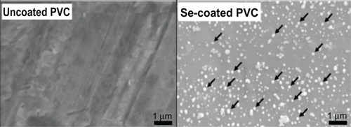
Figure 2 Scanning electron microscopic image, taken at a 45° tilt, of the medium concentration selenium nanoparticles precipitated on polyvinyl chloride.
Note: The hemispherical shape of the selenium nanoparticles on the substrate surface.
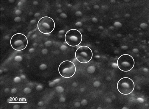
Figure 3 X-ray spectra of an uncoated region on PVC (top) and a region on PVC coated with selenium nanoparticles (bottom). Elemental selenium peaks are detected in PVC coated with selenium nanoparticles, demonstrating that the nanoparticles were in fact selenium. Peaks of carbon (C) and chlorine (Cl) present are from the PVC substrate; peaks of gold (Au) present are from the sputter-coating process.
Abbreviations: PVC, polyvinyl chloride; EDS, energy dispersive spectrum.
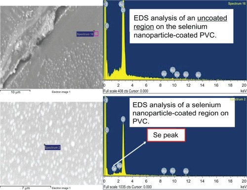
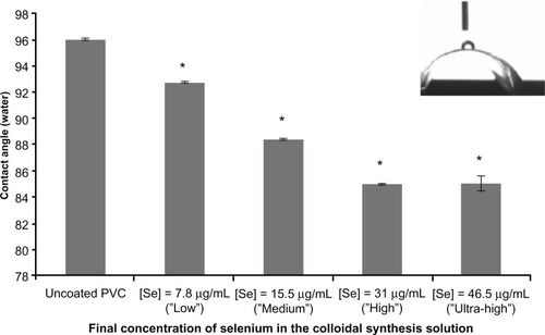
Figure 4 Water contact angles of PVC coated and not coated with selenium nanoparticles.
Notes: *P < 0.01 compared with uncoated PVC. The data are presented as the mean ± standard error of the mean (n = 3).
Abbreviation: PVC, polyvinyl chloride.
Cytotoxicity studies
The viability of fibroblasts as determined by MTT assays after the 4-hour (adhesion) time point showed that the number of live cells on PVC coated with selenium nanoparticles was significantly higher than the number of live cells on PVC not coated with selenium nanoparticles. The number of live cells also increased as the final concentration of selenium in the colloidal synthesis solution increased. Furthermore, the viability of fibroblasts as determined by MTT assays after the 24-hour and 72-hour (proliferation) time points was significantly higher on PVC coated with selenium nanoparticles as compared with the PVC not coated with selenium nanoparticles. The number of cells proliferating on the PVC coated with selenium nanoparticles between the 24-hour and 72-hour time points neither significantly increased nor decreased.
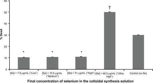
The number of live cells adhering and proliferating on PVC not coated with selenium nanoparticles from 4 hours to 72 hours also did not significantly increase (). Results from the IC50 studies revealed that the concentration of selenium nanoparticles coated on PVC needed to inhibit fibroblast viability by half was approximately 46.5 μg/mL. This is because 52% of the fibroblasts died when exposed to this ultrahigh concentration of selenium nanoparticles, or 2% more than the concentration needed to ascertain the IC50 value ().
Figure 5 Increased fibroblast viability upon exposure to PVC coated with selenium nanoparticles.
Notes:*P < 0.05 for all concentrations at the 4-hour (adhesion) time point as compared with the control; †P < 0.01 for all concentrations at the 24-hour (proliferation) time point as compared with the control; #P < 0.01 for all concentrations at the 72-hour (proliferation) time point as compared with the control. The data are presented as the mean ± standard error of the mean (n = 3).
Abbreviation: PVC, polyvinyl chloride.
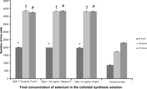
Figure 6 Half maximal inhibitory concentration (IC50 studies show a decrease in fibroblast viability after 72hours when exposed to the ultrahigh concentration of selenium nanoparticles.
Notes: *P < 0.05 for the high, medium, and low concentrations as compared with the control; †P < 0.01 for all concentrations at the ultra-high concentration as compared with the control. The data are presented as the mean ± standard error of the mean (n = 3).
Discussion
Visual characterization by scanning electron microscopy showed that the coating density of PVC coated with selenium nanoparticles as compared with PVC not coated with selenium nanoparticles. The morphology in the high-magnification tilted scanning electron microscopic image strongly suggest that the selenium nanoparticles were bonded to the substrate. Coupled with the fact that cleaning of the PVC samples before coating with the selenium nanoparticles was found to be crucial for the deposition of selenium nanoparticles on the substrate surface, it is speculated that the selenium nanoparticles were formed through a nucleation process. The nucleation sites on the PVC surfaces, eg, defects and impurities, may have also served as high-energy regions for the selenium nucleation process to occur. Determination of the surface coverage of selenium nanoparticles by scanning electron microscopy for all concentrations was approximately 10% (about 300 μg per endotracheal tube), or approximately 100 μg less than the upper tolerable limit. Energy-dispersive x-ray spectra were used to confirm the chemistry of the selenium nanoparticles. Selenium peaks were observed in the area coated with selenium nanoparticles, which confirmed that the nanoparticles were in fact elemental selenium. Contact angle measurements demonstrate the hydrophobicity/hydrophilicity of a medical implant, which is of the utmost importance for potential use in the human body. If the implanted material is water-repelling (ie, hydrophobic), a water drop on the surface of a material will have a contact angle greater than 90°. Otherwise, if the material is water-attracting (ie, hydrophilic), the contact angle will be less than 90°. Contact angle measurements demonstrated that selenium nanoparticles reduced the contact angles, or made the PVC more hydrophilic. This is significant because not only is wettability crucial for ultimate use in a human subject, but hydrophilic surfaces have also been shown to decrease free-swimming bacteria adherence as compared with hydrophobic surfaces.Citation27
The viability of fibroblasts as determined by MTT assays after the 4-hour (adhesion) time point indicates that the number of live cells on PVC coated with selenium nanoparticles was significantly higher than the number of live cells on PVC not coated with selenium nanoparticles. Furthermore, the number of live cells also increased as the final concentration of selenium in the colloidal synthesis solution increased. The viability of fibroblasts as determined by MTT assays after the 24-hour and 72-hour (proliferation) time points was also significantly higher on the PVC coated with selenium nanoparticles as compared with the PVC not coated with selenium nanoparticles. This indicates that fibroblasts may preferentially grow on PVC coated with selenium nanoparticles, indicating that elemental selenium may have a protective, healthy effect on mammalian cells. Results from the IC50 studies revealed that the concentration of selenium nanoparticles coated onto PVC needed to inhibit fibroblast viability by half was approximately 46.5 μg/mL, because 52% of the fibroblasts died when exposed to this ultrahigh concentration of selenium nanoparticles, which is 2% more than the concentration needed to ascertain the IC50 value.
Conclusion
This work demonstrates the potential of using selenium nanoparticles as a nontoxic, antibacterial agent for PVC-related medical devices, particularly endotracheal tubes. Specifically, it has been shown using techniques such as scanning electron microscopy, energy dispersive x-ray spectroscopy, and contact angle measurements, that four different concentrations of elemental selenium nanoparticles can be successfully coated onto samples cut from commercial PVC endotracheal tubes. These aforementioned techniques revealed two critical findings. First, the surface coverage of selenium nanoparticles was found to be approximately 300 μg. This is critical for the ultimate use in an intubated patient because, even if all of the selenium nanoparticles were to elute from the coated endotracheal tube at once, it would not reach the suggested upper tolerable intake level of 400 μg/day. Second, contact angle measurements revealed that coating PVC with selenium nanoparticles made the PVC more hydrophilic. This is significant because hydrophilicity is of the utmost importance for ultimate use in a human subject.
The cytotoxicity of selenium nanoparticles in terms of fibroblast viability was assessed and it has been determined that no cytotoxic effect was elicited after 72 hours. Results from the IC50 studies revealed that a concentration of selenium 1.5 times greater than the high concentration inhibited fibro-blast viability by half. Despite the potential cytotoxicity of selenium nanoparticles, these results are significant, as the concentration of selenium nanoparticles to be used to coat an endotracheal tube for antibacterial purposes could likely be the low or medium concentration, given that both have been shown to reduce S. aureus adhesion and proliferation by over 99%.Citation18 Thus, by using one of these concentrations, the toxic level of selenium would never be reached.
For the first time, to the authors’ knowledge, nanoparticulate elemental selenium has been shown not to have a cytotoxic effect on mammalian cells in vitro, ie, fibroblasts, central to endotracheal tube intubation. These results suggest that selenium nanoparticles have great selectivity for discriminating between bacteria and healthy cells, and display a viable option for coating endotracheal tubes in order to prevent ventilator-associated pneumonia, or any other medical device made of PVC that instigates a hospital-acquired infection.
Acknowledgements
The authors would like to thank the Hermann Foundation for providing the funding for this research.
Disclosure
The authors report no conflicts of interest in this work.
References
- ChastreJFagonJYVentilator-associated pneumoniaAm J Respir Crit Care Med200216586790311934711
- SeckelMImplementing evidence-based practice guidelines to minimize ventilator-associated pneumonia Available from: http://www.aacn.org/WD/CETests/Media/AN0107CE.pdf. Accessed January 7, 2011.
- Centers for Disease Control and PreventionDevice-associated events: VAP2011 Available from: http://www.cdc.gov/nhsn/PDFs/pscManual/6pscVAPcurrent.pdf. Accessed August 20, 2011.
- Office of Quality and PerformanceFY 2008, Q1 technical manual for the VHA performance measurement system2007 http://www.shepherd.org/files/wysiwyg/file/2011%20final%20figures%20for%20media%20-%20device%20infections.pdf.
- DonlanRMBiofilms and device-associated infectionsEmerg Infect Dis2001727728111294723
- EstesRJMeduriGUThe pathogenesis of ventilator-associated pneumonia: I. Mechanisms of bacterial transcolonization and airway inoculationIntensive Care Med1995213653837650262
- SafdarNCrnichCJMakiDGThe pathogenesis of ventilator-associated pneumonia: its relevance to developing effective strategies for preventionRespir Care20055072573915913465
- AlconAFàbregasNTorresAHospital-acquired pneumonia: etiologic considerationsInfect Dis Clin North Am20031767969515008591
- BoneDKDavisJLZuidemaGDCameronJLAspiration pneumonia: prevention of aspiration in patients with tracheostomiesAnn Thorac Surg19741830374834725
- GalTJEffects of endotracheal intubation on normal cough performanceAnesthesiology1980523243297362053
- YahavDRozen-ZviBGafter-GviliALeiboviciLGafterUPaulMAntimicrobial lock solutions for the prevention of infections associated with intravascular catheters in patients undergoing hemodialysis: systematic review and meta-analysis of randomized, controlled trialsClin Infect Dis200847839318498236
- TrerotolaSOJohnsonMSShahHTunneled hemodialysis catheters: use of a silver-coated catheter for prevention of infection: a randomized studyRadiology19982074914969577500
- LoggheCVan OsselCD’HooreWEzzedineHWautersGHaxheJJEvaluation of chlorohexidine and silver-sulfadiazine impregnated central venous catheters for the prevention of bloodstream infection in leukaemic patients: a randomized controlled trialJ Hosp Infect1997371451569364263
- LansdownABSilver in health care: antimicrobial effects and safety in useCurr Probl Dermatol200633173416766878
- YangJHuangKQinSWuXZhaoZChenFAntibacterial action of selenium-enriched probiotics against pathogenicEscherichia coli Dig Dis Sci200954246254
- RatushnayaEVKirovaYISuchkovMADrevkoBIBorodulinVBSynthesis and antibacterial activity of organoselenium compoundsPharm Chem J200236652653 http://www.springerlink.com/content/l784168r2t302436/.
- Pietka-OttlikMWójtowicz-MłochowskaHKołodziejczykKPiaseckiEMłochowskiJNew organoselenium compounds active against pathogenic bacteria, fungi and virusesChem Pharm Bull2008561423142718827382
- RamosJFSelenium nanoparticles for the prevention of polyvinyl chloride-related medical infections Master’s thesisProvidence, RIBrown University School of Engineering2012
- SpallholzJEOn the nature of selenium toxicity and carcinostatic activityFree Radic Biol Med19941745647959166
- SieberFGüntherWHHDazianoJ-PKrieg-KowaldMBousbaaJBulaRJMethod of making, and the use of cytotoxic agents containing elemental selenium United States patent US 7205002. April 17, 2007.
- Navarro-AlarconMCabrera-ViqueCSelenium in food and the human body: a reviewSci Total Environ200840011514118657851
- RaymanMPThe importance of selenium to human healthLancet200035623324110963212
- WangHZhangJYuHElemental selenium at nano size possesses lower toxicity without compromising the fundamental effect on selenoenzymes: comparison with selenomethionine in miceFree Radic Biol Med2007421524153317448899
- PengDZhangJLiuQTaylorEWSize effect of elemental selenium nanoparticles (Nano-Se) at supranutritional levels on selenium accumulation and glutathione S-transferase activityJ Inorg Biochem20071011457146317664013
- BakandSWinderCHayesAComparative in vitro cytotoxicity assessment of selected gaseous compounds in human alveolar epithelial cellsToxicol In Vitro2007211341134717574383
- BakandSHayesADechsakulthornFNanoparticles: a review of particle toxicology following inhalation exposureInhal Toxicol20122412513522260506
- BalazsDJTriandafilluKWoodPInhibition of bacterial adhesion on PVC endotracheal tubes by RF-oxygen glow discharge, sodium hydroxide and silver nitrate treatmentsBiomaterials2004252139215114741629