Abstract
Rectovaginal endometriosis is the most severe form of endometriosis. Clinically, it presents with a number of symptoms including chronic pelvic pain, dysmenorrhea, deep dyspareunia, dyschezia, and rectal bleeding. The gold standard for diagnosis is laparoscopy with histological confirmation; however, there are a number of options for presurgical diagnosis, including clinical examination, transvaginal/transrectal ultrasound, magnetic resonance imagining, colonoscopy, and computed tomography colonography. Treatment can be medical or surgical. Medical therapies include birth control pills, oral progestins, gonadotropin-releasing hormone agonists, danazol, and injectable progestins. Analgesics are often used as well. Surgery improves up to 70% of symptoms. Surgery is either ablative or excisional, and is conducted via transvaginal, laparoscopic, laparotomy, or combined approaches. Common surgical techniques involve shaving of the superficial rectal lesion, laparoscopic anterior discoid resection, and low anterior bowel resection and reanastomosis. Outcomes are generally favorable, but postoperative complications may include intra-abdominal bleeding, anastomotic leaks, rectovaginal fistulas, strictures, chronic constipation, and the need for reoperation. Recurrence of rectal endometriosis is a possibility as well. Other outcomes are improved pain-related symptoms and fertility. Long-term outcomes vary according to the management strategy used. This review will provide the most recent approaches and techniques for the diagnosis and treatment of rectovaginal endometriosis.
Introduction
Rectovaginal endometriosis (RVE) is one of the most severe forms of endometriosis, and is considered stage 4 according to Kirtner’s classification.Citation1,Citation2 It is much less common than ovarian or peritoneal endometriosis and affects between 3.8% and 37% of all patients with endometriosis.Citation3,Citation4 RVE is deep infiltrating endometriosis (DIE) that infiltrates the vagina, rectum, and the rectovaginal septum, and it obliterates the posterior cul-de-sac or the pouch of Douglas.Citation4 Furthermore, in cases where the endometriotic nodule exceeds 30 mm in diameter, ureteral involvement occurs in 17.9% of patients.Citation5 Anywhere from 5.3%–12% of patients are estimated to have bowel endometriosis. The rectosigmoid is the most common site of gastrointestinal involvement affecting 74% of those patients.Citation3,Citation4
Preoperative diagnosis can be challenging. There is a notable absence of agreed upon disease-specific endoscopic and radiological features. However, several diagnostic methods have been proposed and studied in the literature including digital rectovaginal examination, transvaginal/transrectal ultrasounds, magnetic resonance imaging (MRI), colonoscopy, computed tomography (CT) colonography and, ultimately, laparoscopic excision with histological confirmation.Citation6–Citation8
In RVE, medical treatments can be used, but they are often ineffective or only temporarily effective in controlling the associated symptoms. Complete removal of the endometriotic tissue is what provides the most long-term pain relief and improved quality of life.Citation4 Thus, surgery is the definitive treatment of choice for most women.Citation1,Citation4 Surgery is either ablative or excisional, and is conducted via transvaginal, laparoscopic, laparotomy, or combined approaches.Citation4 Common surgical techniques involve shaving of the superficial rectal lesion, laparoscopic anterior discoid resection (ADR), and low anterior bowel resection and reanastomosis.Citation1
Surgical therapy of bowel endometriosis can be technically challenging and lengthy. When bowel surgery is necessary for treatment, complications increase up to 53%.Citation4 Bowel resections occur needlessly in 1.7%–28.6% of cases.Citation4
It is clear that RVE has a number of diagnostic and management options. Long-term outcomes vary according to the management strategy used. This review will provide the most recent approaches and techniques for the diagnosis and treatment of RVE.
Methods
This review aims to describe the state-of-the-art diagnostic and treatment modalities of RVE. The database PubMed (National Center for Biotechnology Information, US National Library of Medicine, Bethesda, MD, USA) was used. The terms used most frequently in the search were: “recto-sigmoid endometriosis, recto-vaginal endometriosis, deep infiltrating endometriosis, and a combinations of these terms with the terms pelvic pain, dyspareunia, ovarian endometrioma, transvaginal ultrasound, trans-vaginal sonography, trans-rectal ultrasound, trans-rectal sonography, colonoscopy, MRI, CT, laparoscopy, anterior discoid resection, segmental bowel resection, and quality of life”. No restrictions were used. If the article was published in another language, the information would still be used if an English abstract was available. The search was limited to primarily the last 10 years, although most of the articles cited were published within the last 5 years. References from articles were reviewed and pertinent articles were used.
Clinical presentation
Symptoms of DIE include chronic pelvic pain, dysmenorrhea, deep dyspareunia, and dyschezia.Citation1 Endometriosis infiltrating the lumen of the intestine can cause obstruction (), rectal bleeding, hemorrhagic ascites, protein loss, intussusception, and edema.Citation9 The degree of pain in relation to the severity of the condition varies among patients. Some cases of mild endometriosis can be associated with significant pain and other cases with severe endometriosis may experience little or no pain. Some estimate that 5% of patients with DIE are pain-free.Citation3,Citation10 Pain can be caused by compression or infiltration of specific nerves by the ectopic endometrial growth, or the anatomical position of the lesion on the organ can prevent or interfere with its function.Citation11 Histological evaluation of RVE with severe pain is associated with a higher proportion of intraneural and perineural infiltration. Nerves were shown to be in close relationship with the endometriotic nodules and the fibrotic tissue, and this was suggestive of the relationship between endometriotic lesions and pain.Citation12 Pain variations can be attributed to several factors.
The cytokines and growth factors (estradiol, prostaglandins, and nerve growth factor) associated with endometriosis have been correlated with pain sensation. In addition, current studies have proposed that endometriosis causes a hyperalgesic state whereby endometriotic lesions could stimulate peripheral nerve fibers to sensitize the central nervous system and lead to phantom pains in the absence of lesions.Citation13 This could impact treatment strategies and help explain the varying relief patients report experiencing from therapy. The variation in symptoms can make the disease challenging to identify clinically.
Dyspareunia is a common symptom of patients with DIE. In an article published in 2013 from the International Society for Sexual Medicine,Citation14 the Sexual Health Outcomes in Women Questionnaire and the Short Form-36 questionnaire evaluating quality of life analyzed the sexual impairment and related quality of life of patients with DIE. The results showed a decrease in sexual satisfaction and sense of well-being. Other analyses showed that RVE had a 67% dyspareunia rate in comparison with a 53% dyspareunia rate among women with endometriosis in other regions, such as in the peritoneum or ovaries. These differences were considered marginal and could be indicative of equal sexual dissatisfaction among patients with all types of endometriosis.Citation15
Some patients with DIE may have problems with fertility and seek reproductive therapy such as in vitro fertilization (IVF). Studies have shown that those patients with this severe form of endometriosis have a lower cumulative pregnancy rate in comparison with patients with less severe forms of endometriosis.Citation16 Those with DIE have a cumulative pregnancy rate of 69.4%, which can increase slightly after several rounds of in vitro fertilization. However, after three rounds of IVF, surgery is suggested.Citation16 Data from other studies revealed no difference in pregnancy rates when comparing patients with different stages of endometriosis.Citation17 The only difference found was an increase in the interval between surgery and pregnancy among patients with DIE.
Risk factors for deep infiltrating endometriosis
The group most affected by DIE includes those who fall in the age group of 21–25 years.Citation18 About 20% of women develop severe endometriosis as either the deep infiltrating type, or as cystic ovarian disease.Citation19 There is a significant delay between the onset of disease and diagnosis (about 10 years). In addition, about 74% of patients receive one false diagnosis before being accurately diagnosed.Citation20
Several markers or clues that point to the diagnosis of DIE can be identified by taking a thorough history. A family history of endometriosis is a primary risk factor. In fact, a range of data reports that there is a two- to tenfold risk of developing endometriosis when a first-degree relative has the disease.Citation13,Citation21,Citation22 Other risk factors include absenteeism from school during menstruation and early and prolonged use of oral contraceptives for the treatment of primary dysmenorrhea. Nonsteroidal anti-inflammatory drugs are the first line of therapy used for primary dysmenorrheal, and oral contraceptive pills (OCPs) are generally the next choice. However, for those women with DIE, birth control pills are generally insufficient therapy.Citation22
A cross-sectional study found that the use of oral contraceptives for primary dysmenorrhea is associated with a surgical diagnosis of DIE, with an adjusted odds ratio of 16.2. This is likely not a causative relationship, but rather, the need for the long-term use of OCPs can be used as a marker for the development of DIE.Citation23 In addition, using combined oral contraceptives preoperatively has been associated with a decreased need for bowel resection. However, the temporary symptom control that oral contraceptives provide might have impacted surgical decision making. Such symptom control includes relieving endometriosis-related dysmenorrhea and preventing the recurrence of ovarian endometriomas. Evidently, more research is needed to assess the effects of hormonal therapies in the management of RVE.Citation3
Ovarian endometriomas have been considered markers for severe and multifocal DIE.Citation24 Ovarian endometriomas are known to coexist with deep infiltrating endometriotic lesions affecting the uterine surface, cul-de-sac, and the uterosacral ligaments.Citation25 In a study evaluating 153 patients undergoing resection for RVE, one-third of patients had concomitant endometriomas.Citation3 Thus, it is important to ensure that all endometriotic lesions are removed during surgery to maximize the relief of pelvic pain symptoms and to minimize the risk of recurrence.Citation25 In addition, infertility is a long-term outcome that results from endometriomas that adhere to the ovaries or fallopian tubes.
Diagnosis
Common presenting symptoms of RVE include dysmenorrhea, dyschezia, dyspareunia, chronic pelvic pain, and bowel symptoms including rectal bleeding, bloating, diarrhea, and constipation.Citation1,Citation26 In 80% of patients with RVE, a triad of dysmenorrhea, bowel ailments, and dyspareunia present upon diagnosis.Citation1 Some studies report that digestive symptoms are not usually indicative of the endometriotic lesion location.Citation27 The symptoms are generally caused by the inflammatory factors irritating the bowel, rather than by infiltration of the disease into the bowel – except in a few cases where stenosis of the bowel lumen results from the associated fibrosis.Citation27 However, other analyses have revealed that some types of pain are indicative of location. Findings showed that dysmenorrhea increased with pouch of Douglas infiltration, dyspareunia increased with uterosacral ligament infiltration, noncyclic pelvic pain increased with bowel involvement, dyschezia during menstruation increased with vaginal infiltration, lower urinary tract symptoms increased with bladder involvement, and gastrointestinal symptoms were associated with bowel and vaginal involvement.Citation28
During a digital rectovaginal exam, a mass in the rectovaginal or rectocervical region can sometimes be detected. Generally, a nodule must be palpated and found to be painful in the cul-de-sac or uterosacral region in order for it to be diagnostically relevant.Citation6 Research has shown that in those regions, the positive predictive value (PPV) of diagnosing endometriosis clinically by tenderness, nodularity, tenderness with nodularity, and tenderness or nodularity is 85.5%, 94%, 94.6%, and 86.7%, respectively.Citation29 In another study, digital vaginal examination in general had a sensitivity of 72%, specificity of 54%, PPV of 63%, negative predictive value (NPV) of 68%, and an accuracy of 63% for the detection of a rectovaginal lesion.Citation6 In addition, this procedure had a sensitivity of 68%, specificity of 46%, PPV of 45%, NPV of 69%, and an accuracy of 55% for the detection of a rectocervical lesion. Although an essential element of the initial assessment, a physical examination is generally of limited value in evaluating DIE because of its poor ability to discriminate whether growths involve the vagina, rectovaginal space, or uterosacral ligaments.Citation30
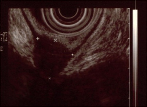
Laparoscopy is the gold standard for the diagnosis of endometriosis, and histological confirmation can be beneficial due to high false-positive rates of visual diagnosis. Due to the invasiveness of the procedure, other methods are often employed to detect the lesion and to aid with preoperative planning and patient counseling. Transvaginal ultrasound (TVS), transrectal ultrasound, CT colonography, and MRI are examples of the preoperative methods available to detect deep infiltrating RVE.Citation6 There is varying data on which offers the highest sensitivity, specificity, PPV, NPV, and accuracy in cases of deep RVE. According to one study, TVS was superior to other modalities in detecting RVE, with a sensitivity of 98%, specificity of 100%, PPV of 100%, and NPV at 98%.Citation6 In contrast, the study found MRI to have values of 83%, 98%, 98%, and 85% respectively. Other studies have shown similar results with sensitivities of 73.9%–98%, and specificities of 87.5%–100% with TVS.Citation31–Citation34 In a cohort study consisting of 422 patients, sensitivities and specificities of TVS were categorized according to the rectovaginal septum, rectum, and sigmoid colon. Results were 52% and 96%, 65% and 99%, and 69% and 98%, respectively.Citation35 There has been data collected on the high degree of false negatives that TVS generates at the uterosacral ligament and when the pouch of Douglas is obliterated ().Citation35
Figure 1 This is an endorectal ultrasound revealing a deep endometriosis nodule involving the muscular layer of the rectum.
According to a study published in 2012, TVS accompanied by the use of saline injection (saline contrast sonovaginography [SCSV]) showed superior sensitivity, specificity, PPV, and NPV. It is believed that the pressure from the saline solution creates a clearer view of the walls of the vagina. It can thus detect the location and invasion of the lesions more accurately.Citation36 In addition, patients in this study were more comfortable with this technique, though the data for this finding were not statistically significant. Although SCSV appeared superior to traditional TVS in this study, the authors did not suggest its use as a first-line strategy unless the physician had extensive training in this approach. In the analysis, MRI had the same results as SCSV in diagnosing rectal endometriosis with matching sensitivities of 66.7%. This value supersedes the 33.3% sensitivity found with the use of TVS alone in this study. Although MRI sensitivity is quantitatively superior to TVS in this analysis, it can be very time consuming and quite costly. SCSV could be a more sensitive and affordable option if physicians are adequately trained.Citation36
In a study from 2003,Citation37 SCSV presented with similar data to those discussed above. The results revealed TVS to have a sensitivity of 90.6%, specificity of 85.7%, PPV of 93.5%, and NPV of 80%. In addition, SCSV was associated with minimal pain and discomfort.
Another variation of TVS includes the “tenderness guided” approach, which uses excess amounts of gel to create an acoustic window of pressure to guide the physician to areas where the patient exhibits more pain, thus identifying the areas of endometriotic tissue. This technique found specificities of 95% and sensitivities of 90%, suggesting another inexpensive yet sensitive approach to diagnosis.Citation38
The uterine sliding sign is a useful diagnostic tool that can be employed during TVS. This technique presses the transducer into the posterior vaginal fornix and withdraws backwards to determine the motion of the rectum against the posterior vaginal fornix and posterior uterine wall in the midsagittal plane. External pressure is also applied to the uterus by placing one’s hand on top of the abdomen. If movement is absent, the uterine sliding sign is considered negative and is suggestive of adhesions.Citation30 In a study that evaluated this approach in regards to DIE located in the pouch of Douglas, a negative uterine sliding sign was indicative of endometriosis with a sensitivity of 83.3%, specificity of 97.1%, PPV of 92.6%, NPV of 93.2%, and an accuracy of 93%. This approach is considered useful in women with a high risk for bowel endometriosis because of its high degree of certainty, especially with regards to the involvement of the pouch of Douglas.Citation39
Both transvaginal and transrectal ultrasound can detect endometriosis as an irregular mass that is hypoechoic with a hyperechoic rim. Transrectal ultrasounds, however, can be quite useful in measuring the distance from the endometriotic lesion to the anal verge. In fact, in a retrospective study,Citation40 rectal endoscopic sonography showed a PPV of 100% in detecting muscularis layer infiltration; however, it was not as accurate in determining infiltration of the submucosal/mucosal layers. Sensitivity, specificity, PPV, and NPV for those layers were 89%, 26%, 55%, and 71%, respectively. It is important to note that there have been results where specificity in the submucosa/mucosa is as high as 94%.Citation34
Colonoscopy can be used to diagnose endometriosis that has invaded into the bowel as well.Citation8 Rectoscopy is not commonly used for lesions because growths generally do not penetrate past the muscularis propria (<10%) or mucosa. Thus, colonic endometriomas are easily missed on colonoscopy.Citation7,Citation8 Because of the clinical symptoms and endoscopic/radiologic findings, RVE can be mistaken for malignancy, and thus must be considered in the differential diagnosis when a patient is undergoing colonoscopy.Citation8
CT-based virtual colonoscopy is useful in determining stenosis of the bowel due to the infiltration of endometriosis. It is particularly efficient in evaluating whether the patient is in need of shaving or full-thickness resection when presenting with DIE in the colon.Citation41
Double-contrast barium enemas have an accuracy of 90% and PPV of 97% in diagnosing colonic involvement of endometriosis.Citation35
Modified virtual colonoscopy, also known as CT colonography, is another useful tool for determining the infiltration of endometriosis into the bowel. CT colonography has a wide range of views expanding into the submucosa and the serosa, unlike traditional colonoscopy. In this modified approach, a large obstetric tampon is inserted high into the vagina, a Foley catheter is inserted into the rectum releasing CO2, and the pelvis is scanned. This technique is noninvasive, of short duration (20 minutes), requires minimal preparation, and does not require sedation. It uses a lower dose of radiation than virtual colonoscopy, pyelogram, and barium enemas. No endoscope or barium is required, and it is approximately 50% less costly than optical colonoscopy. The main advantage is that the bowel is insufflated with CO2, which allows for multiple viewing modalities. Abdominal, retroperitoneal, and pelvic organs can be scanned as well. DIE can be visualized in the bowel, urinary tract, as well as in the rectovaginal and recto-peritoneal regions. This is a good technique for young women who wish to avoid high radiation doses and need a comprehensive presurgical evaluation because it allows for almost all presurgical investigation to be completed in one study ().Citation42,Citation43
Figure 2 This is a modified virtual colonography with transparent views of the rectum and lower sigmoid.
Abbreviations: L, length; S, stricture; D, distance to the anal verge; MURO, describes disseminated endometriosis beyond the rectogenital organs.
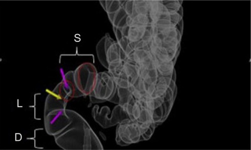
A descriptive imaging classification was proposed using modified virtual colonoscopy in an attempt to quantify the severity of rectogenital disease and disseminated endometriosis. The LSD/MURO classification signifies the severity of the pathology by giving it a numeric evaluation (ie, 0, 1, 2, or 3). Rectogenital disease is described by the LSD component; it identifies and quantifies pathology from the anal verge to the lower sigmoid.
L corresponds to the length of stricture (0 = no stricture or rectogenital nodule with no bowel involvement; 1 = a stricture length of <3 cm; 2 = a stricture length of 3–5 cm; and 3 = a stricture length >5 cm and/or non-distensibility). The stricture (S) is calculated by measuring the smallest stricture diameter and comparing it with the closest normal bowel lumen diameter (0 = no stricture; 1 =<30% stricture; 2 =30%–60% stricture; and 3 =>60% stricture). D is the distance from the anal verge (0 = bowel involvement; 1 =>15 cm; 2 = 8–15 cm; and 3 = <8 cm). The score increases as the pathology gets closer to the anal verge. MURO describes disseminated endometriosis beyond the rectogenital organs; M corresponds to multifocal disease above the lower sigmoid, U corresponds to urinary tract involvement, R corresponds to reproductive organ involvement, and O corresponds to abdominal organs such as the liver.
MRI is often the preoperative, noninvasive method of choice used to diagnose RVE due to its high level of accuracy in predicting specific locations of the infiltrating endometriosis in the intestinal, pelvic, and retroperitoneal regions.Citation3,Citation36,Citation44,Citation45 It should also be noted that MRI has an overall specificity of 90.3% in predicting lesions in the pelvic and retroperitoneal regions.Citation45 MRI allows precise localization of predicted lesions and visualization of multiple planes of multifocal, scattered, and small lesions.Citation36 Though the aforementioned studies Citation3,Citation36,Citation44,Citation45 stated that MRI was useful in the intestinal, pelvic, and retroperitoneal regions preoperatively, a different study revealed it was particularly helpful in detecting lesions invading the bladder region, and less effective in the peritoneal regions.Citation46
The extent of disease infiltration is important for selecting which surgical technique will be employed. Nonetheless, during surgical excision, lesions are occasionally underestimated due to their location, or overestimated secondary to the extent of the fibrotic tissue present.Citation47 MRI may be contraindicated or avoided in patients with significant anxiety or claustrophobia, and CT can be a good option for those patients, with sensitivity, specificity, PPV, NPV, and accuracy values of 87%, 100%, 100%, 77%, and 91%, respectively. It can be especially helpful in detecting the endometriotic bowel implants (98.7% specificity) when combined with a water enema.Citation45
Specificity on MRI is exceptionally high; however, research has been conducted on techniques that further improve specificity. One such technique included using ~100–120 mL of nonsterile ultrasound gel to opacify the vagina and rectum before imaging.Citation48 This allowed for delineation of the pelvic organs, especially of the vagina and rectovaginal septum. The sensitivity increased from 63.1% to 81.7% after opacification. In addition the specificity, though almost perfect prior to opacification (99.28%), reached 100% after opacification. Other techniques involve recognizing a sub center foci and T2 hyperintensity to visualize ectopic endometrial glands within fibrotic masses ().Citation49
Figure 3 This is a preoperative assessment using magnetic resonance imaging, revealing a deep infiltrating endometriosis nodule with an obvious increase in rectal wall thickness.
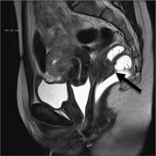
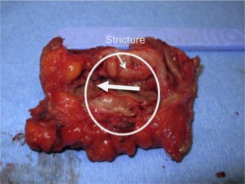
Figure 4 This is an endometriotic stricture >30% managed by segmental resection.
Another analysis found that different modalities work better for different regions. TVS worked best for endometriomas; MRI worked best for uterosacral ligaments, and small lesions on the bladder; and rectal endoscopic sonography worked best for the rectovaginal septum. In conclusion, the study found that all three were complementary for preoperative diagnosis.Citation50
Treatment options
Pain management for RVE is highly variable. There are a number of hormonal therapies, which can partially alleviate pain. These therapies include birth control pills, gonadotropin-releasing hormone agonists, danazol, injectable progestins, and analgesics.Citation13 Oral progestins are an option as well. One study revealed both surgical therapy and medical therapy with progestins equally improved deep dyspareunia in women with RVE.Citation51 These data are suggestive of the fact that if the patients’ only complaints surround dyspareunia, a conservative approach that could be used is hormonal therapy with progestins. Furthermore, low-dose OCPs have shown some benefit in reducing bowel endometriosis-related symptoms of dysmenorrhea, dyspareunia, and dyschezia. In addition to pain relief, therapy with OCPs has been shown to significantly reduce volumetric colorectal plaques when visualized by endoscopic ultrasound.Citation52 Also, OCPs have been shown to reduce the risk for bowel resection and the recurrence of ovarian endometriomas.Citation3
Although not approved for use in endometriosis by the US Food and Drug Administration, the levonorgestrel-releasing intrauterine system used after conservative surgery can be effective in reducing chronic pain.Citation53
Most often, surgery provides the most relief and improves at least 70% of pain-related symptoms.Citation13,Citation54 There are a number of surgical options for patients. Conventional laparoscopic or robotic approaches have been used.Citation55 Laparoscopic shaving resections, also known as superficial partial thickness excisions, are used in less severe cases and are associated with fewer complications of bowel obstruction and nerve damage.Citation47,Citation56 In a retrospective analysis that examined the various resection techniques of rectal endometriosis, the shaving of superficial lesions had a 6% complication rate (CR) in comparison with a 23% CR in laparoscopic discoid resection and a 38% CR with laparoscopic segmental resection. It is important to note that the majority of lesions handled with the shaving technique were smaller than 1 cm; the procedure was less invasive, and thus more easily managed in this manner during surgery. In more extensive cases, segmental and discoid resections were performed. Though complications were less frequent with the shaving approach, pain relief was less effective. This could be due to incomplete excision and recurrence of the endometriotic infiltrate.Citation47 Other studies have compared patients treated conservatively to patients treated with radical excision of DIE and have concluded similarly that radical removal greatly decreased the severity of dysmenorrhea and the recurrence of pelvic pain.Citation57
In addition, several energy sources can be used in treatment, including laser or electrosurgery.Citation58 Laparoscopic laser resection is especially helpful in removing infiltrations of the rectovaginal septum and rectovaginal pouch.Citation56,Citation59 In a study that used a CO2 laser, out of 48 patients, five needed further surgery over a 17.9-month range, and there was a 19% recurrence rate. In addition, extreme fatigue reduced from 58% of patients to 10% of patients after laser resection. Finally, 59% of patients considered improvement “excellent”, and 41% considered improvement “satisfactory.”Citation59
Laparoscopic CO2 laser was used in another study that compared women who had laser excision with and without bowel resection.Citation60 Clinical outcomes for both groups were equal, except for a higher minor CR occurring in women with bowel resection. Laparoscopic laser ablation, in combination with segmental bowel resection, is associated with good clinical outcomes for women with DIE.Citation61
A small retrospective study found that laparoscopic ADR, when feasible, was superior to low anterior resection (LAR) due to decreased operative time, blood loss, and length of hospitalization. In addition, ADR improved symptoms of diarrhea, constipation, daily performance, dyschezia, rectal pain, and dyspareunia more effectively than LAR. These data suggest that ADR – when feasible – should be the initial surgical treatment, except in cases where there is multifocal rectal involvement, large rectal nodules, luminal stenosis, and recurrent disease. In these situations, LAR and reanastomosis may be the patient’s best option.Citation1 When radical resection was compared with the conservative approach of rectal shaving and nodule excision, the conservative approach had better results on the Knowles–Eccersley–Scott Symptom Questionnaire, Gastrointestinal Quality of Life Index, depression/self-perception Fecal Incontinence Quality of Life score, and significantly improved values for postoperative constipation. Once again, this study suggests better outcomes if the rate of colorectal resection is reduced.Citation62
The classical laparoscopic technique is non nerve-sparing and removes parts of the mesentery attached to the posterior colon. This can lead to the damaging of nerves that coordinate bladder and intestinal function, and can result in a loss of vasculature to the bowel. This is often the technique used by colorectal surgeons for colon cancer. However in RVE, a surgeon can use a nerve-sparing technique, which takes advantage of the digital zoom function on the laparoscope to avoid the inferior hypogastric nerve plexus in order to preserve urinary function.Citation35,Citation47,Citation63,Citation64 A key step in the nerve-sparing approach is that it accurately identifies the anatomic structures in the posterior parametrium prior to removal of the lesions. The surgeon should take the time to distinguish the cranial structure (uterosacral ligaments), the caudad structure (rectovaginal ligaments), and the lateral caudad structure (lateral rectal ligaments). These structures hold the autonomic nerves, which innervate the pelvic viscera.Citation64,Citation65 A case series from 2010 found that only three of 16 patients who underwent the nerve-sparing technique needed urinary catheterization that lasted a maximum of 2 days. In comparison, 14 of 55 patients with the classical resection needed catheterization for 3–6 months and two patients had persistent urinary retention.Citation63 A prospective cohort study of 61 patients showed longer mean catheter duration of 39.8 days in patients who underwent the nerve-sparing technique, as opposed to a mean catheter duration of 121 days in patients who underwent the classical resection.Citation66 Evidently, both analyses were in congruence that the non-nerve-sparing group did not show signs of improvement in their urinary dysfunction after 6 months, and that the nerve-sparing technique was the ideal choice for surgical excision.Citation63,Citation66
A recent surgical approach described the use of both laparoscopic and transanal techniques. Laparoscopically, the lesions are shaved and released from the walls of the vagina and surrounding areas. Transanally, a Contour® Transtar™ stapler (Ethicon Endo-Surgery Inc., Cincinnati, OH, USA) both excises and staples the lesion. The study that addressed this technique was limited in size, only reporting the data from six patients.Citation67 Four of the patients did not experience any postoperative digestive pain and had clear margins on excision. The technique is very appealing because it avoids opening the lumen and minimizes the leakage of bowel contaminants into the peritoneum. The device also has the ability to excise nodules as high as 10 cm inward from the anal canal and up to 5 cm in diameter, as opposed to 7 cm inward and 2 cm in diameter, respectively, for other staplers.Citation67
Robot-assisted techniques have been reported in recent years. Results from a cohort study conducted over the past 5 years suggest robotics as a useful technique for complex cases such as those concomitant with hysterectomy and sacrocolpopexy.Citation55 In a limited study utilizing the robot-assisted approach, out of 19 patients with bowel resection, 23 patients with rectovaginal septum resection, and five bladder resections, one anastomotic leakage was reported. No intraoperative complications or conversion to laparotomy occurred in this study.Citation55 More research about this approach and its cost-effectiveness is needed.
Complications and long-term outcomes
According to a retrospective study conducted with 52 patients who had laparoscopic bowel resections from 2002–2009, only four patients (7%) had complications. These complications included intra-abdominal bleeding, rectovaginal fistula, and anastomotic leakage.Citation7 In a similar retrospective study conducted with 23 patients, three patients (13%) experienced major complications including anastomotic stenosis, bowel fistula, and bladder fistula. Other complications included constipation in 23% of patients, dyschezia in 43% of patients, and dysuria in 18% of patients.Citation68 Rectovaginal fistulas are often seen when lesions are removed in the lower rectum accompanied by a hysterectomy. Using an omental flap or imbricating the seromuscularis layer over the staples can sometimes avoid this complication.Citation47 In a comprehensive study of 1,128 full laparoscopic bowel resections for intestinal endometriosis and 19 robot-assisted resections, the overall CR was 8.7% (94 complications out of 1,147 procedures). The major complications were 24 anastomotic leaks (2.1%), 18 rectovaginal fistulas (1.5%), 13 intra-abdominal bleeds (1.1%), eight pelvic fluid collections (0.7%), and two urinary injuries (0.2%). The minor complications included nine transient bowel obstructions (0.8%), four minor rectal bleedings (0.3%), ten wound infections (0.9%), two urinary infections (0.2%), and four transient urinary retentions (0.3%). Recurrence of pain was found in 38 patients. Overall, 99% of symptoms were alleviated from the surgeries.Citation69 Although complications are common, overall improvement in pelvic pain and quality of life is evident with bowel resection. Because some complications are more severe, such as rectal and bladder dysfunctions, patients should be evaluated and counseled carefully before undergoing surgery; various approaches should also be levied according to what optimizes patient satisfaction and outcomes.Citation68 In addition, the recurrence of lesions and symptoms is always a possibility. In patients over 40 years of age with no interest in becoming pregnant, hysterectomy with or without bilateral oophorectomy can be considered.Citation70 The surgical approach and the extent of excision should be tailored carefully to the individual patient and to the presenting symptoms.
Quality of life appears to be markedly improved for patients who chose a surgical option. In a study conducted in Brazil, a Short Form-36 Health Status Questionnaire was given to 151 women who underwent laparoscopic resection between 2002 and 2009, and the researchers found significant improvements in pain-related symptoms (P<0.001) and a significant increase in scores across all domains of physical and mental health (P<0.001).Citation71 A cohort study out of Italy used the same Short Form-36 Health Status Questionnaire; the researchers examined quality of life in patients who underwent segmental bowel resection and those who had nodules shaved instead.Citation72 Results showed a significant increase in quality of life in all patients, which was congruent with the findings from the aforementioned study. It should be noted, however, that surgical resection did not appear to be superior to shaving. Both methods worked equally well at improving quality of life. Other questionnaires such as the EuroQol Group EQ-5D™ (EuroQol Group, Rotterdam, The Netherlands) have been used in the analysis of quality of life after laparoscopic resection, and improvements in quality of life were shown to be statistically significant over the course of 3 years of follow up.Citation73
In addition to the relief of bowel-related symptoms, sexual dysfunction can be alleviated by surgery. Research has shown that endometriotic nodules in the pouch of Douglas in particular are associated with sexual pain, and laparoscopic resection can significantly improve these symptoms.Citation74 It has been shown that 1 year after rectovaginal resection, sexual satisfaction is improved significantly.Citation75,Citation76
Surgery can also improve fertility, even when surgery does not remove all endometriotic lesions.Citation77 Analyzing whether surgery improves spontaneous conception or IVF is difficult to substantiate. In a systematic review using eleven selected studies, patients with RVE who were infertile before surgery were evaluated for their postsurgical pregnancy rates with spontaneous pregnancy and IVF. The average postoperative conception rate in all women seeking pregnancy apart from preoperative fertility status and IVF performance was 39% (223/571), but only 24% (123/510) in infertile women who tried for spontaneous conception. These data suggest that patients with RVE who undergo surgery should know that postoperative conception is a real possibility, but generic overestimation should be avoided.Citation78
Conclusion
RVE is the most severe form of endometriosis. Symptoms vary among patients, and clinical diagnosis can be challenging. A number of modalities are employed in diagnosis, including physical examination, ultrasound, MRI, colonoscopy, and CT colonography. The gold standard for diagnosis is laparoscopy with histological confirmation. Several studies showed TVS as a very specific, sensitive, affordable, and time-efficient modality for the diagnosis of DIE. However, other modalities, like MRI and CT colonography, can be used and provide information such as the depth at which the lesion infiltrates the bowel wall. Once a diagnosis is determined, medical therapy is usually not adequate to relieve symptoms; although birth control pills are associated with a reduced need for bowel resection. Surgery ranges from superficial partial thickness excisions to radical colorectal resection and reanastomosis. Outcomes from the different surgical options vary; most studies showed that the more radical the resection, the greater the postsurgical CR. However, the recurrence rate was higher with the less aggressive approaches. There has been a significant development of surgical equipment. Research is needed to assess the clinical outcomes associated with using various devices. In conclusion, RVE is a serious diagnosis for patients, and a number of options for the diagnosis and treatment of these patients are available. Diagnostic and therapeutic approaches should be tailored to the individual patient, her symptoms and her treatment goals.
Disclosure
The authors report no conflicts of interest in this work.
References
- MoawadNSGuidoRRamanathanRMansuriaSLeeTComparison of laparoscopic anterior discoid resection and laparoscopic low anterior resection of deep infiltrating rectosigmoid endometriosisJSLS201115333133821985719
- RobertsCPRockJAThe current staging system for endometriosis: does it help?Obstet Gynecol Clin North Am200330111513212699261
- TarjanneSSjöbergJHeikinheimoORectovaginal endometriosis-characteristics of operative treatment and factors predicting bowel resectionJ Minim Invasive Gynecol200916330230619269901
- Zanetti-DällenbachRBartleyJMüllerCSchneiderAKöhlerCCombined vaginal-laparoscopic-abdominal approach for the surgical treatment of rectovaginal endometriosis with bowel resection: a comparison of this new technique with various established approaches by laparoscopy and laparotomySurg Endosc2008224995100117705065
- KondoWBrancoAWTrippiaCHRibeiroRZomerMTRetrocervical deep infiltrating endometriotic lesions larger than thirty millimeters are associated with an increased rate of ureteral involvementJ Minim Invasive Gynecol201320110010323312249
- AbraoMSGonçalvesMODiasJAPodgaecSChamieLPBlasbalgRComparison between clinical examination, transvaginal sonography and magnetic resonance imaging for the diagnosis of deep endometriosisHum Reprod200722123092309717947378
- JelencFRibič-PuceljMJuvanRKobalBSinkovecJSalamunVLaparoscopic rectal resection of deep infiltrating endometriosisJ Laparoendosc Adv Surg Tech A2012221666922166117
- SassiSBouassidaMTouinsiHExceptional cause of bowel obstruction: rectal endometriosis mimicking carcinoma of rectum – a case reportPan Afr Med J2011103322187615
- PortaleTRBrancaAScillettaRPesceAPuleoSIleo-colic endometriosis: a rare localization of a frequent disease case reportAnn Ital Chir2013841
- KoninckxPRUssiaAAdamyanLWattiezADonnezJDeep endometriosis: definition, diagnosis, and treatmentFertil Steril201298356457122938769
- FauconnierAFritelXChapronCEndometriosis and pelvic pain: epidemiological evidence of the relationship and implicationsGynecol Obstet Fertil20093715769 French19128998
- AnafVSimonPEl NakadiIRelationship between endometriotic foci and nerves in rectovaginal endometriotic nodulesHum Reprod20001581744175010920097
- FalconeTLebovicDIClinical management of endometriosisObstet Gynecol2011118369170521860303
- MontanariGDi DonatoNBenfenatiAWomen with deep infiltrating endometriosis: sexual satisfaction, desire, orgasm, and pelvic problem interference with sexJ Sex Med20131061559156623551753
- VercelliniPSomiglianaEBuggioLBarbaraGFrattaruoloMPFedeleL“I can’t get no satisfaction”: deep dyspareunia and sexual functioning in women with rectovaginal endometriosisFertil Steril201298615031511. e122910685
- BallesterMOppenheimerAMathieu d’ArgentEDeep infiltrating endometriosis is a determinant factor of cumulative pregnancy rate after intracytoplasmic sperm injection/in vitro fertilization cycles in patients with endometriomasFertil Steril201297236737222177465
- XueQZengCXuYClinical effect on women with different types of endometriosis related infertility treated by conservative surgeryZhonghua Fu Chan Ke Za Zhi20134811619 Chinese23531245
- Guerrero HernándezAOropeza RechyGGómez GarcíaERisk factors associated, diagnostic methods and treatment for endometriosis, used in clinical service endometriosis gynecology Hospital General de Mexico (2009–2011)Ginecol Obstet Mex20128010637643 Spanish23240226
- KoninckxPRMartinDTreatment of deeply infiltrating endometriosisCurr Opin Obstet Gynecol1994632312418038409
- HudelistGFritzerNThomasADiagnostic delay for endometriosis in Austria and Germany: causes and possible consequencesHum Reprod201227123412341622990516
- DalsgaardTHjordt HansenMVHartwellDLidegaardOReproductive prognosis in daughters of women with and without endometriosisHum Reprod20132882284228823696543
- ChapronCLafay-PilletMCMonceauEQuestioning patients about their adolescent history can identify markers associated with deep infiltrating endometriosisFertil Steril201195387788121071024
- ChapronCSouzaCBorgheseBOral contraceptives and endometriosis: the past use of oral contraceptives for treating severe primary dysmenorrhea is associated with endometriosis, especially deep infiltrating endometriosisHum Reprod20112682028203521642638
- ChapronCPietin-VialleCBorgheseBDavyCFoulotHChopinNAssociated ovarian endometrioma is a marker for greater severity of deeply infiltrating endometriosisFertil Steril200992245345718692806
- DaiYLengJHLangJHRelationship of pelvic clinic-pathological features and the pain symptoms in ovarian endometriomaZhonghua Fu Chan Ke Za Zhi2013482118122 Chinese23544493
- LengJHLangJHDaiYLiHJLiXYRelationship between pain symptoms and clinico-pathological features of pelvic endometriosisZhonghua Fu Chan Ke Za Zhi2007423165168 Chinese17537300
- RomanHNessJSuciuNAre digestive symptoms in women presenting with pelvic endometriosis specific to lesion localizations? A preliminary prospective studyHum Reprod201227123440344922962316
- FauconnierAChapronCDubuissonJBVieiraMDoussetBBréartGRelation between pain symptoms and the anatomic location of deep infiltrating endometriosisFertil Steril200278471972612372446
- CheewadhanaraksSPeeyananjarassriKDhanaworavibulKLiabsuetrakulTPositive predictive value of clinical diagnosis of endometriosisJ Med Assoc Thai200487774074415521226
- HudelistGFritzerNStaettnerSUterine sliding sign: a simple sonographic predictor for presence of deep infiltrating endometriosis of the rectumUltrasound Obstet Gynecol201341669269523400893
- PikettyMChopinNDoussetBPreoperative work-up for patients with deeply infiltrating endometriosis: transvaginal ultrasonography must definitely be the first-line imaging examinationHum Reprod200924360260719095669
- BazotMDetchevRCortezAAmouyalPUzanSDaraïETransvaginal sonography and rectal endoscopic sonography for the assessment of pelvic endometriosis: a preliminary comparisonHum Reprod20031881686169212871883
- HudelistGTuttliesFRauterGPucherSKecksteinJCan transvaginal sonography predict infiltration depth in patients with deep infiltrating endometriosis of the rectum?Hum Reprod20092451012101719221097
- GoncalvesMOPodgaecSDiasJAGonzalezMAbraoMSTransvaginal ultrasonography with bowel preparation is able to predict the number of lesions and rectosigmoid layers affected in cases of deep endometriosis, defining surgical strategyHum Reprod201025366567120023291
- FratelliNSciosciaMBassiEMusolaMMinelliLTrivellaGTransvaginal sonography for preoperative assessment of deep endometriosisJ Clin Ultrasound2013412697523233390
- SaccardiCCosmiEBorgheroATregnaghiADessoleSLittaPComparison between transvaginal sonography, saline contrast sonovaginography and magnetic resonance imaging in the diagnosis of posterior deep infiltrating endometriosisUltrasound Obstet Gynecol201240446446922253192
- CosmiESaccardiCLittaPThe sonographic diagnosis of deep endometriosisJ Ultrasound Med200928341041119244084
- GuerrieroSAjossaSGeradaMD’AquilaMPirasBMelisGB“Tenderness-guided” transvaginal ultrasonography: a new method for the detection of deep endometriosis in patients with chronic pelvic painFertil Steril20078851293129717548084
- ReidSLuCCasikarIPrediction of pouch of Douglas obliteration in women with suspected endometriosis using a new real-time dynamic transvaginal ultrasound technique: the sliding signUltrasound Obstet Gynecol201341668569123001892
- RossiLPalazzoLYazbeckCCan rectal endoscopic sonography predict infiltration depth in patients with deep infiltrating endometriosis of the rectum?Ultrasound Obstet Gynecol EpubJune102013
- VassilieffMSuaudOCollet-SavoyeCComputed tomography-based virtual colonoscopy: an examination useful for the choice of the surgical management of colorectal endometriosisGynecol Obstet Fertil2011396339345 French21596608
- van der WatJKaplanMDRomanHDa CostaCThe use of modified virtual colonoscopy to structure a descriptive imaging classification with implied severity for rectogenital and disseminated endometriosisJ Minim Invasive Gynecol EpubJune52013
- van der WatJKaplanMDModified virtual colonoscopy: a noninvasive technique for the diagnosis of rectovaginal septum and deep infiltrating pelvic endometriosisJ Minim Invasive Gynecol200714563864317848328
- BazotMDaraiEHouraniRDeep pelvic endometriosis: MR imaging for diagnosis and prediction of extension of diseaseRadiolog y20042322379389
- Stabile IanoraAAMoschettaMLorussoFRectosigmoid endometriosis: Comparison between CT water enema and video lap-aroscopyClin Radiol201368989590123809266
- KrügerKBehrendtKNiedobitek-KreuterGKoltermannKEbertADLocation-dependent value of pelvic MRI in the preoperative diagnosis of endometriosisEur J Obstet Gynecol Reprod Biol20131691939823478073
- PereiraRMZanattaAPretiCDde PaulaFJda MottaELSerafiniPCShould the gynecologist perform laparoscopic bowel resection to treat endometriosis? Results over 7 years in 168 patientsJ Minim Invasive Gynecol200916447247919573824
- ChassangMNovellasSBloch-MarcotteCUtility of vaginal and rectal contrast medium in MRI for the detection of deep pelvic endometriosisEur Radiol20102041003101019862535
- SiegelmanESOliverERMR imaging of endometriosis: ten imaging pearlsRadiographics20123261675169123065164
- Gauche CazalisCKoskasMMartinBPalazzoLMadelenatPYazbeckCPreoperative imaging of deeply infiltrating endometriosis in: Transvaginal sonography, rectal endoscopic sonography and magnetic resonance imagingGynecol Obstet Fertil20124011634641 French23123282
- VercelliniPSomiglianaEConsonniDFrattaruoloMPDe GiorgiOFedeleLSurgical versus medical treatment for endometriosis-associated severe deep dyspareunia: I. Effect on pain during intercourse and patient satisfactionHum Reprod201227123450345922926841
- FerrariSPersicoPDI PuppoFContinuous low-dose oral contraceptive in the treatment of colorectal endometriosis evaluated by rectal endoscopic ultrasonographyActa Obstet Gynecol Scand201291669970322268632
- Abou-SettaAMAl-InanyHGFarquharCMLevonorgestrel-releasing intrauterine device (LNG-IUD) for symptomatic endometriosis following surgeryCochrane Database Syst Rev2006CD00507217054236
- De CiccoCCoronaRSchonmanRMailovaKUssiaAKoninckxPBowel resection for deep endometriosis: a systematic reviewBJOG2011118328529121040395
- SiestoGIedaNRosatiRVitobelloDRobotic surgery for deep endometriosis: a paradigm shiftInt J Med Robot EpubJune132013
- BerkesEBokorARigóJJrCurrent treatment of endometriosis with laparoscopic surgeryOrv Hetil20101512811371144 Hungarian20570794
- HidakaTNakashimaAHashimotoYSaitoSEffects of laparoscopic radical surgery for deep endometriosis on endometriosis-related pelvic painMinim Invasive Ther Allied Technol201221535536122985066
- RobbinsMLExcision of endometriosis with laparosonic coagulating shearsJ Am Assoc Gynecol Laparosc19996219920310226132
- KristensenJKjerJJLaparoscopic laser resection of rectovaginal pouch and rectovaginal septum endometriosis: the impact on pelvic pain and quality of lifeActa Obstet Gynecol Scand200786121467147117851806
- MeulemanCTomassettiCWolthuisAClinical outcome after radical excision of moderate-severe endometriosis with or without bowel resection and reanastomosis: a prospective cohort studyAnn Surg Epub410 2013
- MeulemanCTomassettiCD’HoogheTMClinical outcome after laparoscopic radical excision of endometriosis and laparoscopic segmental bowel resectionCurr Opin Obstet Gynecol201224424525222729087
- RomanHVassilieffMTuechJJPostoperative digestive function after radical versus conservative surgical philosophy for deep endometriosis infiltrating the rectumFertil Steril20139961695170423465818
- KavallarisABanzCChalvatzasNLaparoscopic nerve-sparing surgery of deep infiltrating endometriosis: description of the technique and patients’ outcomeArch Gynecol Obstet2011284113113520680309
- CeccaroniMClariziaRRoviglioneGRuffoGNeuro-anatomy of the posterior parametrium and surgical considerations for a nerve-sparing approach in radical pelvic surgerySurg Endosc EpubJune202013
- AzaïsHCollinetPDelmasVRubodCUterosacral ligament and hypogastric nerve anatomical relationship. Application to deep endometriotic nodules surgeryGynecol Obstet Fertil2013413179183 French23490276
- CeccaroniMClariziaRBruniFNerve-sparing laparoscopic eradication of deep endometriosis with segmental rectal and parametrial resection: the Negrar method. A single-center, prospective, clinical trialSurg Endosc20122672029204522278102
- BridouxVRomanHKianifardBCombined transanal and laparoscopic approach for the treatment of deep endometriosis infiltrating the rectumHum Reprod201227241842622158086
- BoileauLLaporteSBourgauxJFLaparoscopic colorectal resection for deep pelvic endometriosis: Evaluation of post-operative outcomeJ Gynecol Obstet Biol Reprod (Paris)2012412128135 French22071018
- RuffoGRossiniRThe outcomes of laparoscopic resection of bowel endometriosisCurr Opin Obstet Gynecol201325430230723817230
- BorgheseBSantulliPStreuliILafay-PilletMCde ZieglerDChapronCRecurrence of pain after surgery for deeply infiltrating endometriosis: How does it happen? How to manage?J Gynecol Obstet Biol Reprod (Paris)2012 French
- BassiMAPodgaecSDiasJAD’Amico FilhoNPettaCAAbraoMSQuality of life after segmental resection of the rectosigmoid by laparoscopy in patients with deep infiltrating endometriosis with bowel involvementJ Minim Invasive Gynecol201118673073321930435
- MabroukMMontanariGGuerriniMDoes laparoscopic management of deep infiltrating endometriosis improve quality of life? A prospective studyHealth Qual Life Outcomes201199822054310
- RomanJDSurgical treatment of endometriosis in private practice: cohort study with mean follow-up of 3 yearsJ Minim Invasive Gynecol2010171424620129331
- AnafVSimonPEl NakadiISimonartTNoelJBuxantFImpact of surgical resection of rectovaginal pouch of douglas endometriotic nodules on pelvic pain and some elements of patients’ sex lifeJ Am Assoc Gynecol Laparosc200181556011172115
- KössiJSetäläMMäkinenJHärkkiPLuostarinenMQuality of life and sexual function 1 year after laparoscopic rectosigmoid resection for endometriosisColorectal Dis201315110210822642851
- SetäläMHärkkiPMatomäkiJMäkinenJKössiJSexual functioning, quality of life and pelvic pain 12 months after endometriosis surgery including vaginal resectionActa Obstet Gynecol Scand201291669269822404128
- DonnezJNisolleMAdvanced laparoscopic surgery for the removal of rectovaginal septum endometriotic or adenomyotic nodulesBaillieres Clin Obstet Gynaecol1995947697748821254
- VercelliniPBarbaraGBuggioLFrattaruoloMPSomiglianaEFedeleLEffect of patient selection on estimate of reproductive success after surgery for rectovaginal endometriosis: literature reviewReprod Biomed Online201224438939522377155