Abstract
Background
Breast cancer is a major cause of death; in fact, it is the most common type, in order of the number of global deaths, of cancer in women worldwide. This research seeks to investigate how triptolide, an extract from the Chinese herb Tripterygium wilfordii Hook F, induces apoptosis in MCF-7 human breast cancer cells. Accumulating evidence suggests a role for lysosomal proteases in the activation of apoptosis. However, there is also some controversy regarding the direct participation of lysosomal proteases in activation of key apoptosis-related caspases and release of mitochondrial cytochrome c. In the present study, we demonstrate that triptolide induces an atypical, lysosomal-mediated apoptotic cell death in MCF-7 cells because they lack caspase-3.
Methods
MCF-7 cell death was characterized via cellular morphology, chromatin condensation, 3-(4,5-dimethylthiazol-2-yl)-2,5-diphenyltetrazolium bromide colorimetric cell growth inhibition assay and the expression levels of proapoptotic proteins. Acridine orange and LysoTracker® staining were performed to visualize lysosomes. Lysosomal enzymatic activity was monitored using an acid phosphatase assay and western blotting of cathepsin B protein levels in the cytosolic fraction, which showed increased enzymatic activity in drug-treated cells.
Results
These experiments suggest that triptolide-treated MCF-7 cells undergo atypical apoptosis and that, during the early stages, lysosomal enzymes leak into the cytosol, indicating lysosomal membrane permeability.
Conclusion
Our results suggest that further studies are warranted to investigate triptolide’s potential as an anticancer therapeutic agent.
Introduction
Breast cancer is the second deadliest cancer among women in the United States, and the National Cancer Institute now estimates that one in seven women living in the United States will develop breast cancer in their lifetime.Citation1 With rates of breast cancer increasing, it will continue to be a major cause of cancer deaths in women worldwide.Citation2,Citation3 Treatments such as chemotherapy and radiation are known to have harmful side effects on normal cells. Therefore, it is imperative to develop safer and noninvasive treatments. The use of natural products in the fight against cancer is one possible way of slowing tumor growth and may allow us to design more effective therapies.
Triptolide is an extract from the Chinese herb Tripterygium wilfordii Hook F. Long used in traditional Chinese medicine, the herb is purported to have immunosuppressive and anti-inflammatory properties,Citation4,Citation5 and triptolide has been identified as a major chemical component governing these properties. Triptolide is a diterpenoid triepoxide. Looking at the chemical composition of triptolide, it is a derivative of isoprene, which is part of the terpene family. Triptolide consists of four isoprene molecules.Citation6 Through the biosynthetic processing of the isoprene units in some plants, certain precursors to vitamins A and E are produced.Citation6,Citation7 These strong antioxidants may account for some of triptolide’s anticancer effects. A recent report demonstrated that triptolide covalently binds to a subunit of the general transcription factor TFIIH, and therefore inhibits RNA polymerase II-mediated transcription.Citation8 So far, the mechanisms proposed for the potent anticancer activity of triptolide, at concentrations in the nM range, include caspase activation in U937 human promonocytic cells,Citation9 sensitization of several tumor cells to tumor necrosis factor-alpha,Citation10 and downregulation of X-linked inhibitor of apoptosis protein (XIAP) expression in leukemic cells.Citation11 Based on the cell type, triptolide has been shown to induce different cell death mechanisms, including autophagy and apoptosis.Citation12
Programmed cell death is of major importance in regulating organismal development, tissue homeostasis, and stress response and interconnects with cell survival and proliferation.Citation13 Before the middle of the 20th century, there were various types of cell death observed.Citation14 Despite that, just prior to 1990, apoptosis was synonymous with programmed cell death with the commitment to a physiologically controlled process. Recently, there has been a resurgence in studies using past knowledge of the other types of programmed cell death.Citation14–Citation18 In the case of metamorphosis in insects, it was demonstrated that programmed cell death and perhaps apoptosis can proceed without an early or controlling activation of an endonuclease.Citation16,Citation19 While many stimuli can act as triggers, the activation of lysosomal enzymes is the most likely candidate.Citation16
Lysosomes, discovered over fifty years ago, are the major cell digestive organelles.Citation20,Citation21 They contain a number of hydrolases that are capable of breaking down nucleic acids, proteins, carbohydrates, and lipids.Citation20 Today, it is clear that lysosomes and lysosomal proteases can be involved in apoptosis. Among lysosomal proteases, the role of cathepsins in cancer progression is especially well documented.Citation22,Citation23 Following their release into the cytosol, they cleave Bid and degrade antiapoptotic Bcl-2 proteins, thereby triggering the mitochondrial pathway of apoptosis, with lysosomal membrane permeabilization (LMP) being the critical step in this pathway.Citation24 A recent report supports an apoptosis model involving lysosomal– mitochondrial crosstalk induced by the combination of an epigenetic modulator and a DNA-damaging agent.Citation25 Various insults, including oxidative stress and DNA damage, may lead to the limited release of cathepsins that culminate in the induction of apoptosis. Hsp70 has been implicated in playing an important role for inhibiting LMP to promote the survival of stressed cells.Citation26 However, blocking cathepsins by small molecule inhibitors has been shown to significantly delay cancer progression in a number of mouse cancer models as well as to sensitize tumor cells to other chemotherapeutic agents.Citation27 The release of cathepsins in excess may lead to cellular necrosis.Citation28 Lysosomal-mediated apoptosis is still largely under investigation and not fully understood.
In this study, we demonstrate that triptolide induces morphological features characteristic of apoptosis, namely cell shrinkage and chromatin condensation. Furthermore, we show that cleaved poly (ADP-ribose) polymerase (PARP) and cleaved caspase-7 and cleaved caspase-9 levels were elevated in triptolide-treated cells. Lysosomal enzymatic activity and localization experiments revealed that these organelles participate in the cell death mediated by triptolide. We also show in cell fractionation studies that cytosolic levels of cathepsin B are increased during the early stages of apoptosis. These results suggest that triptolide-induced apoptosis in MCF-7 cells is mediated by lysosomes. To our knowledge, this study is the first to report a link between lysosomal hydrolases and triptolide’s anticancer effects.
Materials and methods
Materials
Triptolide (C20H24O6, molecular weight 360.4, 95% purity) was purchased from Calbiochem (EMD Millipore Corporation, Billerica, MA, USA) and dissolved in dimethyl sulfoxide ([DMSO] Sigma-Aldrich, St Louis, MO, USA). Stock solutions of triptolide in DMSO were stored at −20°C at a concentration of 1 mg/mL. For all control experiments, cells were treated with vehicle (DMSO) as indicated.
Cell culture
MCF-7 cells were purchased from American Type Culture Collection ([ATCC] Manassas, VA, USA). Cells were grown in Minimum Essential Medium ([MEM] Lonza Inc, Allendale, NJ, USA) and supplemented with 100 U/mL penicillin and 100 μg/mL streptomycin (ATCC), and 10% fetal bovine serum (Atlanta Biologicals, Lawrenceville, GA, USA). All cells were grown in T-25 flasks (Life Technologies, Carlsbad CA, USA) and incubated at 37°C in a humidified chamber at 95% O2/5% CO2. Cells were subcultured by washing with a 1× phosphate-buffered solution ([PBS] 137 mM NaCl, 2.7 mM KCl, 4.3 mM Na2HPO4×7H2O, 1.4 mM KH2PO4, pH adjusted to 7.4) and using trypsin (0.25% [w/v] trypsin−0.53 mM ethylene-diaminetetraacetic acid [EDTA] solution; Life Technologies). DU145 prostate cancer cell line was also purchased from ATCC and used as a positive control for the caspase-3 immunoblotting assay. DU145 cells were grown in the same conditions as described above for MCF-7 cells.
Hoechst staining
Cells were treated in the presence or absence of 10 ng/mL (median lethal dose [LD50]) triptolide for 24 hours at 37°C and then with 1 mg/mL of DNA fluorochrome bisbenzimide (Hoechst 33258; Sigma-Aldrich) for 15 minutes at 37°C in the dark. Images were captured using the EVOS® FL fluorescence microscope (Life Technologies). The percentage of apoptotic cells (cells with nuclear condensation) was determined by examining at least 200 cells per condition in five randomly selected fields at 400× magnification.
Assessment of cell viability
To test for apoptosis, the 3-[4,5-dimethylthiazol-2-yl]-2,5-diphenyltetrazolium bromide (MTT) reduction assay (Sigma-Aldrich) was used. Dissolved MTT is converted to an insoluble purple formazan by cleavage of the tetrazolium ring by dehydrogenase enzymes of living cells.Citation29 Stock solutions of MTT were prepared in MEM at a concentration of 5 mg/mL. Cells were treated in 6-well plates with varying concentrations of triptolide (1 ng/mL, 5 ng/mL, 10 ng/mL, 15 ng/mL, and 20 ng/mL) for 24 hours. After this incubation period, MTT (0.2 mL) was added to each well and the cells were incubated for 4 hours at 37°C in the dark. The medium was then removed and the converted dye was solubilized with the addition of acidic isopropanol (0.1 N HCl (hydrochloric acid) in absolute isopropanol). Absorbance, which was proportional to cell viability, was measured at 570 nm with background subtraction at 660 nm. All readings were performed in triplicate.
Acid phosphatase (AP) assay
A biochemical assay (Sigma-Aldrich Procedure No 104) was used to measure the activity of AP, a marker enzyme for lysosomes.Citation19 Cells were treated in the presence or absence of 10 ng/mL triptolide for 24 hours at 37°C. Cell lysates were then transferred to a 0.9% NaCl solution, and centrifuged for 5 minutes at 10,000 × g. The g should be italicized. The reaction mixture consisted of 0.5 mL p-nitrophenol phosphate (substrate), 0.5 mL 90 mM citrate buffer (pH 4.8), and 0.1 mL of lysates and was incubated for 30 minutes at room temperature. The reaction was terminated with the addition of 0.5 mL 0.1 N NaOH. In alkali, liberated p-nitrophenol was measured spectrophotometrically at 410 nm. Units of enzyme activity were determined from the calibration curve obtained by measuring the amount of color produced.
Acridine orange
Cells were grown on coverslips and incubated for 24 hours in the presence or absence of triptolide. Stock solutions of acridine orange were prepared (5 mg/mL). Cells were incubated for 15 minutes in the dark with acridine orange (final concentration, 2.5 μg/mL) at 37°C. Cells were then rinsed with 1 × PBS and coverslips were mounted using Crystal Mount (Sigma-Aldrich). Images were captured using the EVOS FL fluorescence microscope.
LysoTracker® staining
Cells were grown on 6-well plates and incubated for 24 hours in the presence (10 ng/mL) or absence of triptolide. 1 mM stock solutions of LysoTracker Green DND-26 (Molecular Probes; Life Technologies) were prepared to 1 μM working solution with warm culture media. Cells were incubated for 1.5 hours in the dark with LysoTracker working solution at 37°C. Cells were then rinsed with 1 × Tris-buffered saline (TBS) and observed using a Motic AE31 fluorescence microscope (Causeway Bay Hong Kong) and SPOT imaging software (v 4.7.0.35; SPOT Imaging Solutions, Sterling Heights, MI, USA).
Cell lysis and protein quantification
Whole-cell lysate
Cells were treated with varying concentrations of triptolide for 24 hours in 6-well plates. Both culture medium and adherent cells were collected in 15 mL tubes, and samples were centrifuged at 3,000 × g for 5 minutes. Cells were rinsed once with 1 × cold PBS (from 10X PBS; Thermo Fisher Scientific, Waltham, MA, USA); 200 μL of SoluLyse-M (Genlantis, San Diego, CA, USA) with 1% protease inhibitor cocktail and 1% EDTA Solution (Thermo Fisher Scientific) were added to each, and incubation occurred at room temperature for 10 minutes. The lysates were then collected in microcentrifuge tubes and centrifugation occurred at 14,000 × g for 5 minutes. The clarified lysates were transferred into fresh tubes for further analysis.
Cytosolic/particulate separation
Cells were treated with varying concentrations of triptolide for 3 hours (early apoptotic stage) and 24 hours (late apoptotic stage) in T-25 flasks. Both culture medium and adherent cells were collected in 15 mL tubes, and samples were centrifuged at 3,000 × g for 5 minutes. Supernatants were decanted off and precipitates were rinsed with 1 × cold PBS and centrifuged again. After supernatants were decanted off, we followed the manufacturer’s protocol for the Cytosol/Particulate Rapid Separation Kit (BioVision, Milpitas, CA, USA).
The Pierce BCA Protein Assay Kit (Thermo Fisher Scientific) was used to determine protein concentration. Samples were then analyzed at 562 nm using the Nano-Drop 2000 Spectrophotometer (Thermo Fisher Scientific). A standard curve was generated and the protein concentrations of the unknown samples were determined.
Western blot analysis
Lysates were prepared using Laemmli Sample Buffer (Bio-Rad Laboratories, Hercules, CA, USA). Samples were separated by 10% EZRun protein gel solution (Thermo Fisher Scientific) and electrophoresed at 150 volts until the dye front was at the end of the gel. Proteins were transferred onto polyvinylidene difluoride membranes (PVDF) membranes using a Trans-Blot Turbo Blotting System (Bio-Rad Laboratories). After the blocking, membranes were incubated in the appropriate dilution of primary antibodies in 1X Tris-buffered saline/0.1% Tween 20 (TBST) overnight at 4°C with gentle shaking. Anti-caspase-9 and anti-caspase-7 antibodies (cleaved: Cell Signaling, Danvers, MA, USA; full length: Santa Cruz Biotechnology, Dallas, TX, USA), anti-PARP antibodies (Cell Signaling), and anti-cathepsin B antibody (AbCam, Cambridge, MA USA) were used. Anti-β-actin antibody (Santa Cruz Biotechnology) was used as a loading control. Blots were washed three times for 5–10 minutes per wash in 1 × TBST followed by incubation in the appropriately diluted horseradish peroxidase-conjugated anti-rabbit (or anti-mouse) immunoglobulin G secondary antibodies (Cell Signaling or Santa Cruz Biotechnology) at room temperature for 1 hour. The blots were washed three times in 1 × TBST for 5 minutes. The SuperSignal West Pico Chemiluminescent Substrate kit (Thermo Fisher Scientific) was used for antigen detection. Images were obtained using a film processor SRX-101 A (Konica Minolta Medical Imaging USA, Newark, NJ, USA). Band intensities were measured using the Image J 1.46r software (National Institutes of Health [NIH], Bethesda, MD, USA).
Statistical analyses
Data were presented as means ± standard error of the mean. Student’s t-test was performed to evaluate differences between treated and control samples. Values were considered statistically significant at P < 0.05.
Results
Cellular morphology of triptolide-treated MCF-7 breast cancer cells
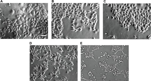
Firstly, we confirmed whether triptolide, which is an apoptotic stimulus in various tumor cell lines,Citation9–Citation11,Citation30 induces apoptosis in MCF-7 cells. Phase-contrast microscopy was used after treatment with various concentrations of triptolide for 24 hours. Uniform normal adhesive and colonized cells were observed in the control group (), whereas significant reduction in colony formation of the experimental groups could be observed in a dose-dependent manner (–). We observed that whole cells shrank and a part of the cells detached with increasing triptolide concentrations, but there were few instances of cellular blebbing. Meanwhile, typical cellular blebbing was observed in triptolide-treated DU145 cells (data not shown). These results suggest that triptolide can induce cell death independent of cytoplasmic blebbing as has been demonstrated with other drugs in MCF-7 cells.Citation31,Citation32
Figure 1 Phase-contrast micrographs of triptolide-treated MCF-7 cells incubated for 24 hours.
Effect of triptolide on chromatin condensation
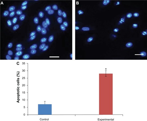
Chromatin condensation is a hallmark of apoptosis. To support the notion that triptolide induces apoptosis in MCF-7 cells, cells were treated in the presence or absence of triptolide and then stained with Hoechst 33258. Normal, flattened nuclear morphology was observed in control cells (), whereas round, shrunken cells displaying fragmented nuclei were detected in experimental cells (). The percentage of apoptotic cells in control and experimental cells was quantified (). These data add supportive evidence that triptolide treatment results in apoptosis in MCF-7 cells.
Figure 2 Effect of triptolide on chromatin condensation.
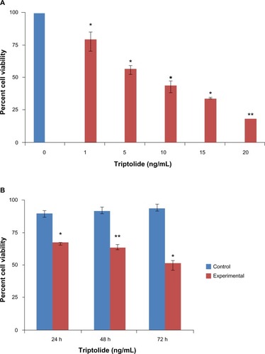
Effect of triptolide on cell viability
A previous study demonstrated that triptolide inhibits cell viability in MCF-7 cells in a dose- and time-dependent manner,Citation33 and we sought to confirm these results. Reduced cell viability was quantitatively determined by MTT assay after the cells were treated with various concentrations of triptolide by spectrophotometric means. MCF-7 cells were treated in the presence or absence of triptolide at varying concentrations to determine if there was a dose-dependent and/or time-dependent relationship in conjunction with triptolide treatments. As seen in , triptolide inhibited proliferation of MCF-7 cells in a dose-dependent manner. The results indicate that cytotoxicity of triptolide in MCF-7 cells was significant when compared with control cells. The MTT assay result shows dose-dependent death of MCF-7 cells with triptolide treatment. From these data, we chose the 10 ng/mL concentration of triptolide as our LD50 for all future experiments. Furthermore, to examine whether triptolide had an effect over an extended period of time, studies were conducted for 24, 48, and 72 hours. The increase in cell death was directly proportional to longer incubation periods (). Taken together, these results show that MCF-7 cells were efficiently killed by triptolide in a dose- and time-dependent manner.
Figure 3 Cell death determination by MTT assay.
Abbreviations: MTT, 3-[4,5-dimethylthiazol-2-yl]-2,5-diphenyltetrazolium bromide; SEM, standard error of the mean.
Effect of triptolide on the expression of pro-apoptotic proteins
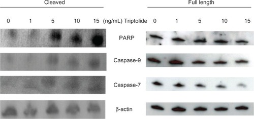
To further elucidate whether apoptosis mediates MCF-7 cell death, we investigated expression levels of pro-apoptotic proteins, PARP, caspase-9, and caspase-7, using western blotting. In general, caspase-3 is a major component of the apoptotic signaling pathway. But MCF-7 has an inherent lack of caspase-3,Citation31,Citation32 and the 35 kDa full-length caspase-3 was not detected in MCF-7 cells, although it was detected in control DU145 prostate cancer cells by western blotting (data not shown). The expression levels of the full-length PARP band decreased in a dose-dependent manner with triptolide concentration, and a band representing the cleaved PARP increased in a dose-dependent manner (). Full-length caspase-9 and caspase-7 protein levels were also downregulated in experimental cells in a dose-dependent manner. In contrast, cleaved caspase-9 and cleaved caspase-7 protein levels increased in a dose-dependent manner (). Taken together, these results indicate that triptolide induces a dose-dependent activation of pro-apoptotic proteins in caspase-3-deficient MCF-7 cells.
Figure 4 Effect of triptolide on pro-apoptotic proteins expression levels.
Abbreviations: PARP, poly(ADP-ribose) polymerase; SDS-PAGE, sodium dodecyl sulfate polyacrylamide gel electrophoresis.
Lysosomal enzymatic activity and localization
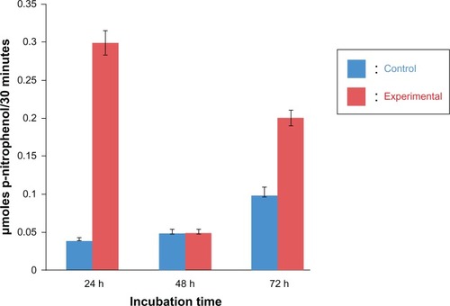
We quantified the activity of lysosomal enzymes by monitoring the activity of AP, a marker enzyme for lysosomes.Citation19 MCF-7 Cells were seeded in 12-well plates for 24 hours and then incubated at various times in the presence or absence of 10 ng/mL triptolide. This concentration was chosen as the LD50 from our MTT experiments. AP activity was elevated after 24 and 72 hours in experimental cells (). This observation has also been demonstrated in the labial glands of Manduca sexta, where lysosomal AP activity increased during early larval to pupal metamorphosis and leveled off during mid-metamorphosis; ultimately, the bulk of the cytoplasm was engulfed by autophagic vacuoles.Citation19
Figure 5 Effect of triptolide on lysosomal enzyme activity.
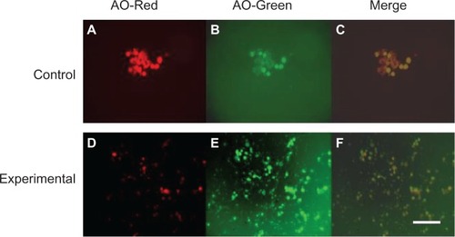
To further demonstrate that triptolide induces atypical apoptosis via acidic organelles (eg, lysosomes), cells were stained with acridine orange, a fluorescent dye that accumulates in acidic organelles. Control cells displayed distinct red fluorescence in an acidic environment, the lysosomes, () and a weak, green fluorescence in a neutral environment, the cytosol, (). In contrast, in experimental cells, red fluorescence was reduced () and green fluorescence was maximally increased (), which might suggest that advanced lysosomal rupture resulted in triptolide-treated cells. Merged images of control and experimental cells are shown in and , respectively.
Figure 6 Effect of triptolide on localization of lysosomes.
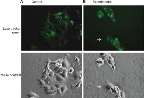
Finally, we observed the morphology of lysosomes using LysoTracker Green, which concentrates in intact lysosomes and emits green fluorescence (). After 24 hours from cell seeding, cells were incubated for 24 hours in the presence or absence of 10 ng/mL triptolide. We found that controls had a majority of healthy and adhesive cells, which displayed a punctate green fluorescence staining pattern (). In contrast, a significant reduction in cell number and increased cell detachment were observed in triptolide-treated cells (). Furthermore, there was increased uptake of LysoTracker Green in experimental cells compared to control cells (). These results suggest that triptolide exposure results in modulation of lysosomal membrane integrity.
Figure 7 Morphological comparison of lysosomes.
Effect of triptolide on the expression of lysosomal proteins
To further confirm our hypothesis that triptolide induces programmed cell death triggered by lysosomal disintegration, the leakage of lysosomal enzymes in the cytosolic fractions was investigated.
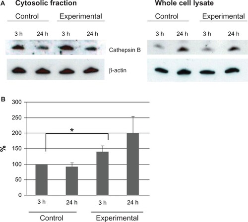
As described in the “Materials and methods” section, MCF-7 cells were seeded for 24 hours and then incubated in the presence or absence of 10 ng/mL triptolide. Each whole-cell lysate was separated into cytosolic and particulate fractions according to the manufacturer’s instructions. We investigated the protein expression levels of the cysteine protease cathepsin B, as an indicator of the leakage, using the MCF-7 cytosolic fractions analyzed by western blotting. Each sample included two time points, 3 hours and 24 hours after drug treatment. In general apoptosis, 3 hours might be an early apoptotic stage and 24 hours might be a late stage. In , mature cathepsin B protein levels were compared between cytosolic and total fractions of untreated MCF-7 cells and 10 ng/mL triptolide-treated MCF-7 cells. The data support the fact that cathepsin B leaked into the cytosol 3 hours after drug treatment and experimental cytosolic fractions contained more cathepsin B than control samples at both time points. Total protein fractions also showed a band corresponding to cathepsin B, which indicates that triptolide does not just increase total cathepsin B. In , an increase in cathepsin B protein levels from 3 hours to 24 hours in the cytosolic fractions was detected by quantitative analysis. These results suggest that triptolide promotes leakage of lysosomal hydrolases into the cytosol. The release of lysosomal enzyme cathepsin B into the cytosol confirmed our LysoTracker Green results that triptolide affects lysosomal integrity.
Figure 8 Lysosomal protein expression levels on cytosolic fraction of MCF-7 cells.
In summary, our results suggest that programmed cell death and atypical apoptosis, in MCF-7 cells, as a result of triptolide exposure, is dependent on LMP.
Discussion
Triptolide, an extract of Tripterygium wilfordii Hook F, has been widely used in the People’s Republic of China for the treatment of inflammatory and autoimmune diseases for centuries. In addition to its anti-inflammatory and immunosuppressive effects, anticancer effects have also been reported in recent years.Citation10,Citation11,Citation34 Triptolide can covalently interact with and inhibit the general transcription factor TFIIH component XPB, explaining its transcriptional effects.Citation8 The expression of metalloproteinase 10 (ADAM10) is increased in several tumors, including leukemia, and is involved in malignant cell growth and cancer progression. ADAM10 is a novel target of triptolide; inhibition of ADAM10 by triptolide might be another mechanism by which triptolide inhibits tumorigenesis.Citation34 Polycystin-2 (PC2) has been identified as a candidate target for the therapeutic action of triptolide and an association between PC2 expression and sensitivity to triptolide-induced growth arrest/cell death has been observed.Citation35 Triptolide has been reported to be the first known inhibitor of dCTP pyrophosphatase 1 (DCTPP1).Citation36 DCTTP1 is postulated to be a gatekeeper enzyme, protecting RNA or DNA against incorporation of noncanonical nucleotide triphosphates. However, it is also possible that the antitumor effects of triptolide may be explained by some other, yet unidentified, mechanisms. The fact that multiple molecular targets participate in triptolide’s pharmacological functions support the notion that triptolide can be an effective anticancer agent against cells and tumors that lack caspase-3.
Among the various in vitro cell line models available for breast cancer, the MCF-7 cell line presents distinctive properties that may help shed new light on the mechanism of action of triptolide. In particular, MCF-7 is an estrogen receptor-positive cell line that lacks caspase-3 and beclin-1;Citation37,Citation38 thus, it represents a cell model with compromised apoptotic machinery and low autophagic activity that might influence cellular response to anticancer drug treatment. We propose that the deletion of caspase-3 in tumors or cells will not limit triptolide’s usefulness because other mechanisms are at work, such as autophagy. Triptolide induces apoptosis in various cancer cells and, thus far, very little is known about the effect of triptolide on MCF-7 cells, except that triptolide exposure results in an increase in p53 expression,Citation33 a decrease in estrogen receptor-α expression,Citation33 a decrease in ADAM10 expression,Citation34 and focal adhesion kinase cleavage.Citation30
A previous study suggested that triptolide may regulate lysosomal-mediated apoptosis in MCF-7 breast cancer cells.Citation39 In the present study, we demonstrate that triptolide’s cytotoxicity is dose- and time-dependent in MCF-7 cells, as examined by morphology (), Hoechst staining (), MTT assay (), and western blot analysis (). Our MTT data are in agreement with a previous report by Liu et al.Citation33 Regarding triptolide-induced morphology, MCF-7 cells displayed less cytoplasmic blebbing (), and the pattern of pro-apoptotic protein expression levels () was similar to that of staurosporine-treated MCF-7 cells.Citation32 PARP and procaspase-9 and -7 protein levels were downregulated in experimental cells in a dose-dependent manner, and cleaved PARP, caspase-9, and caspase-7 protein levels increased in a dose-dependent manner (). Active caspase-9 is an initiator protease that complexes with Apaf-1 and cytochrome c to form the apoptosome.Citation40 Caspase-7 has been shown to cleave PARP-1 in apoptotic MCF-7 cells,Citation41 while a more recent study revealed that caspase-7 uses an exosite to promote PARP proteolysis.Citation42 Therefore, we concluded that MCF-7 cell death as a result of triptolide exposure is atypical apoptosis.
Before the discovery of the caspase family of proteases, most cell deaths were considered to be lysosomalCitation43 or “type II,”Citation15 requiring activation of the lysosomal compartment. In insects, cell death at metamorphosis is typically autophagic, and blocking autophagy is a pupariation lethal.Citation45 This autophagic type of cell death may also be used in situations in which conventional apoptosis is blocked or limited by mutation or other controls, as in MCF7 cells, which lack caspase-3, or in which massive cell death overwhelms the ability of phagocytes to clear the terrain.Citation46 Zakeri et al reported that in metamorphosing, secretory cells and under conditions where the majority of cells die, the bulk of the cytoplasm is consumed by expansion of the lysosomal system well before nuclear collapse is manifested.Citation16 In the current study, we demonstrate that triptolide induces programmed cell death in MCF-7 cells that is presumably characterized by a leakage of lysosomal enzymes into the cytosol.
We report here that triptolide induces lysosomal-mediated apoptosis in MCF-7 cells. illustrates the activity of AP Halaby et al previously used AP to monitor apoptosis in degenerating M. sexta labial glands during the larval to pupal metamorphosis.Citation19 Others have used this enzyme to measure cell death in Drosophila salivary glands as well as insect intersegmental muscles.Citation47,Citation14 AP activity was elevated after 24 and 72 hours in experimental cells (). These results are supported by our previous study examining AP activity in the labial gland of Manduca during the larval to pupal molt.Citation48 The relatively high levels of AP activity in control cells at 72 hours may be explained by the fact that activities of specific lysosomal hydrolases are higher in tumor cells than in the corresponding normal cells.Citation49 These results suggests that triptolide activates lysosomes. Next, experimental cells stained with the vital dye acridine orange displayed neutral cytosolic staining, which did not occur in control cells ( and ). Acridine orange concentrated in lysosomes emits a granular red fluorescence, whereas, in the cytosol, it emits a diffuse green fluorescence.Citation50 These data suggest that triptolide mediated a reduction in red fluorescence while an increased diffuse cytosolic green fluorescence indicated a relocation of acridine orange from the lysosomes to the cytosol following a change in lysosome permeability. This provides supporting evidence for our hypothesis that triptolide modulates LMP. Similarly, exposure of cells to triptolide resulted in an increase in the accumulation of the lysosomotropic probe LysoTracker Green ().
LysoTracker Green staining also demonstrated that triptolide-treated cells displayed diffuse and intense staining, whereas staining was punctate and reduced in control cells ().
Finally, expression levels of cathepsin B in MCF-7 cytosolic fractions supported susceptibility to lysosomal permeabilization. Elevated cathepsin B was detected at the early stage of cell death (3 hours post-triptolide-treatment). Decreased expression of cathepsin B was detected in control cells (). Altogether, these results suggest that triptolide affects LMP. The resulting change in lysosomal membrane permeability in turn leads to leakage of the lysosomal hydrolases into the cytosol, where they can trigger the apoptotic cascade by activating proteins such as Bid.Citation23,Citation24 This notion, that lysosomal rupture may be an upstream event during some instances of apoptosis, is supported by and is also reinforced by work from others.Citation51–Citation53 These findings suggest that lysosomal destabilization might play an integral part in programmed cell death via the intrinsic pathway.
Increasing evidence suggests that lysosomes are important mediators of programmed cell death. In autophagic cell death, lysosomes fuse with autophagosomes to form autophagolysosomes, by which their contents are degraded.Citation54 In apoptosis, cathepsins are released from lysosomes into the cytoplasm and trigger a cascade of intracellular degradation.Citation53,Citation55,Citation56 The involvement of lysosomes in both programmed cell death pathwaysCitation55–Citation57 may suggest an involvement of cathepsins as a functional link between apoptosis and autophagy. This hypothesis was confirmed by results of a recent study showing that inhibition of cathepsins by E64d, an inhibitor of papain-like cysteine proteases, resulted in a significant reduction of apoptotic cell numbers accompanied by an increase in autophagosome formation in MCF-7 cells exposed to camptothecin.Citation58
Conclusion
The nature of triptolide to act as an anti-inflammatory, anticancer, and antiproliferative agent makes it an attractive alternative to current methods of cancer therapy. The untoward side effects of chemotherapy and radiation can be debilitating to the health of the patient and, in some cases, can actually make the cancer worse or bring on a new type of cancer.Citation59,Citation60 For these reasons, novel and safer alternative treatment modalities need to be investigated. A desirable and clean method of killing cancer cells is via programmed cell death. The dying cell dies in a self-contained fashion because the cellular contents remain membrane-bound and then the cell is removed by macrophages. It is often the goal of chemotherapeutic, radiation, and hormonal treatments to induce apoptosis in cancer cells.Citation61,Citation62
In the present study, we demonstrate that triptolide modulates apoptosis in MCF-7 cells by inducing lysosomal membrane permeability, increasing the expression levels of pro-apoptotic proteins, and inhibiting cell growth in a dose-and time-dependent manner. To our knowledge, this is the first report in the literature of triptolide-induced lysosomal membrane permeability as an anticancer treatment. The importance of lysosomal-mediated programmed cell death has been underappreciated, and this study is innovative in the sense that it revisits this historically important organelle. Further studies are warranted to decipher the molecular mechanisms by which triptolide modulates programmed cell death in various cancer cells.
Acknowledgments
This work was supported by the National Institute of General Medical Sciences grant T34 GM079079 to RH. The authors also wish to thank Juan C Sanchez for technical assistance.
Disclosure
The authors report no conflicts of interest in this work.
References
- National Cancer Institute’s Website Available from: http://seer.cancer.gov/statfacts/html/breast.html#incidence-mortality
- ParkinDMBrayFFerlayJPisaniPGlobal cancer statistics, 2002CA Cancer J Clin2005557410815761078
- SiegelRNaishadhamDJemalACancer statistics, 2012CA Cancer J Clin2012621102922237781
- LiFQLuXZLiangXBTriptolide, a Chinese herbal extract, protects dopaminergic neurons from inflammation-mediated damage through inhibition of microglial activationJ Neuroimmunol2004148243114975583
- QiuDKaoPNImmunosuppressive and anti-inflammatory mechanisms of triptolide, the principal active diterpenoid from the Chinese medicinal herb Tripterygium wilfordii Hook. fDrugs R D2003411812568630
- BrinkerAMMaJLipskyPERaskinIMedicinal chemistry and pharmacology of genus TripterygiumPhytochemistry20076873276617250858
- ChanMAKohlmeierJEBrandenMJungMBenedictSHTriptolide is more effective in preventing T cell proliferation and interferon-gamma production than is FK506Phytother Res19991346446710479754
- TitovDVGilmanBHeQLXPB, a subunit of TFIIH, is a target of the natural product triptolideNat Chem Biol2011718218821278739
- ChoiYJKimTGKimYHImmunosuppressant PG490 (triptolide) induces apoptosis through the activation of caspase-3 and down-regulation of XIAP in U937 cellsBiochem Pharmacol20036627328012826269
- LeeKYChangWTQiuDKaoPNRosenGDPG490 (triptolide) cooperates with tumor necrosis factor-α to induce apoptosis in tumor cellsJ Biol Chem1999274134511345510224110
- CarterBZMakDHSchoberWDTriptolide sensitizes AML cells to TRAIL-induced apoptosis via decrease of XIAP and p53-mediated increase of DR5Blood20081113742375018187663
- KroschTCSangwanVBanerjeeSTriptolide-mediated cell death in neuroblastoma occurs by both apoptosis and autophagy pathways and results in inhibition of nuclear factor-kappa B activityAm J Surg201320538739623428154
- JainMVPaczullaAMKlonischTInterconnections between apoptotic, autophagic and necrotic pathways: implications for cancer therapy developmentJ Cell Mol Med201317122923301705
- MaghsoudiNZakeriZLockshinRAProgrammed cell death and apoptosis – where it came from and where it is going: from Elie Metchnikoff to the control of caspasesExp Oncol20123414615223069998
- SchweichelJUMerkerHJThe morphology of various types of cell death in prenatal tissuesTeratology197372532664807128
- ZakeriZFQuaglinoDLathamTLockshinRADelayed internucleosomal DNA fragmentation in programmed cell deathFASEB J199374704788462789
- KitanakaCKatoKIjiriRIncreased Ras expression and caspase-independent neuroblastoma cell death: possible mechanism of spontaneous neuroblastoma regressionJ Natl Cancer Inst20029435836811880474
- de LizRHorstHPizzolattiMGFrödeTSGirardDActivation of human neutrophils by the anti-inflammatory mediator Esenbeckia leiocarpa leads to atypical apoptosisMediators Inflamm2012201219838222649276
- HalabyRZakeriZLockshinRAMetabolic events during programmed cell death in insect labial glandsBiochem Cell Biol1994725976017654333
- de DuveCThe lysosome turns fiftyNat Cell Biol2005784784916136179
- LuzioLPPryorPRBrightNALysosomes: fusion and functionNat Rev Mol Cell Biol2007862263217637737
- LinderSShoshanMCLysosomes and endoplasmic reticulum: Targets for improved, selective anticancer therapyDrug Resist Updat2005819920416055370
- JohanssonAAppelqvistHNilssonCKagedalKRobergKOllingerKRegulation of apoptosis-associated lysosomal membrane permeabilizationApoptosis20101552754020077016
- ČesenMHPeganKSpesATurkBLysosomal pathway to cell death and their therapeutic applicationsExp Cell Res20123181245125122465226
- CheriyathVKuhnsMAKalaycioMEBordenECPotentiation of apoptosis by histone deacetylase inhibitors and doxorubicin combination: cytoplasmic cathepsin B as a mediator of apoptosis in multiple myelomaBr J Cancer201110495796721364585
- KirkegaardTRothAGPetersenNHHsp70 stabilizes lysosomes and reverts Niemann-Pick disease-associated lysosomal pathologyNature201046354955320111001
- ShreeTOlsonOCElieBTMacrophages and cathepsin proteases blunt chemotherapeutic response in breast cancerGenes Dev2011252465247922156207
- PaquetCSanéATBeaucheminMBertrandRCaspase- and mitochondrial dysfunction-dependent mechanisms of lysosomal leakage and cathepsin B activation in DNA damage-induced apoptosisLeukemia20051978479115759029
- MosmannTRapid colorimetric assay for cellular growth and survival: application to proliferation and cytotoxicity assaysJ Immunol Methods19836555636606682
- TanBJTanBHChiuGNEffect of triptolide on focal adhesion kinase and survival in MCF-7 breast cancer cellsOncol Rep2011261315132121805042
- JänickeRUSprengartMLWatiMRPorterAGCaspase-3 is required for DNA fragmentation and morphological changes associated with apoptosisJ Biol Chem1998273935793609545256
- TaimenPKallajokiMNuMA and nuclear lamins behave differently in Fas-mediated apoptosisJ Cell Sci200311657158312508117
- LiuJJiangZXiaoJEffects of triptolide from Tripterygium wilfordii on ERalpha and p53 expression in two human breast cancer cell linesPhytomedicine2009161006101319524422
- SoundararajanRSayatRRobertsonGSMarignariPATriptolide: an inhibitor of a disintegrin and metalloproteinase 10 (ADAM10) in cancer cellsCancer Biol Ther200982054206219783906
- LeuenrothSJOkuharaDShotwellJDTriptolide is a traditional Chinese medicine-derived inhibitor of polycystic kidney diseaseProc Natl Acad Sci U S A20071044389439417360534
- CorsonTWCavgaHAberleNCrewsCMTriptolide directly inhibits dCTP pyrophosphataseChembiochem2011121767177321671327
- SemenovDVAronovPAKuliginaEVPotapenkoMORichterVAOligonucleosome DNA fragmentation of caspase 3 deficient MCF-7 cells in palmitate-induced apoptosisNucleosides Nucleotides Nucleic Acids20042383183615560068
- MizushimaNTransgenic models of autophagyDereticVAutophagy in Immunity and Infection: A Novel Immune EffectorWeinheimWiley-VCH20065565
- MessinaMEJrHalabyRDoes triptolide induce lysosomal-mediated apoptosis in human breast cancer cells?Med Hypotheses201177919321486687
- BrattonSBSalvesenGSRegulation of the Apaf-1-caspase-9 apoptosomeJ Cell Sci20101233209321420844150
- GermainMAffarEBD’AmoursDDixitVMSalvesenGSPoirierGGCleavage of automodified poly(ADP-ribose) polymerase during apoptosis. Evidence for involvement of caspase-7J Biol Chem1999274283792838410497198
- BoucherDBlaisVDenaultJBCaspase-7 uses an exosite to promote poly(ADP ribose) polymerase 1 proteolysisProc Natl Acad Sci U S A20121095669567422451931
- LockshinRAProgrammed cell death. Activation of lysis by a mechanism involving the synthesis of proteinJ Insect Physiol196915150515165348113
- SchweichelJUMerkerHJThe morphology of various types of cell death in prenatal tissuesTeratology197372532664807128
- JuhászGCsikósGSinkaRErdélyiMSassMThe Drosophila homolog of Aut1 is essential for autophagy and developmentFEBS Lett200354315415812753924
- LockshinRAZakeriZApoptosis, autophagy, and moreInt J Biochem Cell Biol2004362405241915325581
- JochováJZakeriZLockshinRARearrangement of the tubulin and actin cytoskeleton during programmed cell death in Drosophila salivary glandsCell Death Differ1997414014916465220
- HalabyRMartinezMLLockshinRZakeriZ20-Hydroxyecdysone induces apoptosis in the labial glands of Manduca sextaJ Res Lepid200337310
- PooleARTumour lysosomal enzymes and invasive growthDingleJTLysosomes in Biology and Pathology3AmsterdamNorth-Holland Publishing Company1973303337
- YuHLouJRDingWQClioquinol independently targets NF-kappaB and lysosome pathways in human cancer cellsAnticancer Res2010302087209220651355
- IvanovaSRepnikUBojicLPetelinATurkVTurkBLysosomes in apoptosisMethods Enzymol200844218319918662570
- BoyaPAndreauKPoncetDLysosomal membrane permeabilization induces cell death in a mitochondrion-dependent fashionJ Exp Med20031971323133412756268
- CirmanTOresićKMazovecGDSelective disruption of lysosomes in HeLa cells triggers apoptosis mediated by cleavage of Bid by multiple papain-like lysosomal cathepsinsJ Biol Chem20042793578358714581476
- KimJKlionskyDJAutophagy, cytoplasm-to-vacuole targeting pathway, and pexophagy in yeast and mammalian cellsAnnu Rev Biochem20006930334210966461
- ZhaoMAntunesFEatonJWBrunkUTLysosomal enzymes promote mitochondrial oxidant production, cytochrome c release and apoptosisEur J Biochem20032703778378612950261
- BrunkUTNeuzilJEatonJWLysosomal involvement in apoptosisRedox Rep20016919711450988
- LeistMJäätteläMTriggering of apoptosis by cathepsinsCell Death Differ2001832432611550083
- Lamparska-PrzybyszMGajkowskaBMotylTCathepsins and BID are involved in the molecular switch between apoptosis and autophagy in breast cancer MCF-7 cells exposed to camptothecinJ Physiol Pharmacol20055615917916077201
- ReddingSWHavemanCWTreating the discomfort of oral ulceration resulting from cancer chemotherapyCompend Contin Educ Dent19992038939239439611692345
- LorigKRSobelDSRitterPLLaurentDHobbsMEffect of a self management program on patients with chronic diseaseEff Clin Pract2001425626211769298
- XueLYChiuSMOleinickNLStaurosporine-induced death of MCF-7 human breast cancer cells: a distinction between caspase-3-dependent steps of apoptosis and the critical lethal lesionsExp Cell Res200328313514512581734
- BrownJMWoutersBGApoptosis, p53, and tumor cell sensitivity to anticancer agentsCancer Res1999591391139910197600