
Abstract
Uterine fibroids are a major cause of morbidity in women of a reproductive age (and sometimes even after menopause). There are several factors that are attributed to underlie the development and incidence of these common tumors, but this further corroborates their relatively unknown etiology. The most likely presentation of fibroids is by their effect on the woman’s menstrual cycle or pelvic pressure symptoms. Leiomyosarcoma is a very rare entity that should be suspected in postmenopausal women with fibroid growth (and no concurrent hormone replacement therapy). The gold standard diagnostic modality for uterine fibroids appears to be gray-scale ultrasonography, with magnetic resonance imaging being a close second option in complex clinical circumstances. The management of uterine fibroids can be approached medically, surgically, and even by minimal access techniques. The recent introduction of selective progesterone receptor modulators (SPRMs) and aromatase inhibitors has added more armamentarium to the medical options of treatment. Uterine artery embolization (UAE) has now been well-recognized as a uterine-sparing (fertility-preserving) method of treating fibroids. More recently, the introduction of ultrasound waves (MRgFUS) or radiofrequency (VizAblate™ and Acessa™) for uterine fibroid ablation has added to the options of minimal access treatment. More definite surgery in the form of myomectomy or hysterectomy can be performed via the minimal access or open route methods. Our article seeks to review the already established information on uterine fibroids with added emphasis on contemporary knowledge.
Introduction
Uterine fibroids (also known as leiomyomas or myomas) are the commonest benign uterine tumors, with an estimated incidence of 20%–40% in women during their reproductive years.Citation1,Citation2 They are monoclonal tumors of the uterine smooth muscle cells and consist of large amounts of extracellular matrix that contain collagen, fibronectin, and proteoglycan.Citation3,Citation4 Even though their pathogenesis is not clearly known, there is considerable evidence that estrogens and progestogens proliferate tumor growth,Citation5,Citation6 as the fibroids rarely appear before menarcheCitation7 and regress after menopause.Citation8 They are classified by their location relative to the layers of the uterus (as subserous, intramural, or submucous) and can be single or multiple.
Epidemiology and etiology
Race and age
A study carried out in the USA with randomly selected women between the ages of 35 and 49 years (who were screened by self-report, medical record, and sonography) showed that the incidence of uterine fibroids by age 35 was 60% among African-American women, increasing to >80% by age 50, whereas Caucasian women showed an incidence of 40% by age 35, and almost 70% by age 50.Citation9
The cumulative incidence (based both on ultrasonographic detection of fibroids in women with an intact uterus and evidence of prior fibroids among women who have had hysterectomies) increases with age, but the rate of increase slows at older ages. This suggests that the older premenopausal uterus is less susceptible to fibroid development.Citation10
Early menarche
Most of the older studies (reviewed by Schwartz)Citation11 had reported an increased risk of fibroids with earlier age of menarche and the newer data confirm these findings. Early age of menarche is also a risk factor for other hormonally mediated conditions such as endometrial and breast cancers.Citation12,Citation13 The biological mechanisms are not understood, and they may or may not be the same for the different hormonally mediated conditions.
Parity and pregnancy
Parity has been inversely associated with a risk of fibroid development in the earlier studies,Citation14 and the newer studies confirm these findings.Citation15,Citation16 Although a direct protective effect of pregnancy has been demonstrated, little is known of the mechanism. There have been some suggestions that during postpartum uterine remodeling, there could be selective apoptosis of small lesions.Citation14 Ischemia during parturition has also been proposed as a mechanism.Citation17 Thus, it may be implied that fibroid tissue could be highly susceptible to ischemia during both parturition and remodeling.Citation10
Caffeine intake
There has been recent evidence suggesting a relationship between alcohol and caffeine intake with a risk of developing fibroids, especially with evidence gathered from the Black Women’s Health Study.Citation18 Current drinkers had significantly higher risks than women who had never consumed alcohol, and there appears to be a dose response for both duration of alcohol consumption and number of drinks per day. With regards to caffeine, among women <35 years of age, the highest categories of caffeinated coffee (≥3 cups/day) and caffeine intake (≥500 mg/day) were both associated with increased fibroid risk.Citation10
Other possible factors
There is contemporary interest in the influence of dynamics encompassing the likes of uterine infection, hormonal, metabolic, dietary, stress, and environmental factors. The underlying biological mechanism of infection-related oncogenesis proposed is that injury caused by infection or inflammation proceeds through several possible pathways, leading to increased extracellular matrix, cell proliferation, and decreased apoptosis, apropos of abnormal tissue repair.Citation19–Citation22 The upregulation of extracellular matrix proteins that is consistently seen in gene profiling studies of fibroids compared with normal myometriumCitation23 is consistent with such a mechanism. As luteinizing hormone (LH) shares a receptor with human chorionic gonadotropin, the hormone that stimulates uterine growth during early pregnancy, it is hypothesized that peri-menopausal increases in LH would stimulate fibroid growth.Citation24 Metabolic factors like diabetes, polycystic ovaries, and hypertension have been examined. Dietary factors have looked at the intake of soy which tends to have anti-estrogenic effects when endogenous estrogens are high (ie, premenopausal women), thus hypothesizing that soy intake might reduce fibroid risk.Citation10 A possible mechanism of the impact of stress involves the effects of stress on adrenal activity that could raise progesterone levels,Citation25 and thus increase fibroid development. All these areas of interest are currently hypothetical and need further study to clarify their exact role in the etiology of fibroids.
Presentation
Uterine fibroids are the cause for some of the most common gynecological problems among women presenting to gynecology emergency and outpatient departments in the UK. They are often asymptomatic but they can cause a multitude of symptoms () such as abnormal uterine bleeding, a feeling of pelvic pressure, urinary incontinence or retention, or pain. They may also be associated with reproductive problems such as infertility and miscarriage.Citation26
Table 1 The clinical presentation of uterine leiomyomas
The presenting symptoms play an important role in deciding the appropriate form of treatment for the affected woman. Management strategies are usually individualized based on the severity of the symptoms, the size and location of the fibroid, the patient’s age and their chronological proximity to menopause, and the patient’s desire for future fertility.Citation27 The majority of women with uterine fibroids are asymptomatic, and consequently get less clinical attention; fibroid tumors often remain undiagnosed.Citation28,Citation29 The most common presenting factor that symptomatic women typically complain about is abnormal uterine bleeding, specifically in terms of heavy and prolonged bleeding.Citation1 In a study by Wegienka et al, women with myomas were more likely to report a ‘gushing’ type of bleeding and higher pad/tampon use than women without myomas.Citation30 In addition, women with uterine fibroids may suffer more often from dyspareunia and non-cyclic pelvic pain.Citation31 Although bleeding and pelvic pain symptoms are frequently reported in literature as the main symptoms related to uterine fibroids, the number of systematic studies on fibroid symptoms is limited.Citation32
Diagnosis
Precise uterine fibroid mapping (localization, measurement, and characterization) is essential for research into clarifying the natural history of these tumors and for evaluating therapeutic responses to investigational agents.Citation33 The optimal selection of patients for medical therapy, noninvasive procedures, or surgery depends on an accurate assessment of the size, number, and position of myomas. Imaging techniques available for confirming the diagnosis of myomas include sonography, saline-infusion sonography, and MRI.Citation34
Ultrasonography
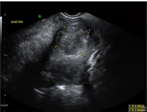
Ultrasonography using the transabdominal and transvaginal routes has been employed most frequently, due to its accessibility and relatively low cost.Citation35 While a cost-effective instrument, ultrasound has been criticized for its significant operator-dependence, resulting in inferior reproducibility when compared to MRI.Citation36–Citation39 Ideally, both transabdominal and transvaginal scans should be performed. Transvaginal scans are more sensitive for the diagnosis of small fibroids. However, when the uterus is bulky or retroverted, the uterine fundus may lie outside of the field of view. Transabdominal views are often of limited value if the patient is obese. Ultrasonography in skilled hands can detect fibroids as small as 5 mm on transvaginal ultrasounds. Typically, fibroids appear as well-defined, solid masses with a whorled appearance (). These are usually of similar echogenicity to the myometrium, but sometimes may be hypoechoic. They cause the uterus to appear bulky or may cause an alteration of the normal uterine contour. Even non-calcified fibroids often show a degree of posterior acoustic shadowing, though this is of course more marked in calcified fibroids. Degenerate fibroids may have a complex appearance, with areas of cystic change.Citation40 Doppler ultrasound typically shows circumferential vascularity. However, fibroids which are necrotic or have undergone torsion will show absence of flow.Citation41 Submucous fibroids are usually clearly visible separate from the endometrium under transvaginal ultrasound, but can be difficult to differentiate from polyps.Citation40 Sonography may be inadequate for determining the precise number and position of myomas, although transvaginal sonography is reasonably reliable for uteri <375 mL in total volume or containing four myomas or fewer.Citation42 Large fibroids can occasionally cause obstruction of the ureters, with secondary hydronephrosis. Therefore, ultrasound examination should include the urinary tract whenever a large pelvic mass is identified. The diagnosis of fibroids on ultrasound is usually reasonably straightforward though focal adenomyosis can mimic a fibroid and a pedunculated uterine fibroid can sometimes be mistaken for an adnexal mass.Citation43 When there is doubt about the origin of a pelvic mass at ultrasound, further evaluation with MRI should be performed.Citation40
Figure 1 A 37-year-old Afro-Caribbean woman with a history of menorrhagia. Transvaginal ultrasound image showed a bulky fibroid uterus with multiple intramural fibroids. The largest fibroid (pictured above) is at the fundus on the posterior wall measuring 4.5 × 4 cm.
Saline infusion sonohysterography
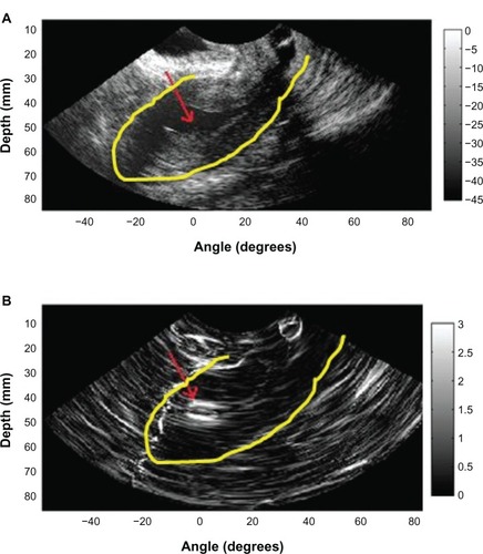
Saline infusion sonohysterography-based imaging is usually used as a supplementary or adjunct imaging modality for characterization of focal uterine masses diagnosed on B-mode ultrasound images. During transvaginal ultrasound, a uterine mass may appear as an area of increased echogenicity bulging into the endometrial cavity with echogenicity similar to that of the myometrium. In addition, it is difficult to distinguish a leiomyoma from a blood clot or a polyp, and leiomyomas also may obscure the endometrium on imaging or cause an overestimation of endometrial thickness. Transvaginal sonography may be used initially in the detection of endometrial polyps, which may appear as hyperechoic masses surrounded by a hypoechoic endometrium.Citation44 However, it may be difficult to detect some polyps because they may appear as a diffusely thickened endometrium.Citation45 Nevertheless, polyps may be better visualized during saline infusion sonohysterography, in which the saline pushes apart the uterine cavity, and the polyps appear as smoothly margined focal lesions that protrude into the endometrial cavity (). Saline infusion sonohysterography is also effective in distinguishing diffuse endometrial changes and focal intracavitary protuberances.Citation44 However, it is limited in its ability to differentiate between endometrial hyperplasia (premalignant polyps) and endometrial carcinoma.Citation46
Figure 2 B-mode image (A) obtained during saline infusion sonohysterography in a 46-year-old woman with a diagnosis of an endometrial polyp and corresponding strain image (B) obtained using the 2-dimensional multilevel hybrid algorithm. Red arrows indicate the location of the polyp, and yellow contours indicate the outer uterine wall.
The quality of the image obtained is dependent on the amount of deformation induced by the saline injection into the uterine cavity. If the amount of saline injected is too low, the deformation induced is too small to provide images with a reasonable signal-to-noise ratio. This situation has led to generation of suboptimal strain images in some instances because of the minimal deformation of the tissue. The amount of deformation applied via saline injection is dependent on the ability of the patient to tolerate the discomfort induced due to saline injection into the uterus; in other cases, insufficient deformation is due to the presence of saline from a previous infusion and the inability to withdraw sufficient fluid before starting a new infusion. Patient comfort is of paramount importance during the data acquisition because saline injection can be quite painful and distressing to the patient. Care should be taken to induce a sufficient deformation of the uterine wall that is tolerated by the patient to obtain high-quality strain images. The use of saline infusion sonohysterography-based strain imaging as a standalone imaging technique for the detection of uterine masses requires additional validation on a larger number of patients and at multiple clinical sites.Citation44
In conclusion, there seems to be limited diagnostic accuracy with saline infusion sonohysterography when better tolerated and more accurate imaging exists with ultrasonography (above) and MRI imaging (see below).
Magnetic resonance imaging (MRI)
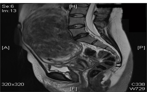
Magnetic resonance imaging, while more costly, has been touted as the most sensitive modality for evaluating uterine myomas (), particularly for the detection of small fibroids.Citation42 MRI is accurate in diagnosing a leiomyoma with a sensitivity of 88%–93% and a specificity of 66%–91%,Citation47,Citation48 and in differentiating leiomyoma from focal adenomyosis. Thus, MRI is more sensitive in identifying uterine fibroids than ultrasound, does not involve the use of ionizing radiation, and it can readily demonstrate the uterine zonal anatomy.Citation37,Citation42,Citation49 Submucosal, intramural, and subserosal fibroids are usually easily differentiated with MRI, and fibroids as small as 5 mm in diameter can be demonstrated. Fibroids in relatively unusual locations, such as within the cervix, can also be identified.Citation40 MRI can be very helpful when investigating suspected acute fibroid complications when the patient presents to the emergency department,Citation50 and it is also a valuable tool that can be used to both predict and assess the response of fibroids to uterine artery embolization (UAE).Citation40 Uterine fibroids are composed of a combination of smooth muscle cells and fibrous connective tissue. As these masses enlarge, they commonly outgrow their blood supply and undergo varying degrees of necrosis, which accounts for their variable signal intensities on MRI.Citation51 Typically, leiomyomas are low in signal intensity relative to their surrounding myometrium on T2W imaging and are isointense to myometrium on T1W imaging.Citation52,Citation54 These signal characteristics are related to the most common form of degeneration (60%), which is hyalinization throughout the leiomyoma.Citation53,Citation54 Weinreb et al defined diagnostic criteria for a leiomyoma to include a uterine mass that is predominantly hypointense compared to the myometrium on T2W imaging and predominantly hypointense on T1W imaging.Citation43
Figure 3 A 47-year-old woman with a hugely enlarged uterus. The fundus is at the level of L3. It contains a solitary large intramural fibroid which is mostly right sided, displacing the cavity to the left. This fibroid measures approximately 14.2 × 10.9 × 8.8 cm.
In some instances, it can be difficult to differentiate a large exophytic, subserosal leiomyoma from a solid adnexal mass such as an ovarian neoplasm. Differentiation is clinically very important because of differences in treatment and prognostic implications. MRI in combination with the patient’s clinical findings can be invaluable in making this distinction and can avoid unnecessary laparoscopy and/or exploratory surgery.Citation51 Location is an important distinguishing characteristic. If the mass can be definitively separated from the ovaries or is contiguous with the round ligament, then an ovarian etiology is unlikely. A well-described MRI feature that is helpful in the evaluation of large pelvic masses has been referred to as the ‘bridging vascular sign,’Citation55 which consists of vessels and/or signal voids that extend from the uterus to supply a pelvic mass. The identification of the bridging vascular sign increases the diagnostic confidence that a large pelvic mass is a uterine leiomyoma.Citation55 The bridging vessels can be identified as enhancing tubular structures on contrast-enhanced T1W imaging or as flow voids on a T2W fast spin-echo sequence.Citation51 In a study by Kim et al, the bridging vascular sign was present in 20 out of 26 exophytic leiomyomas and was absent in all other adnexal masses, resulting in a diagnostic accuracy of 80%.Citation55
Fibroids that have undergone acute degeneration show great diversity in their MRI appearances with cystic change and areas of non-enhancement. In cases of red degeneration, the patient often presents with an acute abdomen. The use of multiplanar views can enable localization of fibroids and can make it possible to distinguish fibroids from acute presentations of ovarian masses. MRI appearances show high signal intensity centrally within the fibroid on T1-weighted images consistent with blood, with reduced signal at the periphery on T2-weighted images secondary to hemosiderin deposition.Citation50 There may be heterogeneous signal intensity on T2, with no enhancement post-gadolinium administration (although gadolinium is not given to pregnant patients).Citation54,Citation56
Fibroids that demonstrate high signal on T1W images prior to embolization are likely to have a poor response to UAE as they may already have outgrown their blood supply and undergone hemorrhagic necrosis.Citation40 Conversely, high signal on T2W images prior to embolization has been shown to be a predictor of good response.Citation57 The vascularity of a fibroid is demonstrated by gadolinium enhancement and this is also a predictor of good response to UAE. Post-UAE fibroids typically show high signal on T1W images due to hemorrhagic necrosis.Citation40
Management
Conservative
Management of a patient with uterine fibroids is highly dependent on the presentation and patient wishes. Other causes of abnormal uterine bleeding, pelvic pressure/pain, and subfertility usually need to be ruled out beforehand. In many cases, the management of the fibroids carries surgical risks, and in some women, the fibroids are best left alone. Women with symptoms who have small fibroids but are close to menopause or who are trying to conceive should be offered conservative treatment with analgesics and hematinics.Citation58
Because fibroids are so common in women, it is almost a disservice to call leiomyomata a pathologic entity.Citation59 In several studies, women with minimal or no symptoms had no change in quality of life or symptomatology when they were conservatively managed; thus, prophylactic treatment is unwarranted.Citation60,Citation61
Medical
At present, medical treatments are only used for short-term therapy because of the significant risks with long-term therapy, or lack of evidence regarding the benefits and risks of long-term therapy with the newer medical agents. They may be used or are used in the following situations:
as ‘stand-alone’ treatment for temporary relief of symptoms for short periods. This application is suitable in women with symptomatic fibroids in the peri-menopausal years or in patients not suitable for surgery due to medical reasons;
as a pre-operative adjunct to reduce the size of fibroids, to control bleeding, and to improve hemoglobin levels. The reduction in fibroid size may also convert a technically difficult procedure to an easier procedure (eg, abdominal hysterectomy to vaginal hysterectomy). They can be used before myomectomy, hysterectomy, and hysteroscopic submucous resection of fibroids. At present, gonadotrophin-releasing hormone analogs (GnRHa) are mainly used for this purpose; and
in research, as part of an evaluation of new potential therapies.Citation3
Antifibrinolytics agents (tranexamic acid)
Used only during the menstrual cycle, tranexamic acid has been associated with up to a 50% decrease in bleeding in women with menorrhagia,Citation62 but there are few studies on women with leiomyomas. One study found no difference in blood loss with tranexamic acid therapy but had a sample size of 12 women with leiomyomas.Citation63 Thrombotic risk was found to be similar to that of other menorrhagia treatments.Citation64 There is retrospective evidence of increased necrosis in leiomyomas among tranexamic acid users.Citation65
Combined oral contraceptive (COC) and progestins
Combined oral contraceptive (COC) hormones have been widely trialed by physicians to reduce the blood loss associated with uterine fibroids.Citation66 A large prospective study including more than 3,000 patients with fibroids found a positive correlation between the early use of COCs (before 17 years) and the incidence of fibroids.Citation67 Other controlled trials showed no association between the use of COCs and the development of uterine fibroids.Citation68,Citation69 The effect of COCs and progestins in reducing the size of fibroids is not documented.Citation70–Citation72
Mirena® IUS
The levonorgestrel intra-uterine device (LNG-IUS) (Mirena) is effective in reducing menstrual blood loss and should be considered as an alternative to surgical treatment.Citation73 The LNG-IUS has been shown to reduce menstrual blood loss by 94% over 3 months, and is well accepted by most women when used in the general population of women with menorrhagia.Citation74
At present, there are no randomized controlled trials (RCTs) on the effect of LNG-IUS in menorrhagic women with uterine myomas. There are, of course, reports of its use in these women, with striking reductions in menorrhagia being reported.Citation75 Although some women with large intramural myomas had spontaneous expulsion of the LNG-IUS at various intervals, they wanted re-insertion of the device because of remarkable reduction in menorrhagia.Citation76 Significant increases in hemoglobin levels in blood were observed after insertion of the LNG-IUS, but no significant differences were noted in myoma volume and uterine volume, as assessed by MRI between pre-treatment and 12 months of use.Citation75,Citation76 There is therefore an obvious need for further research in this area.Citation77 It would be interesting to establish whether expulsion of the device is dependent upon the size, number, or location of the fibroids; it is reasonable to assume that submucous fibroids, or large intramural fibroids distorting the cavity, are likely to be associated with an increased risk, but definitive studies need to be undertaken in order to provide the evidence base to better advise patients.
GnRH analogs (GnRHa)
GnRHa alone or more commonly with ‘add-back therapy’, are frequently used as temporizing measures in peri-menopausal women, or pre-operatively to reduce fibroid size and render surgery safer/easier. A single injection of GnRHa produces an initial stimulation of pituitary gonadotrophins, resulting in increased secretion of follicle-stimulating hormone (FSH) and LH and the expected gonadal response. However, continuous or repeated administration of GnRHa in a continuous (non-pulsatile) fashion or administration of supraphysiological doses ultimately produces inhibition of the pituitary–gonadal axis.
GnRHa undoubtedly induce fibroid tumor shrinkage, the degree of which has been shown to be directly proportional to the percentage of cells that are estrogen receptor positive, thus implicating estrogen as a major effector of tumor growth and its reduction as the central mechanism of fibroid shrinkage with GnRHa therapy.Citation78 Chegini et al found evidence of suppression of signal transduction pathways involving growth factors, ovarian steroids, and adhesion molecules with a resulting decrease in DNA synthesis, cell proliferation, and production of transforming growth factor-b.Citation79,Citation80 They found that medical treatment causes altered regulation in a number of genes involved in the regulation of cell growth, signal transduction, transcription factors, and cell structures.Citation81 Therapy with leuprolide acetate has been associated with hyalization of leiomyomataCitation82 and decreases in uterine or tumor arterial size and blood flow variables.Citation83–Citation85
A Cochrane Systematic ReviewCitation86 to evaluate the role of GnRHa prior to either hysterectomy or myomectomy showed that pre- and postoperative hemoglobin and hematocrit were improved significantly by the use of GnRHa prior to surgery. Uterine volume and size, fibroid volume and pelvic symptoms were all reduced. Hysterectomy was rendered easier, with reduced operating time, and a greater proportion of hysterectomy patients were able to have a vaginal rather than an abdominal procedure. Blood loss and rate of vertical incision were reduced for both myomectomy and hysterectomy. Duration of hospital stay was also reduced. The disadvantages of GnRHa include cost, menopausal symptoms, and with prolonged therapy, bone demineralization. In addition, smaller fibroids may be overlooked at the time of surgery, only to recur once GnRHa is discontinued (thereby increasing the apparent risk or recurrence of fibroids following myomectomy). GnRHa cannot be used as long-term standalone therapies for fibroid disease because of the rapid rebound growth of the fibroids upon cessation of therapy.
Selective estrogen receptor modulators (SERMs)
SERMs are non-steroidal estrogen receptor ligands that display tissue-specific agonist/antagonist estrogenic actions.Citation66 They are most commonly used in the treatment and prevention of estrogen receptor-positive carcinoma of the breast; well-known examples being tamoxifen and raloxifene.
The most probable hypothesis that explains SERMs’ mechanism of action is that they induce changes in estrogen receptors, which result in differential expression of specific estrogen-regulated genes in different tissues.Citation87 Any molecule that blocks estrogen activity has the potential for therapeutic activity against fibroids, since estrogen is known to influence fibroid growth. SERMs are therefore within this category of molecule. However, because of its endometrial hyperplastic effect and case reports of fibroid growth following treatment, the potential of tamoxifen in the treatment of fibroids has not been investigated in RCTs. Raloxifene has been showed to enhance the shrinkage of uterine fibroids in postmenopausal women.Citation88,Citation89 However, a recent report from Italy that addressed the effect of raloxifene on uterine leiomyoma showed that the leiomyoma size in premenopausal women who were administered daily 60 mg doses of raloxifene over a 2-year period exhibited no change in leiomyoma size.Citation90
Selective progesterone receptor modulators (SPRMs)
The effects of progesterone on target tissues are mediated via the progesterone receptor (PR), which belongs to the nuclear receptor family.Citation91 Progesterone has dual actions on fibroid growth. It stimulates growth by upregulating epidermal growth factor (EGF) and Bcl-2 and downregulating tumor necrosis factor-alpha expression while it inhibits growth by downregulating insulin-like growth factor-1 (IGF-1) expression. While it has long been established that estrogen promotes fibroid growth, recent biochemical and clinical studies have suggested that progesterone and the PR may also enhance proliferative activity in fibroids.Citation92,Citation93 These observations have therefore raised the possibility that anti-progestins and agents or molecules that modulate the activity of the PR could be useful in the medical management of uterine fibroids (). Since the emergence of mifepristone (RU-486), the first PR antagonist, more than 25 years ago, hundreds of steroidal as well as non-steroidal compounds displaying progesterone antagonist (PA) or mixed agonist/antagonist activity have been synthesized. Collectively, they are known as progesterone receptor modulators (PRMs). Some of the PRMs which have been the subject of recent clinical trials or research studies in relation to fibroid treatment include mifepristone, CDB-4124 (telapristone), CP-8947, J867 (asoprisnil), and CDB-2914 (ulipristal acetate).Citation94
Figure 4 Mechanisms of action of progesterone receptor modulators on uterine fibroids.
Abbreviations: ER, estrogen receptors; PR, progesterone receptors; AR, androgen receptors; TRAIL, TNF-related apoptosis-inducing ligand; MMP, matrix metalloproteinases; TIMP, tissue inhibitors of metalloproteinases.
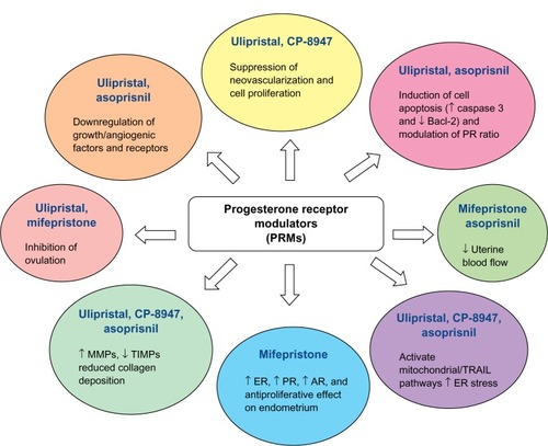
A number of clinical trials have established the potential of SPRMs in the treatment of uterine fibroids. They are associated with a reduction in pain, bleeding, size of fibroids, and overall improvement in quality of life. Unlike long-acting GnRH analogs, they do not have the drawbacks of profound estrogen deficiency and decrease in bone mineral density.Citation94
Mifepristone
Early reports of the use of mifepristone for the treatment of fibroids date back to 2002, when De Leo et al used doses ranging from 12.5 to 50 mg daily and reported a reduction in uterine/fibroid volume of 40%–50%, with amenorrhea in most subjects.Citation95 This report was corroborated by a paper a year later from a group who used mifepristone at a dose of 5 or 10 mg per day for 1 year, and found that it was effective in decreasing mean uterine volume by 50%, while amenor-rhea occurred in 40%–70% of the subjects.Citation96 Endometrial hyperplasia was seen in some women on mifepristone which limited the long-term use of this drug among those desiring a medical therapeutic alternative. The apparent effectiveness of mifepristone, however, in reducing myoma volume and improving fibroid-related symptoms and quality of life, and the minimal side effects, all point to a need for a large RCT with sufficient power to define its true place in the medical management of uterine fibroids. A combination of mifepristone and the LNG-IUS could prove especially useful as the IUS would obviate the development of endometrial hyperplasia while also promoting a reduction in menstrual flow.Citation94 In another RCT, 100 women were assigned to mifepristone 5 or 10 mg daily for 3 months without a placebo group: with both doses, there were equivalent reductions in fibroid and uterine volumes and symptomatic improvements.Citation97
Ulipristal acetate
Ulipristal acetate (UA) has recently successfully completed two Phase III clinical trials (PEARL [PGL 4001’s Efficacy Assessment in Reduction of symptoms due to uterine Leiomyomata] I and II) in Europe, demonstrating its efficacy and safety for the treatment of symptomatic uterine fibroids in patients eligible for surgery.Citation94 PEARL I compared treatment with oral UA for up to 13 weeks at a dose of 5 mg per day (96 women) or 10 mg per day (98 women) with a placebo (48 women) in patients with fibroids, menorrhagia, and anemia. Treatment with UA for 13 weeks effectively controlled excessive bleeding due to uterine fibroids and reduced the size of the fibroids.Citation98 PEARL II was a double-blind, non-inferiority trial, which randomly assigned 307 patients with symptomatic fibroids and excessive uterine bleeding to receive three months of daily therapy with oral UA (at a dose of either 5 mg or 10 mg) or once monthly intramuscular injections of the GnRH analog leuprolide acetate (at a dose of 3.75 mg). Uterine bleeding was controlled in 90% of patients receiving 5 mg of UA, in 98% of those receiving 10 mg, while the figure for leuprolide acetate was 89%. Both UA doses were non-inferior to once monthly leuprolide acetate in controlling uterine bleeding and were significantly less likely to cause hot flushes.Citation99
Clinicians detecting endometrial thickening in women treated with SPRMs need to be aware that administration of SPRMs for longer than 3 months may lead to endometrial thickening. This is related to cystic glandular dilation, not endometrial hyperplasia, and pathologists need to be aware of PRM-associated endometrial changes (PAEC) and avoid misclassifying the appearance as hyperplasia. It is also important to consider the limitations of the current data while describing the effects of SPRMs on the endometrium. Most existing studies have described the endometrial changes over short periods (months) of follow-up, but atypical hyperplasia and possibly malignant change take years to develop. Long-term studies are therefore necessary to evaluate such outcomes.Citation94
Aromatase inhibitors
Aromatase inhibitors (AI) significantly block both ovarian and peripheral estrogen production within 1 day of treatment.Citation100 The underlying mechanism is the inhibition of the aromatase enzyme, an enzyme that catalyzes the conversion of androgenic substances into estrogens.Citation101 Recent reports have suggested that aromatase is expressed to a greater extent in uterine leiomyoma tissues of African-American women compared to Caucasian women, which may contribute to the higher incidence of fibroids in African-American women.Citation102 Aromatase inhibitors have been shown to be effective against fibroids in limited short-term studies with dosing regimens that included 2.5 mg per day of letrozole and 1 mg per day of anastrozole.Citation103 One of the major concerns with the use of aromatase inhibitors is the reported bone loss with prolonged use, which necessitates the concomitant use of oral contraceptive pills or progesterone.Citation104 A recently published RCT compared the effects of 3 months of aromatase inhibitor (letrozole) to that of 3 months of gonadotropin-releasing hormone agonist (triptorelin) on uterine leiomyoma volume and hormonal status. The results showed an advantage of the rapid onset of action of AIs in addition to the avoidance of the flare-ups that initially occur with GnRHa. Both treatment options induced significant shrinkage of the uterine fibroids and improvement in leiomyoma-associated symptoms.Citation105 The mean reduction of leiomyoma volume with 3 months’ use of anastrazole is 55.7%.Citation106 Several RCTs are underway that would hopefully add to our understanding of the potential promising role of AIs in the treatment of uterine leiomyomas.Citation107
Somatostatin analogs
Increasing evidence has demonstrated a role for growth factors, such as insulin growth factor I (IGF-I) and IGF-II, in the initiation and progression of uterine fibroids.Citation108–Citation111 Leiomyoma tissue expresses higher levels of IGF-I/IGF-II receptors compared to normal adjacent myometrium.Citation110,Citation112 It has recently been reported that acromegalic patients have a higher prevalence of uterine fibroids than the general population.Citation113 Lanreotide, which is a long-acting somatostatin analog that has been shown to reduce growth hormone secretion, has also recently been evaluated in seven women with uterine fibroids in Italy.Citation114 Interestingly, lanreotide induced a 42% mean myoma volume reduction within a 3-month period. These results show that somatostatin analogs may potentially be a new therapy for uterine fibroids.Citation115 The treatment with somatostatin analogs for diseases other than leiomyoma appears to be safe and is usually well-tolerated with some reports of gallstone formation.Citation116,Citation117 However, the lack of clinical trials which test the long-term use of somatostatin analogs along with the severe and adverse health implications such as decreased life expectancy due to accelerated heart disease which was observed in adults with growth hormone deficiency may hinder its future use for leiomyoma treatment.Citation66
Other hormonal agents
A preliminary study published in 2007 favored the use of cabergoline as a medical treatment of uterine fibroids. The authors reported a volume reduction of about 50% with 6 weeks’ use.Citation118 The same group performed a subsequent study that compared cabergoline with diphereline, which is a gonadotropin-releasing hormone agonist. They reported comparable results in terms of the shrinkage of the fibroids and improvement in the sonographic, clinical, and intraoperative outcomes.Citation119
Gestrinone is a steroid that possesses anti-estrogen receptor and anti-progesterone receptor properties in various tissues, including the endometrium. A recent report from Italy evaluated the use of gestrinone in the treatment of premenopausal women with uterine fibroids. The authors reported a 32%±10% reduction in uterine volume.Citation120 A subsequent study reported up to 60% leiomyoma shrinkage in size.Citation121 Gestrinone is a contraceptive agent and also exhibits several unfavorable side effects, such as mild androgenicity, weight gain, seborrhea, acne, hirsutism, and occasional hoarseness.Citation66
Vitamin D
There is evidence that Vitamin D is an anti-fibrotic factor and inhibits growth and induces apoptosis in cultured human leiomyoma cells through the downregulation of proliferating cell nuclear antigen (PCNA), cyclin-dependent kinase 1 (CDK1), and B-cell lymphoma 2 (BCL-2) and suppresses catechol-O-methyl transferase (COMT) expression and activity in human leiomyoma cells.Citation122,Citation123 A study observed that 1,25-dihydroxyvitamin D3 is significantly lower in women with fibroids compared to normal healthy controls; additionally, lower levels of total serum 25-hydroxyvitamin D3 have been detected in women with fibroids compared to healthy controls. The aim of this study was to determine whether serum levels of Vitamin D correlated with disease severity in women with symptomatic uterine fibroids. The study population consisted of 67 patients who had detailed repeated pelvic ultrasound evaluations over a 2-year period with specific measurements of the total uterine volume and the volume of the individual leiomyoma lesions. The patients also had detailed laboratory analysis including serum 25-hydroxyvitamin D3 levels. Preliminary results have suggested a strong dose-response correlation between lower serum vitamin D levels and increased severity of uterine fibroids. This presents an opportunity for the potential use of vitamin D or its potent analogs as novel treatment options or for the prevention of uterine fibroids.Citation124,Citation125
Epigallocatechin gallate (EGCG) – green tea extract
A typical green tea beverage, prepared in a proportion of 1 g leaf to 100 mL water with a 3-minute brew, usually contains 250–350 mg tea solids, and catechins account for 30%–42% of the dry weight of the solids.Citation126 The major green tea catechins are epigallocatechin-3-gallate (EGCG), epigallocatechin (EGC), epicatechin-3-gallate (ECG), and epicatechin. Catechins are a group of bioflavonoids that exhibit antioxidant and anti-inflammatory capacity.Citation127 EGCG appears to block each stage of tumorgenesis by modulating signaling pathways involved in cell proliferation, transformation, inflammation, and oxidative stress, which are clearly involved in pathogenesis of various tumors including uterine fibroids.Citation128 The potential effect and mechanisms of EGCG action on human leiomyoma (HuLM) cells was studied and it was found that EGCG inhibits the proliferation of HuLM cells and induces apoptosis. These results suggest that EGCG may be a potential anti-uterine fibroid agent acting through multiple signal transduction pathways.Citation129
Minimally invasive techniques
Even women without a desire for future pregnancies might not wish to lose their uterus for various reasons. In the past years, several nonsurgical and minimally invasive treatment options have been introduced and studied, which not only preserve the uterus and thereby fertility, but also reduce morbidity and recovery time in comparison with hysterectomy. Although hysterectomy is the only definitive solution, many women and doctors seek the possibility of minimally invasive uterus-sparing alternatives for the treatment of uterine fibroids.Citation130 Some of these therapies include uterine artery embolization (UAE), MRgFUS (magnetic resonance-guided focused ultrasound surgery) and ultrasound-guided ablation (VizAblate™ [Gynesonics, Redwood City, CA, USA] and Acessa™ [Halt Medical, Inc., Brentwood, CA, USA] Procedures).
Uterine artery embolization (UAE)
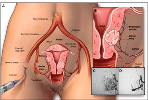
UAE is a percutaneous, image-guided procedure which is performed by a properly trained and experienced interventional radiologist.Citation131 In the past, embolization was used to reduce pelvic bleeding.Citation132 Only since 1995 has UAE been seen as a potential treatment for menorrhagia related to uterine fibroids.Citation133,Citation134 In essence, it involves the placement of an angiographic catheter into the uterine arteries via a common femoral artery approach and injection of embolic agents (in most cases, polyvinyl alcohol particles or trisacryl gelatin microspheres) into both uterine arteries until the flow becomes sluggish ().Citation135–Citation141 The proposed mechanism of UAE’s action is by occluding or markedly reducing uterine blood flow at the arteriolar level to produce an irreversible ischemic injury to the fibroids, causing them to undergo necrosis and shrink, while the normal myometrium is able to recover.Citation142,Citation143
Figure 5 The UAE procedure. Drawings (A and B) illustrate the path of the catheter to deliver embolization particles to occlude the uterine arteries. Angiograms show the fibroid blood supply before (C) and after (D) UAE.
Abbreviation: UAE, uterine artery embolization.
The procedure, which is usually performed under local anesthetic or intravenous conscious sedation, generally requires 1 hour to complete. With operator experience and proper technique, patient radiation exposure during UAE is comparable to that received during routine diagnostic imaging procedures.Citation144–Citation147
Immediately after the end of the procedure, most patients experience moderate-to-severe ischemic pain for 8–12 hours – usually requiring parenteral analgesia (narcotics and non-steroidal, anti-inflammatory drugs), but pain severity gradually decreases after 12 hours,Citation148 and a day-case or overnight stay is required at most. Recovery is typically brief and relatively mild, with 4–5 days of recurrent uterine cramping and constitutional embolization symptoms (generalized malaise, fatigue, nausea, and low-grade fever). Patients can usually return to normal activities within 8–14 days.Citation139,Citation149–Citation152
UAE is not without its own set of complications. They can be divided into immediate (occurring during the procedure), early (within the first 30 days), and late (beyond the first 30 days). Immediate complications include hematoma formation, thrombosis, or pseudo-aneurysm. Reaction to the contrast media is unusual. Failure to cannulate the uterine artery can occur in a very small percentage of patients. Early complications include ‘post-embolization syndrome’ which can occur commonly, although it is only severe in a minority of cases. It consists of pain, nausea, a flu-like illness, mild pyrexia, and raised inflammatory markers. If the symptoms are prolonged or the patient is becoming very ill then infection must be suspected. Significant infection is less common than was originally predicted, which is fortunate as hysterectomy performed soon after uterine artery embolization tends to be a very difficult procedure. Vaginal discharge is common and usually settles down within a few days. Late complications include chronic vaginal discharge, adverse changes in sexual function, and temporary amenorrhea. The incidence of amenorrhea is related to the age of the woman and is particularly common in those over 45 years of age. Fibroid expulsion can occur in 10% of cases and is particularly common with submucous fibroids. Occasionally, operative intervention is required but usually if the patient is warned that this might occur then no further treatment is required.Citation154
A recently published Cochrane reviewCitation155 has looked at the benefits and risks of UAE versus other medical or surgical interventions for symptomatic uterine fibroids. It found that there was no significant difference in satisfaction rates between the two groups in spite of a higher rate of post-procedural complications and further re-interventions in the UAE arm of the study. There was only one study which specifically looked at the impact of UAE versus myomectomy on fertility, an important parameter as both are uterine-sparing procedures.Citation156,Citation157 The findings for live birth were of borderline statistical significance, favoring myomectomy, and there were significantly more pregnancies in the myomectomy group. Not all the women in the study were trying to conceive: 26 after UAE and 40 after myomectomy. The pregnancy rate after UAE was 50%, delivery rate 19%, and miscarriage rate 53%, while these percentages were 78%, 48%, and 23%, respectively, after myomectomy. The differences in all these parameters were statistically significant (P<0.05). UAE was advantageous over hysterectomy or myomectomy with regards to a shorter hospital stay, a quicker return to full activity, and reduced likelihood of a need for blood transfusion. However, UAE was associated with an increased short-term risk of minor complications. The REST (Multicentre Randomised controlled trial comparing uterine artery Embolisation with Surgical Treatment for uterine fibroids) studyCitation158 looked at complications over five years and again the rate of minor complications was higher in the UAE group. No study found a significant difference between the groups in major complications in either short- or long-term follow-up.
In the previous version of this Cochrane review, the authors had concluded that the women opting for UAE should be counseled about a higher surgical intervention rate in the longer term. This has been supported by the 5-year follow-up data from the EMMY (EMbolization versus hysterectoMY)Citation159 and REST trials. There is a five-fold increase in the likelihood of needing further intervention after UAE. However, satisfaction rates were similar in both the groups. Although UAE initially appears to be a cost-effective alternative to hysterectomy, the long-term follow-up at 5 years shows that this advantage is lost due to a subsequent higher rate of re-intervention.Citation158,Citation159
The effect of UAE on ovarian reserve and pregnancy outcome is less well established.Citation130 FSH levels as an indicator of ovarian failure after 2 years were not significantly different between UAE and surgery.Citation155 Another systematic reviewCitation160 showed that miscarriage rates were higher in post-UAE pregnancies (35.2%) compared with pregnant women with a non-treated fibroid uterus, matched for age, and fibroid location (16.5%). The UAE pregnancies were more likely to be delivered by cesarean section and to experience postpartum hemorrhage. Rates of preterm delivery, intrauterine growth restriction, and malpresentation were similar in UAE pregnancies and in control pregnancies with fibroids.
Magnetic resonance-guided focused ultrasound surgery (MRgFUS)
MRgFUS is a relatively new method of thermal ablation for treating fibroids, which uses high-intensity focused ultrasound that passes through the anterior abdominal wall and converges into a precise target point within the fibroid to cause a temperature rise (55°C–90°C) sufficient to induce coagulative necrosis within a few seconds. Concurrent MRI allows accurate tissue targeting and real-time temperature feedback, thereby achieving controlled localized thermal ablation.Citation161–Citation163 The advantages of MRgFUS are its completely non-invasive character and continuous imaging of fibroids and adjacent structures, which optimizes fibroid ablation and prevents injury to adjacent tissues.Citation164 The disadvantage is that relatively few patients are eligible – only those with fibroids located immediately beneath the anterior abdominal wall without bowel interposition or scars in the region of interest. The average treatment time is however, long and the patient has to lie still for the duration of the treatment time.Citation165
The results of a prospective studyCitation166 into pregnancy outcomes after MRgFUS showed that the spontaneous miscarriage rate was 26%, which was comparable with the rate in women of similar age without fibroids.Citation167 Live births occurred in 41% of pregnancies, with an 11% rate of elective pregnancy termination, and 20% ongoing pregnancies beyond 20 gestational weeks the moment the study ended. The vaginal delivery rate was 64%.Citation166
At present, only a minority of women with uterine fibroids seems to meet the inclusion criteria for this new technique. Due to the lack of randomized data, MRgFUS should still be regarded as an experimental treatment.Citation130
Other ablation procedures (VizAblate™ and Acessa™)
With MRgFUS not being that widely available and having a long treatment time, a new transcervical device (VizAblate™) has recently been introduced that combines real-time intrauterine sonography with radiofrequency (RF) ablation for the treatment of fibroids. The fibroid ablation takes place under real-time visualization as an intrauterine sonography probe is built into the device. This built-in imaging removes the need for the physician to coordinate more than one device. The graphical interface delineates the boundaries of both ablation and thermal spread, enabling the gynecologist to avoid thermal injury to the serosa with its potential for adhesiogenesis and injury to bowel or bladder. Overall, the procedure is well-tolerated with low pain scores, allowing it to be considered for future use in the outpatient setting with local anesthetic or conscious sedation. Procedure time is in the range of 30 minutes.Citation168
The novel use of radiofrequency ablation under laparoscopic and intra-abdominal ultrasound guidance to treat patients with symptomatic myomas was first introduced in 2002.Citation169 This has prompted the development of the study device (Acessa™) which carries out laparoscopic ultrasound-guided radiofrequency volumetric thermal ablation of uterine myomas in symptomatic women.Citation170 The key feature to using laparoscopic ultrasound lies in the inherent and immediate proximity of the transducer to the target (fibroid), allowing the use of higher frequencies with significantly increased resolution. In addition, the versatility and mobility of laparoscopic ultrasound permits direct imaging from multiple directions and angles.Citation171 Based on a recent study, radiofrequency volumetric thermal ablation of uterine myomas safely achieved a high rate of patient satisfaction, a low re-intervention rate, and significant improvements in menstrual blood loss, symptom severity, and quality of life through 12 months of follow-up.Citation170
Surgical
The standard treatment for symptomatic uterine fibroids has always been surgical, either hysterectomy or, in women who wish to preserve their fertility, the more conservative procedure of myomectomy.Citation172 Fibroids represent one of the most frequent indications for major surgery in pre-menopausal women and as such, they constitute a major public health cost.Citation173 Myomectomy can be carried out via hysteroscopy, laparoscopy, or classically as an abdominal procedure.
Hysteroscopic myomectomy
Submucosal fibroids can often be removed by hysteroscopic myomectomy. This can be performed under general or regional analgesia, and in some centers it is performed as an office procedure, depending on the type and the size of the fibroid(s). Although it is widely applied and its effect on bleeding complaints is well-known, there is surprisingly little randomized evidence to support this.Citation130 A recent retrospective analysis of 105 patients that underwent hysteroscopic myomectomy for submucosal fibroids, showed the disappearance of bleeding symptoms in 90% of cases after a mean follow-up of 17 months.Citation174 In a RCT, GnRH pre-treatment was found not to increase the number of complete resections.Citation175
The most common perioperative complications associated with hysteroscopic myomectomy are hemorrhage (2.4%), uterine perforation (1.5%), cervical laceration (1%–11%), and fluid overload by intravasation of distension fluid. Delayed complications from hysteroscopic surgery may include intrauterine adhesions and infertility.Citation130 The average reported incidence is around 10% at second-look hysteroscopy, but it seems to be higher in certain conditions, for instance in the resection of multiple, opposing fibroids.Citation176
A systematic review summarizing the effects of surgery on fertility showed that submucous fibroids or intramural fibroids with a submucosal component decreased clinical pregnancy and implantation rates, and removal of submucous fibroids led to a significant increase in pregnancy rate (from 27.2% to 43.3%) and a decrease in miscarriage rate (from 50% to 38.5%).Citation177
Laparoscopic myomectomy
Laparoscopic myomectomy has long been the minimally invasive therapy of choice for symptomatic uterine fibroids, before the introduction of UAE and other minimally invasive therapies. It is still widely used for symptomatic subserosal fibroids and can even be used for intramural fibroids, depending on the position of the fibroid and the skills of the surgeon.Citation130 A prospective study with 235 patients undergoing laparoscopic myomectomy for symptomatic fibroids showed no conversions to laparotomy and in 3 years, only 1.2% of patients had a second laparoscopic myomectomy for recurrent fibroids. By 48 hours after surgery, 86.3% of the patients were discharged.Citation178 A systematic review compared women with subserosal fibroids to women without fibroids, and found no difference for fertility outcomes. In contrast, women with intramural fibroids had significantly lower clinical pregnancy rates and ongoing pregnancy/live birth rates and significantly higher spontaneous miscarriage rates compared with women without fibroids. The same review could not identify a significant effect of the removal of intramural fibroids on fertility outcomes, as compared between women with intramural fibroids and women that underwent removal of their intramural fibroids.Citation177,Citation179
Blood loss is an important clinical problem during myomectomy. A Cochrane review found significant reductions in blood loss with misoprostol, vasopressin, bupivacaine and epinephrine, tranexamic acid, peri-cervical tourniquet, and gelatin–thrombin matrix. There was no evidence of an effect on blood loss with oxytocin.Citation180
Abdominal myomectomy
A cohort study compared perioperative morbidity between women who underwent abdominal myomectomy or abdominal hysterectomy. It reported no significant difference in overall morbidity between the two groups (39% versus 40%, odds ration [OR]: 0.93; 95% confidence interval [CI]: 0.63–1.40). There was significantly lower prevalence of hemorrhage and performance of an unintended procedure in the myomectomy group than in the hysterectomy group. Abdominal myomectomy was a lengthier procedure but was associated with significantly less blood loss. The average hospital stay was significantly shorter in the myomectomy group. Overall, no clinically significant difference in perioperative morbidity between myomectomy and hysterectomy was detected. Myomectomy may be considered a safe alternative to hysterectomy.Citation181
A recent observational study has suggested that abdominal myomectomy might improve reproductive outcomes in patients with myomas. The reproductive performance was particularly good when the patients were younger and had previous pregnancies prior to the surgery. Myomectomy is associated with lower miscarriage rate after pregnancy, compared to those prior to the surgery.Citation182
Hysterectomy
The first successful selected hysterectomy operation was performed in 1813 by Conrad Langenbeck via the vaginal approach,Citation183 and today, after nearly two centuries, hysterectomy is the second most frequent surgery in women of reproductive age, with the first being cesarean section.Citation184 Approximately 55,000 hysterectomies are performed each year in the UK, and over 600,000 in the USA.Citation185,Citation186 The vast majority of these numbers (more than 70%) are for benign indications such as menorrhagia (21%), fibroids (33%), pelvic pain (3%), and uterine prolapse (28%).Citation186
Hysterectomy is the definitive procedure and carries an outstandingly good outcome and guarantees complete cessation of periods with no risk of fibroid recurrence. Hysterectomy can be done via the vaginal, abdominal, or laparoscopic (total or laparoscopic-assisted vaginal) route. Each carries its own advantages and disadvantages. The traditional surgical approach to hysterectomy involves a large abdominal incision, 2–5 day hospital stay, and significant requirements for postoperative analgesia. Laparoscopic techniques have many advantages over laparotomy, including reduced postoperative pain, shorter length of hospital stay, better cosmesis, and quicker resumption of regular activity.Citation183
National Institute for Health and Care Excellence (NICE) guidelines recommend that hysterectomy should be considered only when other treatment options have failed, are contraindicated or are declined by the woman, there is a wish for amenorrhea, the woman (who has been fully informed) requests it, and the woman no longer wishes to retain her uterus and fertility.Citation187 Most importantly, hysterectomy carries some serious risks including damage to the bowel, bladder and/or ureter, hemorrhage requiring blood transfusion, return to theatre because of bleeding/wound dehiscence, pelvic abscess/infection, and venous thrombosis or pulmonary embolism.Citation188
Leiomyosarcomas of the uterus
Uterine leiomyosarcoma is a rare neoplasm with an annual incidence of 0.64 per 100,000 women ().Citation189 It accounts for <5% of all uterine malignancies and approximately 30% of all uterine sarcomas.Citation190 The disease is characterized by a propensity for early hematogenous spread, leading to high local and distant failure rates.Citation191–Citation193 In contrast to the much more common uterine adenocarcinoma, lymphatic spread is a rare event, and the overall prognosis is poor.Citation194 They are considered neoplasms of high metastatic potential with 5-year overall survival rates varying between 0% and 73%.Citation195–Citation197
Figure 6 A 40-year-old woman with a large abdomino-pelvic mass, which turned out to be an aggressive leiomyosarcoma.
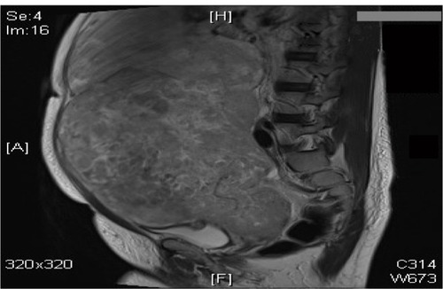
Most occur in women over 40 years of age who usually present with abnormal vaginal bleeding (56%), palpable pelvic mass (54%), and pelvic pain (22%). Signs and symptoms resemble those of the far more common leiomyoma and preoperative distinction between the two tumors may be difficult.Citation198 Nevertheless, malignancy should be suspected by the presence of certain clinical behaviors, such as tumor growth in menopausal women who are not on hormonal replacement therapy.Citation199 Occasionally, the presenting manifestations are related to tumor rupture (hemoperitoneum), extra-uterine extension (one-third to one-half of cases), or metastases.Citation198 Most leiomyosarcomas arise de novo, but an apparent minority of leiomyosarcomas may represent leiomyomas that have undergone malignant transformation.Citation200
Treatment of leiomyosarcomas includes total abdominal hysterectomy and debulking of tumor if present outside the uterus. Removal of the ovaries and lymph node dissection remain controversial as metastases to these organs occur in a small percentage of cases and are frequently associated with intra-abdominal disease.Citation201 Ovarian preservation may be considered in premenopausal patients with early-stage leiomyosarcomas.Citation201 Lymph node metastases have been identified in 6.6% and 11% of two series of patients with leiomyosarcoma who underwent lymphadenectomy.Citation201,Citation202 In the first series, the 5-year disease-specific survival rate was 26% in patients who had positive lymph nodes compared with 64.2% in patients who had negative lymph nodes (P<0.001).Citation202 The influence of adjuvant therapy on survival is uncertain. Radiotherapy may be useful in controlling local recurrences and chemotherapy with doxorubicin or docetaxel/gemcitabine is now used for advanced or recurrent disease, with response rates ranging from 27% to 36%.Citation203,Citation204 Some patients may respond to hormonal treatment.Citation205
Conclusion
Although they are essentially benign, uterine fibroids are associated with significant morbidity to nearly 40% of women during their reproductive years and sometimes even after menopause. Therefore, there is considerable interest in discovering any etiological clues in factors including dietary, stress, and environmental influences. The most practical investigative modality has always been gray-scale ultrasonography, but with the developments of innovative methods of treatments, it is not just enough to diagnose the presence of the fibroid, but also its dynamic relation with surrounding tissue as well. With more and more patients demanding a nonsurgical approach to their symptoms, there is a developing market for selective estrogen and especially progesterone receptor modulators, and exploring the role of aromatase inhibitors, vitamin D, and green tea extract.
Gone are the days when the only surgical approach for fibroids was offering a myomectomy or hysterectomy. The concept of vascular embolization has altered the playing field and allowed women to preserve their fertility status without resorting to surgery. This is being further investigated by the FEMME (A randomised trial of treating Fibroids with either Embolisation or Myomectomy to Measure the Effect on quality of life among women wishing to avoid hysterectomy) trial (to measure and compare the changes in the quality of life women experience when their fibroids are treated by either myomectomy or uterine artery embolization).
Minimally invasive management of fibroids has been even further enriched by the development and introduction of novel techniques (ie, MRgFUS, VizAblate™, and Acessa™).
Disclosure
A T Khan reports no conflicts of interest in this work. M Shehmar has conducted training workshops for Ethicon, while J K Gupta has conducted training workshops for Ethicon and is a principal investigator for the FEMME study.
References
- RyanGLSyropCHVan VoorhisBJRole, epidemiology, and natural history of benign uterine mass lesionsClin Obstet Gynecol20054831232415805789
- WallachEEVlahosNFUterine myomas: an overview of development, clinical features, and managementObstet Gynecol200410439340615292018
- SankaranSManyondaITMedical management of fibroidsBest Pract Res Clin Obstet Gynaecol200822465567618468953
- ParkerWHEtiology, symptomatology, and diagnosis of uterine myomasFertil Steril200787472573617430732
- ReinMSBarbieriRLFriedmanAJProgesterone: a critical role in the pathogenesis of uterine myomasAm J Obstet Gynecol19951721 Pt114187847524
- AndersenJGrowth factors and cytokines in uterine leiomyomasSemin Reprod Endocrinol19961432692828885057
- FieldsKRNeinsteinLSUterine myomas in adolescents: case reports and a review of the literatureJ Pediatr Adolesc Gynecol1996941951988957774
- CramerSFPatelAThe frequency of uterine leiomyomasAm J Clin Pathol19909444354382220671
- Day BairdDDunsonDBHillMCCousinsDSchectmanJMHigh cumulative incidence of uterine leiomyoma in black and white women: ultrasound evidenceAm J Obstet Gynecol2003188110010712548202
- LaughlinSKSchroederJCBairdDDNew directions in the epidemiology of uterine fibroidsSemin Reprod Med201028320421720414843
- SchwartzSMEpidemiology of uterine leiomyomataClin Obstet Gynecol200144231632611344995
- PurdieDMGreenACEpidemiology of endometrial cancerBest Pract Res Clin Obstet Gynaecol200115334135411476557
- ColditzGAEpidemiology of breast cancer. Findings from the nurses’ health studyCancer199371Suppl 4148014898431884
- BairdDDDunsonDBWhy is parity protective for uterine fibroids?Epidemiology200314224725012606893
- ParazziniFRisk factors for clinically diagnosed uterine fibroids in women around menopauseMaturitas200655217417916533580
- WiseLAPalmerJRHarlowBLReproductive factors, hormonal contraception, and risk of uterine leiomyomata in African-American women:a prospective studyAm J Epidemiol2004159211312314718211
- BurbankFChildbirth and myoma treatment by uterine artery occlusion: do they share a common biology?J Am Assoc Gynecol Laparosc200411213815215200765
- WiseLAPalmerJRHarlowBLRisk of uterine leiomyomata in relation to tobacco, alcohol and caffeine consumption in the Black Women’s Health StudyHum Reprod20041981746175415218005
- StewartEANowakRANew concepts in the treatment of uterine leiomyomasObstet Gynecol1998924 Pt 16246279764641
- LeppertPCCatherinoWHSegarsJHA new hypothesis about the origin of uterine fibroids based on gene expression profiling with microarraysAm J Obstet Gynecol2006195241542016635466
- CramerSFMannLCalianeseEDaleyJWilliamsonKAssociation of seedling myomas with myometrial hyperplasiaHum Pathol200940221822518799191
- RogersRNorianJMalikMMechanical homeostasis is altered in uterine leiomyomaAm J Obstet Gynecol2008198447418395046
- CatherinoWSalamaAPotlog-NahariCLeppertPTsibrisJSegarsJGene expression studies in leiomyomata: new directions for researchSemin Reprod Med2004222839015164303
- BairdDDKesnerJSDunsonDBLuteinizing hormone in premeno-pausal women may stimulate uterine leiomyomata developmentJ Soc Gynecol Investig2006132130135
- WirthMMMeierEAFredricksonBLSchultheissOCRelationship between salivary cortisol and progesterone levels in humansBiol Psychol200774110410716904811
- National Institute for Health and Clinical ExcellenceUterine Artery Embolisation for FibroidsLondonNICE2010 Available from: http://www.nice.org.uk/nicemedia/live/11025/51706/51706.pdfAccessed on October 21, 2013
- SabryMAl-HendyAInnovative oral treatments of uterine leiomyomaObstet Gynecol Int2012201294363510.1155/2012/94363522518167
- SchwartzSMMarshallLMBairdDDEpidemiologic contributions to understanding the etiology of uterine leiomyomataEnviron Health Perspect2000108Suppl 582182711035989
- OkoloSIncidence, aetiology and epidemiology of uterine fibroidsBest Pract Res Clin Obstet Gynaecol200822457158818534913
- WegienkaGBairdDDHertz-PicciottoISelf-reported heavy bleeding associated with uterine leiomyomataObstet Gynecol2003101343143712636944
- LippmanSAWarnerMSamuelsSOliveDVercelliniPEskenaziBUterine fibroids and gynecologic pain symptoms in a population-based studyFertil Steril20038061488149414667888
- ZimmermannABernuitDGerlingerCSchaefersMGeppertKPrevalence, symptoms and management of uterine fibroids: an international internet-based survey of 21,746 womenBMC Womens Health201212622448610
- SomiglianaEVercelliniPDaguatiRPasinRDe GiorgiOCrosignaniPGFibroids and female reproduction:a critical analysis of the evidenceHum Reprod Update20071346547617584819
- ParkerWHEtiology, symptomatology, and diagnosis of uterine myomasFertil Steril200787472573617430732
- LevensEDWesleyRPremkumarABlockerWNiemanLKMagnetic resonance imaging and transvaginal ultrasound for determining fibroid burden: implications for research and clinical careAm J Obstet Genecol20092005537. e1e7
- VolkersNAHehenkampWJSpijkerboerAMMR reproducibility in the assessment of uterine fibroids for patients scheduled for uterine artery embolizationCardiovasc Intervent Radiol20083126026818057985
- ZawinMMcCarthySScouttLMComiteFHigh-field MRI and US evaluation of the pelvis in women with leiomyomasMagn Reson Imaging199083713762202877
- DueholmMLundorfEHansenESLedertougSOlesenFEvaluation of the uterine cavity with magnetic resonance imaging, transvaginal sonography, hysterosonographic examination, and diagnostic hysteroscopyFertil Steril20017635035711476785
- DueholmMLundorfESorensenJSLedertougSOlesenFLaursenHReproducibility of evaluation of the uterus by transvaginal sonography, hysterosonographic examination, hysteroscopy and magnetic resonance imagingHum Reprod20021719520011756387
- WildeSScott-BarrettSRadiological appearances of uterine fibroidsIndian J Radiol Imaging200919322223119881092
- RoyCBierryGEl GhaliSBuyXRossiniAAcute torsion of uterine leiomyoma: CT featuresAbdom Imaging20053012012315647881
- DueholmMLundorfEHansenESLedertougSOlesenFAccuracy of magnetic resonance imaging and transvaginal ultrasonography in the diagnosis, mapping, and measurement of uterine myomasAm J Obstet Gynecol200218640941511904599
- WeinrebJCBarkoffNDMegibowADemopoulosRThe value of MR imaging in distinguishing leiomyomas from other solid pelvic masses when sonography is indeterminateAJR Am J Roentgenol19891542952992105017
- OmariEAVargheseTKliewerMAA novel saline infusion sonohysterography-based strain imaging approach for evaluation of uterine abnormalities in vivo: preliminary resultsJ Ultrasound Med201231460961522441918
- DavidsonKGDubinskyTJUltrasonographic evaluation of the endometrium in postmenopausal vaginal bleedingRadiol Clin North Am20034176978012899491
- CohenJRLuxmanDSagiJYovelIWolmanIDavidMPSonohysterography for distinguishing endometrial thickening from endometrial polyps in postmenopausal bleedingUltrasound Obstet Gynecol1994422723012797187
- ByunJYKimSEChoiBGDiffuse and focal adenomyosis: MR imaging findingsRadioGraphics199919Suppl 1s161s17010517452
- AscherSMArnoldLLPattRHAdenomyosis: Prospective comparison of MR imaging and transvaginal sonographyRadiology19941908038068115630
- DudiakCMTurnerDAPatelSKArchieJTSilverBNorusisMUterine leiomyomas in the infertile patient: preoperative localization with MR imaging versus US and hysterosalpingographyRadiology19881676276633283833
- RocheOChavanNAquilinaJRockallARadiological appearances of gynaecological emergenciesInsights Imaging20123326527522696088
- LivermoreJAAdusumilliSMRI of benign uterine conditionsAppl Radiol2007369818
- BrosensJJde SouzaNMBarkerFGUterine junctional zone: function and diseaseLancet19953465585607658784
- MuraseESiegelmanESOutwaterEKUterine leiomyomas: histopathologic features, MR imaging findings, differential diagnosis, and treatmentRadiographics1999191179119710489175
- UedaHTogashiKKonishiIUnusual appearances of uterine leiomyomas: MR imaging findings and their histopathologic backgroundsRadiographics199919Suppl 1s131s14510517450
- KimJCKimSSParkJY“Bridging vascular sign” in the MR diagnosis of exophytic uterine leiomyomaJ Comput Assist Tomogr200024576010667660
- DohkeMWatanabeYOkumuraAComprehensive MR Imaging of acute gynecologic diseasesRadiographics20002061551156611112810
- BurnPRMcCallJMChinnRJVashishtASmithJRHealyJCUterine fibroleiomyoma: MR imaging appearance before and after embolization of uterine arteriesRadiology200021472973410715038
- Simms-StewartDFletcherHCounselling patients with uterine fibroids: a review of the management and complicationsObstet Gynecol Int2012201253936510.1155/2012/53936522272207
- PrittsEAOliveDLWhen should uterine fibroids be treated?Curr Obstet Gynecol Rep2012127180
- CarlsonKJMillerBAFowlerFJThe Maine Women’s Health Study: I Outcomes of hysterectomyObstet Gynecol1994835568134066
- LefebvreGVilosGAllaireCThe management of uterine leiomyomasJ Obstet Gynaecol Can20032539612738981
- LaughlinSKStewartEAUterine leiomyomas: individualizing the approach to a heterogeneous conditionObstet Gynecol20111172 Pt 139640321252757
- LakhaniKPMarshMSPurcellWHardimanPUterine artery blood flow parameters in women with dysfunctional uterine bleeding and uterine fibroids: the effects of tranexamic acidUltrasound Obstet Gynecol1998112832859618854
- SundströmASeamanHKielerHAlfredssonLThe risk of venous thromboembolism associated with the use of tranexamic acid and other drugs used to treat menorrhagia: a case-control study using the General Practice Research DatabaseBJOG2009116919719016686
- IpPPLamKWCheungCLTranexamic acid-associated necrosis and intralesional thrombosis of uterine leiomyomas: a clinicopathologic study of 147 cases emphasizing the importance of drug-induced necrosis and early infarcts in leiomyomasAm J Surg Pathol2007311215122417667546
- Abdel-RahmanMYSabryMAl-HendyAMedical Treatment of Fibroid to Decrease Rate of HysterectomyAl-HendyAHysterectomyRejekaInTech2012115128
- MarshallLMSpiegelmanDGoldmanMBA prospective study of reproductive factors and oral contraceptive use in relation to the risk of uterine leiomyomataFertil Steril19987034324399757871
- ChiaffarinoFParazziniFLa VecchiaCMarsicoSSuraceMRicciEUse of oral contraceptives and uterine fibroids: results from a case-control studyBr J Obstet Gynaecol1999106885786010453838
- ParazziniFNegriELa VecchiaCFedeleLRabaiottiMLuchiniLOral contraceptive use and risk of uterine fibroidsObstet Gynecol19927934304331738528
- FriedmanAJDalyMJuneau-NorcrossMGleasonRReinMSLeBoffMLong-term medical therapy for leiomyomata uteri: a prospective, randomized study of leuprolide acetate depot plus either estrogen-progestin or progestin ‘add-back’ for 2 yearsHum Reprod199499161816257836510
- OrsiniGLaricchiaLFanelliMLow-dose combination oral contraceptives use in women with uterine leiomyomasMinerva Ginecol2002543253261 Italian12063441
- VenkatachalamSBagrateeJSMoodleyJMedical management of uterine fibroids with medroxyprogesterone acetate (Depo Provera): a pilot studyJ Obstet Gynaecol200424779880015763792
- The management of menorrhagia in secondary careEvidence-based clinical guideline No. 5LondonRoyal College of Obstetricians and Gynaecologists1999
- IrvineGACampbell-BrownMBLumsdenMARandomised comparative trial of the levonorgestrel intrauterine system and norethisterone for treatment of idiopathic menorrhagiaBr J Obstet Gynaecol19981055925989647148
- MaruoTLaoag-FernandezJBPakarinenPEffects of the levonorgestrel-releasing intrauterine system on proliferation and apoptosis in the endometriumHum Reprod2001162103210811574499
- MaruoTOharaNMatsuoHEffects of levonorgestrel-releasing IUS and progesterone receptor modulator PRM CDB-2914 on uterine leiomyomasContraception200775S99S10317531625
- National Collaborating Centre for Women’s and Children’s HealthHeavy menstrual bleeding: clinical guidelineLondonNational Institute for Clinical Excellence2007
- BozziniNRodriguesCJPettiDAEffects of treatment with gonadotropin releasing hormone agonist on the uterine leiomyomata structureActa Obstet Gynecol Scand20038233033412716317
- CheginiNKornbergLGonadotropin releasing hormone analogue therapy alters signal transduction pathways involving mitogen-activated protein and focal adhesion kinases in leiomyomaJ Soc Gynecol Investig2003102126
- CheginiNMaCTangXMEffects of GnRH analogues, ‘add-back’ steroid therapy, antiestrogen and antiprogestins on leiomyoma and myometrial smooth muscle cell growth and transforming growth factor-beta expressionMol Hum Reprod2002121071107812468639
- CheginiNVeralaJLuoXGene expression profile of leiomyoma and myometrium and the effect of gonadotropin releasing hormone analogue therapyJ Soc Gynecol Investig200310161171
- DeligdischLHirschmannSAltchekSPathologic changes in gonadotropin releasing hormone agonist analogue treated uterine leiomyomataFertil Steril1997678378419130887
- ReinschRCMurphyAAMoralesAJThe effects of RU 486 and leuprolide acetate on uterine artery blood flow in the fibroid uterus: a prospective, randomized studyAm J Obstet Gynecol1994170162316288203418
- MesiaAFGahrDWildMImmunohistochemistry of vascular changes in leuprolide acetate treated leiomyomasAm J Obstet Gynecol1997176102610299166163
- RutgersJLSpongCYSinowRLeuprolide acetate treatment and myoma arterial sizeObstet Gynecol1995863863887651647
- LethabyAVollenhovenBSowterMPre-operative GnRH analogue therapy before hysterectomy or myomectomy for uterine fibroidsCochrane Database Syst Rev20002CD00054710796723
- MitlakBHCohenFJIn search of optimal long-term female hormone replacement: the potential of selective estrogen receptor modulatorsHorm Res19974841551639378461
- PalombaSSammartinoADi CarloCAffinitoPZulloFNappiCEffects of raloxifene treatment on uterine leiomyomas in postmenopausal womenFertil Steril2001761384311438317
- PalombaSOrioFJrRussoTAntiproliferative and proapoptotic effects of raloxifene on uterine leiomyomas in postmenopausal womenFertil Steril200584115416116009171
- PremkumarAVenzonDJAvilaNGynecologic and hormonal effects of raloxifene in premenopausal womenFertil Steril20078861637164417662283
- GiangrandePHMcDonnellDPThe A and B isoforms of the human progesterone receptor: two functionally different transcription factors encoded by a single geneRecent Prog Horm Res19995429131310548881
- SpitzIMClinical utility of progesterone receptor modulators and their effect on the endometriumCurr Opin Obstet Gynecol20092131832419602929
- MaruoTMatsuoHSamotoTEffects of progesterone on uterine leiomyoma growth and apoptosisSteroids20006558559211108863
- TalaulikarVSManyondaIProgesterone and progesterone receptor modulators in the management of symptomatic uterine fibroidsEur J Obstet Gynecol Reprod Biol201216513514022901974
- De LeoVMorganteGLa MarcaAA benefit-risk assessment of medical treatment for uterine leiomyomasDrug Safety20022575977912222988
- EisingerSHMeldrumSFiscellaKle RouxHDGuzickDSLow-dose mifepristone for uterine leiomyomataObstetr Gynecol2003101243250
- Carbonell EsteveJLAcostaRHerediaBPérezYCastañedaMCHernándezAVMifepristone for the treatment of uterine leiomyomas: a randomized controlled trialObstet Gynecol20081121029103618978102
- DonnezJTatarchukTFBouchardPPEARL I Study GroupUlipristal acetate versus placebo for fibroid treatment before surgeryN Engl J Med201236640942022296075
- DonnezJTomaszewskiJVázquezFPEARL II Study GroupUlipristal acetate versus leuprolide acetate for uterine fibroidsN Engl J Med201236642143222296076
- FiscellaKEisingerSCDB-2914 for uterine leiomyomata treatment: a randomized controlled trialObstet Gynecol20081123707 author reply 707–70818757682
- SmithIEDowsettMAromatase inhibitors in breast cancerN Engl J Med2003348242431244212802030
- IshikawaHReierstadSDemuraMHigh aromatase expression in uterine leiomyoma tissues of African-American womenJ Clin Endocrinol Metab20099451752175619240151
- WilliamsARCritchleyHOOseiJThe effects of the selective progesterone receptor modulator asoprisnil on the morphology of uterine tissues after 3 months treatment in patients with symptomatic uterine leiomyomataHum Reprod20072261696170417339234
- BedaiwyMALuiJLong-term management of endometriosis: medical therapy and treatment of infertilitySex Reprod Menopause2010835
- BairdDTBrownAChengLMifepristone: a novel estrogen-free daily contraceptive pillSteroids20036810–131099110514668004
- VarelasFKPapanicolaouANVavatsi-ChristakiNMakedosGAVlassisGDThe effect of anastrazole on symptomatic uterine leiomyomataObstet Gynecol2007110364364917766612
- American University of Beirut Medical CenterAromatase inhibitors for treatment of uterine leiomyomas Available from: http://clinicaltrials.gov/show/NCT00945360 NLM identifier: NCT00945360Accessed September 7, 2010
- HöppenerJWMosselmanSRohollPJExpression of insulin-like growth factor-I and -II genes in human smooth muscle tumorsEMBO J198875137913853409868
- GloudemansTPrinsenIVan UnnikJALipsCJDen OtterWSus-senbachJSInsulin-like growth factor gene expression in human smooth muscle tumorsCancer Res19905020668966952208134
- NorstedtGLevinovitzAErikssonHRegulation of uterine insulin-like growth factor I mRNA and insulin-like growth factor II mRNA by estrogen in the ratActa Endocrinol (Copenh)198912044664722718698
- ReinMSFriedmanAJPandianMRHeffnerLJThe secretion of insulin-like growth factors I and II by explant cultures of fibroids and myometrium from women treated with a gonadotropin-releasing hormone agonistObstet Gynecol1990763 Pt 13883942116610
- BoehmKDDaimonMGorodeskiIGSheeanLAUtianWHIlanJExpression of the insulin-like and platelet-derived growth factor genes in human uterine tissuesMol Reprod Dev1990272931011979007
- CohenOSchindelBHomburgRUterine leiomyomata – a feature of acromegalyHum Reprod1998137194519469740454
- NilssonCGLuukkainenTDiazJAllonenHIntrauterine contraception with levonorgestrel: a comparative randomised clinical performance studyLancet198118220 Pt 15775806110819
- De LeoVla MarcaAMorganteGSeveriFMPetragliaFAdministration of somatostatin analogue reduces uterine and myoma volume in women with uterine leiomyomataFertil Steril200175363263311239556
- FeeldersRAHoflandLJvan AkenMOMedical therapy of acromegaly: efficacy and safety of somatostatin analoguesDrugs200969162207222619852525
- DaviesPHStewartSELancranjanLSheppardMCStewartPMLong-term therapy with long-acting octreotide (Sandostatin-LAR®) for the management of acromegalyClin Endocrinol (Oxf)19984833113169578821
- MelliMSFarzadiLMadarekEOComparison of the effect of gonadotropin releasing hormone analog (Diphereline) and Cabergoline (Dostinex) treatment on uterine myoma regressionSaudi Med J200728344545017334477
- Sayyah-MelliMTehrani-GadimSDastranj-TabriziAComparison of the effect of gonadotropin-releasing hormone agonist and dopamine receptor agonist on uterine myoma growth. Histologic, sonographic, and intra-operative changesSaudi Med J20093081024103319668882
- La MarcaAGiuliniSVitoGOrvietoRVolpeAJasonniVMGestrinone in the treatment of uterine leiomyomata: effects on uterine blood supplyFertil Steril20048261694169615589885
- CoutinhoEMTreatment of large fibroids with high doses of gestrinoneGynecol Obstet Invest199030144472227612
- SharanCHalderSKThotaCJaleelTNairSAl-HendyAVitamin D inhibits proliferation of human uterine leiomyoma cells via catechol-O-methyltransferaseFertil Steril201195124725320736132
- HalderSKGoodwinJSAl-HendyA1,25-Dihydroxyvitamin D3 reduces TGF-beta3-induced fibrosis-related gene expression in human uterine leiomyoma cellsJ Clin Endocrinol Metab2011964E754E76221289245
- HalderSKGoodwinSAl-HendyAVitamin D exhibits antiestrogenic effects in human uterine leiomyoma cellsFertil Steril2010944S219S220
- HalderSKSharanCGoodwinSAl-HendyA1, 25 dihydroxyvitamin D3 disorganizes actin fibers in human immortalized uterine leiomyoma cellsFertil Steril2009923S127S128
- KhanNMukhtarHTea polyphenols for health promotionLife Sci200781751953317655876
- ChungJEKurisawaMKimYJUyamaHKobayashiSAmplification of antioxidant activity of catechin by polycondensation with acetaldehydeBiomacromolecules20045111311814715016
- KhanNAfaqFSaleemMAhmadNMukhtarHTargeting multiple signaling pathways by green tea polyphenol (−)-epigallocatechin-3-gallateCancer Res20066652500250516510563
- ZhangDAl-HendyMRichard-DavisGMontgomery-RiceVRajaratnamVAl-HendyAAntiproliferative and proapoptotic effects of epigallocatechin gallate on human leiomyoma cellsFertil Steril20109451887189319819432
- van der KooijSMAnkumWMHehenkampWJReview of nonsurgical/minimally invasive treatments for uterine fibroidsCurr Opin Obstet Gynecol201224636837523014141
- SpiesJBSacksDCredentials for uterine artery embolizationJ Vasc Interv Radiol20041511111314963175
- GreenwoodCGlickmanMSchwartzPMorseSSDennyDFObstetric and non-malignant gynecologic bleeding: treatment with angiographic embolization radiologyRadiology198716411551593495816
- GoodwinSVendanthamSMcLucasBFornoAPerellaRPreliminary experience with uterine fibroid embolization for uterine fibroidsJ Vasc Interv Radiol199785175269232565
- RavinaJHBouretJMCiraru-VigneronNRecourse to particular arterial embolization in the treatment of some uterine leiomyomaBull Acad Natl Med19971812233243 French9235224
- RavinaJHHerbreteauDCiraru-VigneronNArterial embolisation to treat uterine myomataLancet19953466716727544859
- HutchinsFLJrWorthington-KirschRBerkowitzRPSelective uterine artery embolization as primary treatment for symptomatic leiomyomata uteriJ Am Assoc Gynecol Laparosc1999627928410459027
- SpiesJBScialliARJhaRCInitial results from uterine fibroid embolization for symptomatic leiomyomataJ Vasc Interv Radiol1999101149115710527190
- PelageJPLe DrefOSoyerPFibroid-related menorrhagia: treatment with superselective embolization of the uterine arteries and midterm follow-upRadiology200021542843110796920
- WalkerWJPelageJPUterine artery embolisation for symptomatic fibroids: clinical results in 400 women with imaging follow-upBr J Obstet Gynecol200210912631272
- PronGBennettJCommonAWallJAschMSnidermanKThe Ontario Uterine Fibroid Embolization Trial. Part 2. Uterine fibroid reduction and symptom relief after uterine artery embolization for fibroidsFertil Steril20037912012712524074
- SpiesJBAllisonSFlickPPolyvinyl alcohol particles and tris-acryl gelatin microspheres for uterine artery embolization for leiomyomas: results of a randomized comparative studyJ Vasc Interv Radiol20041579380015297582
- AitkenEKhaundAHamidSAMillanDCampbellSThe normal human myometrium has a vascular spatial gradient absent in small fibroidsHum Reprod2006212669267816807279
- BanuNSGazeDCBruceHCollinsonPOBelliAMManyondaITMarkers of muscle ischemia, necrosis, and inflammation following uterine artery embolization in the treatment of symptomatic uterine fibroidsAm J Obstet Gynecol2007196213. e1e517346524
- AndrewsRTBrownPHUterine artery embolization: factors influencing patient radiation exposureRadiology200021771372211110933
- NikolicBAbbaraSLevyEInfluence of radiographic technique and equipment on adsorbed ovarian dose associated with uterine artery embolizationJ Vasc Interv Radiol2000111173117811041474
- ZupiEPocekMDauriMSelective uterine artery embolization in the management of uterine myomasFertil Steril20037910711112524072
- WhiteAMBanovacFSpiesJBPatient radiation exposure during uterine fibroid embolization and the dose attributable to aortographyJ Vasc Interv Radiol20071857357617446550
- Worthington-KirschRKollerNETime course of pain after uterine artery embolization for fibroid diseaseMedscape Womens Health200272412142860
- SpiesJBAscherSARothARKimJLevyEBGomez-JorgeJUterine artery embolization for leiomyomataObstet Gynecol200198293411430952
- PronGMocarskiEBennetJTolerance, hospital stay, and recovery after uterine artery embolization for fibroids: the Ontario Uterine Fibroid Embolization TrialJ Vasc Interv Radiol2003141243125014551270
- BrunoJSterbisKFlickPRecovery after uterine artery embolization for leiomyomas: a detailed analysis of its duration and severityJ Vasc Interv Radiol20041580180715297583
- Worthington-KirschRSpiesJBMyersERThe Fibroid Registry for Outcomes Data (FIBROID) for uterine embolization: short term outcomesObstet Gynecol2005106525915994617
- TropeanoGAmorosoSScambiaGNon-surgical management of uterine fibroidsHum Reprod Update200814325927418344356
- LumsdenMAModern management of fibroidsObstet Gynaecol Reprod Med20102038286
- GuptaJKSinhaALumsdenMHickeyMUterine artery embolization for symptomatic uterine fibroidsCochrane Database Syst Rev20125CD00507322592701
- MaraMFucikovaaZMaskovaJKuzelaJHaakovaLUterine fibroid embolization versus myomectomy in women wishing to preserve fertility: preliminary results of a randomized controlled trialEur J Obstet Gynecol Reprod Biol200612622623316293363
- MaraMMaskovaJFucikovaZKuzelDBelsanTSosnaOMidterm clinical and first reproductive results of a randomized controlled trial comparing uterine fibroid embolization and myomectomyCardiovasc InterventRadiol2008317385
- MossJGCooperKGKhaundARandomised comparison of uterine artery embolization (UAE) with surgical treatment in patients with symptomatic uterine fibroids (REST trial): 5-year resultsBJOG201111893694421481151
- van der KooijSMHehenkampWJVolkersNAUterine artery embolization vs hysterectomy in the treatment of symptomatic uterine fibroids: 5-year outcome from the randomized EMMY trialAm J Obstet Gynecol2010203105. e1e1320579960
- HomerHSaridoganEUterine artery embolization for fibroids is associated with an increased risk of miscarriageFertil Steril20109432433019361799
- StewartEAGedroycWMTempanyCMFocused ultrasound treatment of uterine fibroid tumors:safety and feasibility of a noninvasive thermoablative techniqueAm J Obstet Gynecol2003189485412861137
- TempanyCMStewartEAMcDannoldNQuadeBJJoleszFAHynynenKMR imaging-guided focused ultrasound surgery of uterine leiomyomas: a feasibility studyRadiology200322689790512616023
- HindleyJGedroycWMReganLMRI guidance of focused ultrasound therapy of uterine fibroids: early resultsAJR Am J Roentgenol20041831713171915547216
- StewartEARabinoviciJTempanyCMClinical outcomes of focused ultrasound surgery for the treatment of uterine fibroidsFertil Steril200685222916412721
- VoogtMJTrillaudHKimYSVolumetric feedback ablation of uterine fibroids using magnetic resonance-guided high intensity focused ultrasound therapyEur Radiol20122241141721901565
- RabinoviciJDavidMFukunishiHPregnancy outcome after magnetic resonance-guided focused ultrasound surgery (MRgFUS) for conservative treatment of uterine fibroidsFertil Steril20109319920919013566
- MaconochieNDoylePPriorSSimmonsRRisk factors for first trimester miscarriage; results from a UK-population-based case– control studyBJOG200711417018617305901
- Garza-LealJGToubDHernández LeónITranscervical, intrauterine ultrasound-guided radiofrequency ablation of uterine fibroids with the VizAblate System: safety, tolerability, and ablation results in a closed abdomen settingGynecol Surg201183327334
- LeeBBRadiofrequency ablation of uterine leiomyomata: a new minimally invasive hysterectomy alternativeObstet Gynecol20029949S
- ChudnoffSGBermanJMLevineDJHarrisMGuidoRSBanksEOutpatient procedure for the treatment and relief of symptomatic uterine myomasObstet Gynecol201312151075108223635746
- SchirmerBDIntra-operative and laparoscopic ultrasoundHolzheimerRGMannickJASurgical Treatment: Evidence-based and Problem-orientedMunichZuckschwerdt2001
- LethabyAVollenhovenBSowterMCPre-operative GnRH analogue therapy before hysterectomy or myomectomy for uterine fibroidsCochrane Database Syst Rev20012CD00054711405968
- VesseyMPVillard-MackintoshLMcPhersonKCoulterAYeatesDThe epidemiology of hysterectomy: findings in a large cohort studyBr J Obstet Gynaecol1992994024401622913
- Malek-MellouliMBen AmaraFYoussefAHysteroscopic myomectomyTunis Med20129045846222693086
- MavrelosDBen-NagiJDaviesAThe value of preoperative treatment with GnRH analogues in women with submucous fibroids: a double-blind, placebo-controlled randomized trialHum Reprod2010252264226920663795
- GambadauroPGudmundssonJTorrejónRIntrauterine adhesions following conservative treatment of uterine fibroidsObstet Gynecol Int2012201285326922190959
- PrittsEAParkerWHOliveDLFibroids and infertility: an updated systematic review of the evidenceFertil Steril2009911215122318339376
- TinelliAHurstBSHudelistGLaparoscopic myomectomy focusing on the myoma pseudocapsule: technical and outcome reportsHum Reprod20122742743522095838
- DesaiPPatelPFibroids, infertility and laparoscopic myomectomyJ Gynecol Endosc Surg20112364222442534
- KongnyuyEJWiysongeCSInterventions to reduce haemorrhage during myomectomy for fibroidsCochrane Database Syst Rev2011CD00535522071823
- SawinSWPilevskyNDBerlinJAComparability of perioperative morbidity between abdominal myomectomy and hysterectomy for women with uterine leiomyomasAm J Obstet Gynecol200018361448145511120509
- MachupalliSNorkusEPMukherjeeTKReillyKDAbdominal Myo-mectomy Increases Fertility OutcomeGynecol Obstet20133144
- SalamaSSKılıçGSUterine fibroids and current clinical challengesJ Turkish-German Gynecol Assoc2013144045
- TaranFABrownHLStewartEARacial diversity in uterine leiomyoma clinical studiesFertil Steril2010941500150319969296
- hysterectomy-association.org.uk [homepage on the Internet]Latest hysterectomy statistics in UK for the year 2011 to 20122013 [updated January 2, 2013; cited June 24, 2013]. Available from: http://www.hysterectomy-association.org.uk/index.php/research/latest-hysterectomy-statistics-in-uk-for-the-year-2011-to-2012/Accessed June 24, 2013
- OkoloSIncidence, aetiology and epidemiology of uterine fibroidsBest Pract Res Clin Obstet Gynaecol20082257158818534913
- NICE Clinical GuidelineHeavy Menstrual Bleeding National Collaborating Centre for Women’s and Children’s HealthLondonRCOG Press2007
- Royal College of Obstetricians and GynaecologistsConsent Advice 4: Abdominal Hysterectomy for Benign ConditionsLondonRCOG Press2009
- HarlowBLWeissNSLoftonSThe epidemiology of sarcomas of the uterusJ Natl Cancer Inst1986763994023456457
- EchtGJepsonJSteelJTreatment of uterine sarcomasCancer19906635392354406
- RosePGPiverMSTsukadaYLauTPatterns of metastasis in uterine sarcoma. An autopsy studyCancer1989639359382914299
- UedaSMKappDSCheungMKTrends in demographic and clinical characteristics in women diagnosed with corpus cancer and their potential impact on the increasing number of deathsAm J Obstet Gynecol2008198218. e1e618226630
- ZivanovicOLeitaoMMIasonosAStage-specific outcomes of patients with uterine leiomyosarcoma:a comparison of the International Federation of Gynecology and Obstetrics and American Joint Committee on Cancer staging systemsJ Clin Oncol2009272066207219255317
- LeitaoMMSonodaYBrennanMFBarakatRRChiDSIncidence of lymph node and ovarian metastases in leiomyosarcoma of the uterusGynecol Oncol20039120921214529683
- YannopoulosKStoutAPSmooth muscle tumors in childrenCancer19621595814008985
- WeissSWGoldblumJRLeiomyosarcomaEnzinger and Weiss’s Soft Tissue Tumors4th edPhiladelphiaMosby-Harcourt2001p727p748
- de SaintAubainSomerhausenNFletcherCDLeiomyosarcoma of soft tissue in children: clinicopathologic analysis of 20 casesAm J Surg Pathol237755763
- D’AngeloEPratJUterine sarcomas :a reviewGynecol Oncol2010116113113919853898
- PerriTKorachJSadetzkiSObermanBFridmanEBen-BaruchGUterine leiomyosarcoma: does the primary surgical procedure matterInt J Gynecol Cancer20091925726019396005
- van MeursHSDielesJJStelHVA uterine leiomyoma in which a leiomyosarcoma with osteoclast-like giant cells and a metastasis of a ductal breast carcinoma are presentAnn Diagn Pathol2012161677021216642
- GiuntoliII RLMetzingerDSDiMarcoCSRetrospective review of 208 patients with leiomyosarcoma of the uterus: prognostic indicators, surgical management, and adjuvant therapyGynecol Oncol20038946046912798712
- KappDSShinJYChanJKPrognostic factors and survival in 1396 patients with uterine leiomyosarcomas: emphasis on impact of lymphadenectomy and oophorectomyCancer200811282083018189292
- HensleyMLBlessingJAMannelRRosePGFixed-dose rate gemcitabine plus docetaxel as first-line therapy for metastatic uterine leiomyosarcoma: a Gynecologic Oncology Group phase II trialGynecol Oncol200810932933418534250
- HensleyMLIshillNSoslowRAdjuvant gemcitabine plus docetaxel for completely resected stages I-IV high grade uterine leiomyosarcoma: results of a prospective studyGynecol Oncol200911256356719135708
- HardmanMPRomanJJBurnettAFSantinADMetastatic uterine leiomyosarcoma regression using an aromatase inhibitorObstet Gynecol200711051852017666649