
Abstract
Background
Lead (Pb) is a widespread environmental neurotoxin and its exposure even in minute quantities can lead to compromised neuronal functions. A developing brain is particularly vulnerable to Pb mediated toxicity and early-life exposure leads to permanent alterations in brain development and neuronal signaling and plasticity, culminating into cognitive and behavioral dysfunctions and elevated risk of neuropsychiatric disorders later in life. Nevertheless, the underlying biochemical mechanisms have not been completely discerned.
Methods
Because of their ability to fulfill high energy needs and to act as calcium buffers in events of high intensity neuronal activity as well as their adaptive regulatory capability to match the requirements of the dynamicity of synaptic signaling, synapse-specific or synaptic mitochondria (SM) are critical for synaptic development, function and plasticity. Our aim for the present study hence was to characterize the effects of early-life Pb exposure on the functions of SM of prepubertal rats. For this purpose, employing a chronic model of Pb neurotoxicity, we exposed rat pups perinatally and postnatally to Pb and used a plethora of colorimetric and fluorometric assays for assessing redox and bioenergetic properties of SM. In addition, taking advantage of its ability as an antioxidant and as a metal chelator, we employed ascorbic acid (vitamin C) supplementation as an ameliorative therapeutic strategy against Pb-induced neurotoxicity and dysfunction of SM.
Results
Our results suggest that early-life exposure to Pb leads to elevated oxidative stress in cortical SM with consequent compromises in its energy metabolism activity. Ascorbate supplementation resulted in significant recovery of Pb-induced oxidative stress and functional compromise of SM.
Conclusion
Alterations in redox status and bioenergetic properties of SM could potentially contribute to the synaptic dysfunction observed in events of Pb neurotoxicity. Additionally, our study provides evidence for suitability of ascorbate as a significant ameliorative agent in tacking Pb neurotoxicity.
Introduction
Lead (Pb) is a naturally occurring toxic heavy metal, and its widespread use in recent decades has resulted in extensive environmental contamination and increased risk of exposure.Citation1,Citation2 Pb exposure can occur through contaminated air, water as well as food. Moreover, exposure to almost all forms of Pb (metallic, organic and inorganic) can lead to toxicity making it a high-risk environmental toxin of significant concern.Citation2 Despite widespread efforts to contain its exposure, environmental Pb contamination is still a concern especially in urban areas of developing countries with emerging industrial production.Citation1 Importantly, studies have shown that exposure to even low levels of Pb, previously thought to be permissible, can be neurotoxic and have deleterious cognitive and psychological outcomes.Citation3,Citation4 Thus, while blood Pb levels below 10 µg/dL was considered safe, recent studies have provided evidence suggesting that exposure to Pb resulting in blood Pb levels even below this limit can account for irreversible neurotoxicity and detrimental behavioral and cognitive outcomes.Citation5–Citation7
While Pb affects virtually all organs and organ systems in the body including cardiovascular, renal and reproductive systems,Citation2 its ability to act as a potent and pervasive neurotoxin is well documented.Citation8–Citation10 Pb is permeable to the blood–brain barrier, possibly through the action of Ca2+-ATPase, and rapidly accumulates in neurons and astrocytes.Citation11 In fact, the nervous system is regarded as among the most vulnerable target of Pb toxicity, which is evident by behavioral abnormalities, neuromuscular disabilities and cognitive deficits in events of Pb exposure.Citation2,Citation12 Along similar lines, several groups have shown impairment of brain functions upon Pb exposure in both animal and human studies.Citation9 Proper maintenance, function and plasticity of synaptic signaling are critical factors for sensory-motor, cognitive and behavioral functions.Citation13 Not surprisingly, Pb has been shown to induce alterations in both pre-synaptic and post-synaptic functions of neurons.Citation5,Citation9,Citation14 While studies have implicated oxidative stress, interference with calcium signaling and gene expression defects in synaptic dysfunction induced by Pb,Citation15 a complete understanding of molecular mechanisms and targets involved has remained elusive.
Immature nervous system is particularly susceptible to prenatal and postnatal lead exposure because of the presence of a still developing blood–brain barrier.Citation16 Alterations in normal synaptic functions early in brain development can critically affect development, function and plasticity of the brain,Citation5,Citation16,Citation17 in turn leading to permanent compromises in higher order brain functions. Not surprisingly then, developmental Pb neurotoxicity leads to permanent alterations in brain development culminating into irreparable deficits in sensory-motor, cognitive and behavioral functions as well as elevated risk of psychological disorders and stress.Citation4,Citation18–Citation20 Hence, understanding the molecular and cellular factors involved in synaptic dysfunction induced by Pb in the developing nervous system and devising therapeutic strategies against it are of prime importance.
Synapse-specific mitochondria (or synaptic mitochondria [SM]) play essential regulatory roles in the development and physiology of synapses because of their ability to fulfill high energy demands and to regulate redox and calcium signaling.Citation21–Citation24 Additionally, SM fractions are capable of adaptive reconfiguration to suit with the dynamic changes associated with synaptic activity and plasticity.Citation22,Citation25–Citation27 Because of these reasons, although sharing the same origin, SM are not identical to free (non-synaptic) mitochondria.Citation21,Citation23 These differences are in part governed by dissimilarities in their physical sizes, proteome, lipidome and enzyme profiles.Citation21,Citation24 Moreover, synaptic mitochondrial pool is more vulnerable to pathological insults and calcium overload and swelling, and its dysfunction is implicated in synaptic deficits commonly observed in pathologies like neurodegenerative conditions.Citation21–Citation23,Citation26,Citation28 Importantly, SM are important regulators of synaptic morphogenesis during brain development and neuronal plasticity not only because of their bioenergetic functions but also because they modulate synapse pruning and immunoinflammatory functions.Citation17,Citation21,Citation26,Citation27,Citation29 In light of these evidences, alterations in proper functioning of cortical SM upon exposure to early-life stressors like Pb could potentially lead to critical disruptions in synaptic signaling, which could in turn have irreversible detrimental effect on high-order brain functions.
Ascorbic acid or vitamin C is a water-soluble vitamin with known neuromodulatory and neuroprotective properties in events of neurotoxicity and excitotoxicityCitation30–Citation33 as well as in neuropsychiatric diseases.Citation34 Of note, neuroprotective effects of ascorbate supplementation in heavy metal toxicity is also well documented.Citation35,Citation36 Neuroprotective effects of ascorbate are thought to involve a multifaceted array of therapeutic mechanisms involving metal chelation, antioxidant and anti-apoptotic actions and modulation of neurotransmitter signaling.Citation34,Citation35 In fact, therapeutic effects of ascorbate in alleviation of lead toxicity have been documented by a number of groups.Citation37–Citation41
Our aims for this study were 1) to analyze deleterious effects of early-life (prenatal and postnatal) Pb exposure on SM of prepubertal rat cerebral cortices, and 2) to observe if oral supplementation of ascorbic acid could ameliorate these effects.
Materials and methods
Chemicals and reagents
All chemicals were of analytical grade and procured from EMD Millipore or Sigma-Aldrich Co.
Animals and experimental paradigms
All experiments involving animals were carried out in accordance with the institutional guidelines for animal care and use for scientific research and were approved by the Institutional Review Board of Imam Abdulrahman Bin Faisal University. Female Wistar rats were housed in cages with sexually mature males (2:1, male to female ratio) under a light-dark 12/12 regime in rooms with a controlled temperature of 25°C with free access to food chow and drinking water. After separation from the males, each pregnant female was housed in a separate cage. Pregnant females (gestation day GD15) were separated and divided into four groups: Ctrl (control), Pb (lead), Asc (ascorbic acid) and Pb+Asc (lead and ascorbic acid). While dams of the Ctrl and Asc groups received normal drinking water, Pb and Pb+Asc groups were provided with 0.2% lead acetate (2,000 ppm or 6.15 mM) in drinking water from GD15 until weaning of pups at postnatal day P21.Citation42,Citation43 Drinking water to all dams was provided ad libitum. Mothers of Asc and Pb+Asc dams were administered with 500 mg/kg body weight of ascorbic acid using an oral gavage from GD15 until weaning of pups at P21.Citation37 After weaning (at P21), lead acetate and ascorbic acid treatments were stopped for all pups, and male and female pups of each group were separated and maintained with free access to food chow and normal drinking water. Only male pups were selected for experiments and were sacrificed on P30. This paradigm of oral route of Pb administration in drinking water has been used previously and was chosen because it emulates environmental exposure to this heavy metal.Citation42–Citation44 No significant difference in the volume of water consumed was observed between the groups.
Isolation of synapse-specific mitochondrial fraction
Rats were decapitated under anesthesia and brain cortices were dissected out. Synapse-specific mitochondria from cerebral cortices of P30 rats were isolated using Ficoll gradient-based ultracentrifugation as described previously.Citation45 Briefly, cerebral cortices were homogenized in 10 times volume of isolation buffer (10 mM Tris, 1 mM EDTA(K), 320 mM sucrose, pH 7.4) using a Potter Elvehjem pestle and glass tube on ice. The homogenate was then centrifuged at 1,300× g for 10 min at 4°C and resultant supernatant (post-nuclear supernatant [PNS]) was further centrifuged at 17,000× g for 10 min at 4°C to obtain supernatant (post-mitochondrial supernatant [PMS]) and crude mitochondrial pellet (CMP). CMP was resuspended in isolation buffer and loaded onto a discontinuous 7.5%–10% Ficoll medium gradient (ratio of CMP suspension: 7.5% Ficoll: 10% Ficoll was 5:7:7) and centrifuged at 99,000× g for 30 min at 4°C in a Sorvall MX150 micro-ultracentrifuge (Thermo Fisher Scientific, Waltham, MA, USA). This separated CMP into myelin (at first interface), synaptosomes (at second interface) and free (non-synaptic) mitochondria as a pellet. Synaptosomal fraction was then collected and lysed by homogenization in 6 mM Tris pH 8.1 and the homogenate was centrifuged at 11,800× g for 10 min at 4°C. The pellet obtained was washed with 6 mM Tris pH 8.1 and resuspended in 3% Ficoll medium and layered on top of a discontinuous 4.5%–6% Ficoll medium gradient (ratio of 3% Ficoll suspension: 4.5% Ficoll: 6% Ficoll was 3.3:5:10). The gradient tube was centrifuged at 11,300× g for 30 min at 4°C and the pellet obtained was enriched in synapse-specific mitochondrial (SM) fraction and was washed twice with isolation buffer at 10,000× g for 10 min at 4°C. SM were resuspended in isolation buffer, aliquoted and stored at −80°C. Purity and integrity of SM fractions were assessed by enrichment of succinate dehydrogenase (SDH) activity and fumarase activity in presence and absence of Triton X-100, respectively (details are presented in following sections).
Mitochondrial enrichment assay
Enrichment of mitochondria was determined by assaying for the activity of SDH, a well-known mitochondrial marker protein. SDH activity in homogenate, PNS, PMS, CMP and enriched SM fractions was measured spectrophotometrically to verify enrichment at each stage of subcellular fractionation. Assay for SDH activity was performed essentially as described.Citation46 Briefly, SM fraction or other subcellular fractions (0.1 mg protein each) were suspended in 50 mM (K) phosphate buffer, pH 7.4 containing 3 mM potassium ferricyanide (III) acting as an exogenous electron acceptor. Decrease in absorption (420 nm) upon addition of 50 mM succinate was followed to measure the rate of reduction of potassium ferricyanide to potassium ferrocyanide (II) at 30°C for 2 min (ε420 for potassium ferricyanide =1,040 M−1·cm−1). Reaction rate was calculated as nmol ferrocyanide formed per min per mg protein and is represented with respect to reaction rate from homogenate. SM fraction pretreated with 500 mM malonate, a competitive inhibitor of SDH, was used as a negative control. All spectroscopic measurements for this study were performed on an Infinite M200 PRO plate-reader (Tecan, Grodig, Austria).
Mitochondrial integrity assay
Integrity of inner membrane of SM pool isolated from rat cerebral cortices was measured using activity of fumarase, an enzyme localized in mitochondrial matrix as described previously.Citation47 The assay is based upon formation of fumarate from L-malate (fumarase catalyzes the reversible reaction of conversion of malate to fumarate). The enzymatic reaction was monitored as increase in absorbance at 240 nm upon addition of 50 mM malate to SM fraction (0.1 mg protein) resuspended in isolation buffer in presence or absence of 0.2% Triton X-100. The linear part of the kinetic curve was used to calculate the slope of fumarase activity and expressed as µmol fumarate formed/min/mg protein (ε240 for fumarate =2.44 mM−1·cm−1). Integrity of SM pool was calculated as 1-(ν−TX/ν+TX), where ν−TX and ν+TX represent fumarase reaction rates in absence and presence of Triton X-100 and is expressed as a percentage.
Assays for reactive oxygen species
Reactive oxygen species (ROS) levels in SM samples isolated from cerebral cortical tissue of rat pups of the Ctrl, Pb and Pb+Asc groups were measured using a fluorometric method employing 2′,7′-dichlorofluorescein diacetate (DCFH-DA). In presence of ROS, non-fluorescent DCFH-DA is converted to fluorescent dichlorofluorescein (DCF). For the assay, mitochondrial sample (0.5 mg protein) was incubated with 10 µM DCFH-DA for 15 min at 30°C. Formation of DCF was monitored fluorometrically (excitation at 495 nm; emission at 530 nm). A standard curve of DCF was always used in all experiments, and ROS levels were calculated as µM DCF formed per mg protein and is represented with respect to Ctrl samples.
Assay for reactive nitrogen species
Nitric oxide (NO•) end products, total nitrites and nitrates (NOx), were measured using Griess reagent (0.2% N-(1-naphthyl) ethylenediamine dihydrochloride, 2% sulfanilamide and 10% phosphoric acid).Citation48 SM samples (0.5 mg protein) were diluted to a final volume of 100 µL in isolation buffer and incubated with an equal volume of Griess reagent at room temperature in dark for 10 min. Absorbance was measured at 548 nm, and concentration of NOx levels was determined using a sodium nitrite standard curve and is represented as pmol/µg protein.
Oxidative damage assays
Oxidative damage to cortical SM pool was assessed by assays for lipid peroxidation, protein sulfhydryl oxidation and protein carbonylation using thiobarbituric acid (TBA), 5,5′-dithio-bis-(2-nitrobenzoic acid) (DTNB, Ellman reagent) and 2,4-dinitrophenylhydrazine (DNPH), respectively.Citation48,Citation49
Because sucrose present in isolation buffer can interfere with TBA assay,Citation50 synaptic mitochondrial samples (1 mg protein) were washed twice with and resuspended in 100 µL of phosphate-buffered saline (PBS), pH 7.4. SM were then incubated with 200 µL of TBA-TCA-HCl reagent (15% trichloroacetic acid [TCA], 0.375% TBA and 0.25 M HCl) at 80°C for 20 min. A blank comprising the reaction mixture without mitochondria was always included. The samples were subsequently centrifuged at 17,000× g for 5 min and absorbance of the supernatant was measured at 535 nm. Lipid peroxides were determined as pmol thiobarbituric acid reactive substances (TBARS) per µg protein using an ε535 value of 1.56×10⁵ M−1·cm−1 after subtraction of the absorbance of the blank sample.
Protein thiol oxidation was estimated indirectly using an assay that measures free protein thiols (reduced thiol groups).Citation49 In brief, SM fractions (1 mg of protein) were treated with TCA (final concentration 10% w/v) for 10 min on ice, and the protein pellet was obtained after centrifugation at 17,000× g for 3 min. The pellets were then washed twice with 5% TCA and solubilized in 100 mM Tris, pH 8.8, containing 5% SDS. Re-solubilized protein pellet was incubated with 10 mM DTNB for 30 min in dark at 30°C. 2-Nitro-5-thiobenzoate (TNB) ion formed upon reaction of thiols with DTNB provided an estimate of free thiols and is estimated at 412 nm (ε412 for TNB =14,150 M−1·cm−1). Free (reduced) thiol groups in SM preparations were represented as µmol per µg protein after subtraction of the absorbance of the blank sample (lacking any mitochondria).
For quantification of protein carbonyls as another parameter of protein oxidation, SM fractions (1 mg protein; diluted in isolation buffer to a final volume of 100 µL) were incubated with 100 µL of 10 mM DNPH (prepared in 2N HCl) with intermittent shaking in dark at room temperature for 1 h. Buffer blanks without mitochondria were always used in all experiments. Proteins in the reaction mixture were precipitated by TCA (final concentration of 10% w/v) and pelleted by centrifugation at 17,000× g for 5 min. Protein pellets were washed thrice with acetone and dissolved in 8 M urea, 20 mM KH2PO4 (pH 2.3). Protein carbonyls formed were measured at 370 nm and expressed as pmol per µg protein using an ε370 of 2.2×10⁻⁴ M−1·cm−1.
Measurement of mitochondrial antioxidant capacity
Antioxidant power of SM preparations was analyzed following reduction (and consequent decolorization) of the radical cation form of ABTS (2,2′-azino-bis(3-ethylbenzothiazoline-6-sulfonic acid)) (ABTS+•). The ABTS+• decolorization method has been used previously for analyzing the antioxidant power of biological samples including mitochondria.Citation51,Citation52 ABTS+• was produced by incubation of 7 mM ABTS with 2.45 mM potassium persulfate for 12–16 h at room temperature in dark. ABTS+• formed by this method is stable for at least 2 days in dark at room temperature and has absorption maxima at 420, 660 and 734 nm.Citation51 Rate of ABTS+• decolorization at 734 nm upon addition of SM samples was measured for 20 min at 30°C. A blank without mitochondria was used in all experiments. Antioxidant capacities of SM samples are represented with respect to Ctrl group of animals.
Safranin uptake assay
Cationic lipophilic dyes like safranin O have been used as a tool for measurement of mitochondrial membrane potential (MMP, ΔΨm).Citation47,Citation53 The assay is based upon the principle that mitochondrial uptake of safranin O depends on its ΔΨm. For the assay, SM preparations (1 mg protein) were incubated in isolation buffer containing 10 µM safranin O for 15 min at 30°C in dark. Subsequently, the mitochondria were pelleted by centrifugation at 17,000× g for 5 min and fluorescence of mitochondrial pellet and supernatant was measured (excitation at 520 nm; emission at 570 nm). The ratio F(520 nm/570 nm) intramitochondrial/F(520 nm/570 nm) extramitochondrial was calculated to estimate relative mitochondrial uptake of safranin O and was used as an estimation of MMP.Citation54 MMP of SM fractions is represented with respect to values of samples from Ctrl animals.
Mitochondrial MMP generation
MMP (ΔΨm) generation in presence of energizing substrates (malate and glutamate) was assessed using the principle of reduction of fluorescence (quenching) of safranin O in presence of energized mitochondria with a repolarized ΔΨm.Citation53 Briefly, SM preparations (0.5 mg protein) were suspended in isolation buffer containing 10 mM KCl, 1 mM MgCl2 and 10 µM safranin O. ΔΨm generation was monitored by assessing the change in fluorescence of safranin O (excitation at 520 nm; emission at 570 nm) before and 15 min after addition of 50 mM malate and 50 mM glutamate at 30°C. Extent of decrease in safranin O fluorescence upon mitochondrial energization was calculated and used for estimation of MMP repolarization. ΔΨm generation of SM samples is represented with respect to values from Ctrl samples.
Mitochondrial complex assays
Complex I (NADH dehydrogenase) assay
Complex I activity assay was performed as described.Citation55 Briefly, SM (0.1 mg protein) were suspended in isolation buffer containing 3 mM potassium ferricyanide (III). Reaction was initiated by addition of 60 mM NADH, and rate was monitored as decrease in absorption at 420 nm at 30°C. Reaction rate was calculated as nmol ferrocyanide formed per min per mg protein using the linear part of the curve. A negative control employing mitochondria pretreated with 1 mM rotenone was used for each sample to obtain the rotenone-sensitive NADH oxidation, which is considered to represent the actual complex I activity.Citation56 Complex I activity is represented with respect to Ctrl values.
Complex II (succinate dehydrogenase) assay
Assay for complex II or SDH was performed essentially as described in the Mitochondrial enrichment assay section. Reaction rate of SM samples is represented with respect to rate of SM from Ctrl animals.
Complex III (coenzyme Q: cytochrome c oxidoreductase) assay
Assay for complex III activity was carried out as described elsewhere.Citation56 SM samples (0.1 mg protein) suspended in isolation buffer were preincubated with 5 mM KCN to block cytochrome oxidase activity. Reaction was initiated by addition of 5 mM cytochrome c, and the rate of reaction (increase in amounts of reduced cytochrome c) was followed at 550 nm at 30°C (ε550 for reduced cytochrome c =21 mM−1·cm−1). Reaction rate was calculated as µmol cytochrome c reduced per min per mg protein and is represented with respect to rate of complex III activity of SM from Ctrl animals.
Complex IV (cytochrome c oxidase) assay
Reduction of cytochrome c and activity assay of complex IV (cytochrome c oxidase) was performed as described previously.Citation56 Cytochrome c was reduced by addition of a few crystals of sodium dithionite, and reduction of cytochrome c was confirmed by change in color of the solution from blood red to pink. Purification of reduced cytochrome c was performed by desalting using a Sephadex G-25 column (Amersham, Uppsala, Sweden). For the activity assay of complex IV, SM samples (0.1 mg protein) were resuspended in isolation buffer and incubated with 0.3 mM reduced cytochrome c. The rate of oxidation of cytochrome c was followed at 30°C as decrease in absorption at 550 nm.Citation46 Reaction rate was calculated as µmol cytochrome c oxidized per min per mg protein and is represented with respect to rate of SM from Ctrl animals.
ATP synthesis assay
ATP synthesis was measured indirectly by following consumption of inorganic phosphate (Pi) in presence of ADP and substrates malate and glutamate, employing a glucose-hexokinase trap method.Citation46 Reaction mixture contained SM preparations (1 mg protein) resuspended in isolation buffer containing 50 mM glucose, 25 mM KH2PO4, 2 mM MgCl2, 5 units/mL of hexokinase (EC units, 1 µmol substrate/min), 2 mM ADP and 50 mM each of malate and glutamate. The reaction was allowed to proceed for 20 min at 30°C and terminated by addition of TCA (final concentration 10% w/v). The supernatant obtained after centrifugation at 10,000× g for 1 min was assayed for Pi using a phosphomolybdic acid-based protocol employing ascorbic acid as the reducing agent.Citation57 A standard curve (1–50 mM KH2PO4) was used in all experiments, and consumption of Pi is represented with respect to Ctrl values.
Statistical analysis
All graphical representation of data and statistical analyses were performed using GraphPad Prism software. Data are represented as mean ± SEM as indicated in the figure legends. Statistical comparisons were made by using unpaired two-tailed Student’s t-test. One-way analyses of variance followed by post hoc tests with Newman–Keuls correction was used to compare multiple groups. Data were considered significant if p<0.05.
Results
Purified synaptic mitochondrial fraction obtained by Ficoll gradient-based ultracentrifugation is enriched in SDH activity and has high integrity
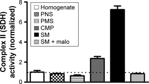
Activity of SDH, a mitochondrial marker enzyme, was measured in various cellular fractions to analyze the level of enrichment of the SM preparations isolated by the discontinuous gradient Ficoll medium-based ultracentrifugation protocol. SDH activity in synaptic mitochondrial preparation was found to be almost 7 times more than that in the starting homogenate material, indicating robust enrichment of mitochondria (). Additionally, we assayed fumarase activity in presence or absence of detergent Triton X-100 to measure the integrity of SM fraction.Citation47 Integrity of SM fraction was found to be 93.6%±3.2% (mean ± SD).
Figure 1 Isolation of purified synapse-specific mitochondrial fraction from rat brain cortices.
Abbreviations: SDH, succinate dehydrogenase; SM, synapse-specific mitochondria; malo, malonate; PNS, post-nuclear supernatant; PMS, post-mitochondrial supernatant; CMP, crude mitochondrial pellet.
Ascorbic acid supplementation ameliorates oxidative damage and reduction in antioxidant capacity of SM induced by early-life Pb exposure
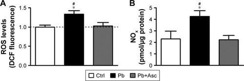
Oxidative stress in SM of rat cerebral cortices was measured using both DCFH-DA and Griess assays for the presence of ROS and reactive nitrogen species (RNS), respectively. Increase in ROS and NOx levels were observed in SM from rats of Pb group when compared to Ctrl group, indicating elevated oxidative stress. Pb-induced increase in oxidative stress was rescued by supplementation with ascorbate ().
Figure 2 SM of rats developmentally exposed to Pb have increased levels of ROS and RNS and the increase is ameliorated by ascorbate.
Abbreviations: SM, synapse-specific mitochondria; Pb, lead; ROS, reactive oxygen species; RNS, reactive nitrogen species; DCFH-DA, 2′,7′-dichlorofluorescein diacetate; Ctrl, control; Asc, ascorbic acid; SEM, standard error of the mean; NOx, total nitrites and nitrates.
Increase in oxidative stress in SM of rats of Pb group was accompanied by elevated levels of lipid and protein oxidation markers, as measured by the presence of TBARS, and protein carbonyls and oxidized protein thiols, respectively. Moreover, ascorbic acid treatment prevented the increase in lipid and protein oxidation in Pb-exposed rats ().
Figure 3 SM from cerebral cortices of rat pups exposed to Pb show increased oxidative damage to lipids and proteins and have reduced antioxidant capacity and their redox status is rescued by ascorbic acid supplementation.
Abbreviations: SM, synapse-specific mitochondria; Pb, lead; TBARS, thiobarbituric acid reactive substances; ABTS, 2,2′-azino-bis(3-ethylbenzothiazoline-6-sulfonic acid); Ctrl, control; Asc, ascorbic acid; SEM, standard error of the mean.
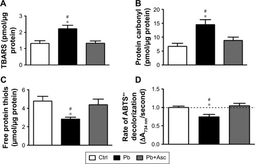
Antioxidant capacity of SM fractions of rat cortices was measured by ABTS radical scavenging assay. Ability to scavenge ABTS+• was significantly reduced in SM fraction of rats from Pb group when compared to Ctrl rats. Antioxidant power of SM from prepubertal rats developmentally exposed to Pb was recovered in rats from Pb+Asc group, highlighting the rescue effects of ascorbate supplementation ().
The data suggest that increase in oxidative stress in SM fraction from prepubertal rats developmentally exposed to Pb is due to both increase in generation of ROS and RNS levels as well as reduction in the capacity to scavenge free radicals. Moreover, high levels of free radicals in SM of Pb-exposed rats resulted in increased protein and lipid damage. Interestingly, elevated levels of RNS/ROS, reduced radical scavenging and increased protein and lipid oxidation are prevented by supplementation with ascorbic acid.
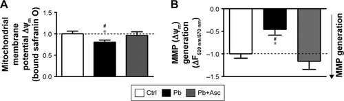
Pb induced alterations in MMP of SM and its repolarization are rescued by ascorbate
Transmembrane potential of mitochondria (inside negative), also called as MMP (ΔΨm), is established by the proton pump of the mitochondrial respiratory chain and is a measure of functional state of mitochondria. ΔΨm is a critical regulator of several mitochondrial functions including bioenergetics (ATP production), mitochondrial dynamics (fusion and fission), redox homeostasis and apoptotic signaling.Citation58 Using the cationic lipophilic probes, safranin O, we found that ΔΨm of SM isolated from Pb-exposed prepubertal rats was slightly but significantly reduced when compared to SM of aged matched controls (). Repolarization of energized SM (in presence of substrates glutamate and malate) was also assayed and found to be significantly reduced in SM isolated from rats developmentally exposed to Pb in comparison to Ctrl rats (), indicating compromises in mitochondrial bioenergetic enzyme functions. Deficits in both basal ΔΨm and repolarization of energized SM of Pb-exposed rats were recovered by supplementation with ascorbic acid (). However, ascorbic acid alone did not alter the basal or energized ΔΨm ().
Figure 4 SM from cerebral cortices of rat pups exposed to Pb show alterations in basal mitochondrial membrane potential as well as energization-induced generation of mitochondrial membrane potential and ascorbic acid abolishes these alterations.
Abbreviations: SM, synapse-specific mitochondria; Pb, lead; MMP, mitochondrial membrane potential; Ctrl, control; Asc, ascorbic acid.
Ascorbic acid supplementation rescues compromised activities of electron transport chain oxidoreductases and ATP synthesis ability of cortical SM from rats developmentally exposed to Pb
To observe any dysregulation in the bioenergetic properties of SM of Pb-exposed prepubertal rats, we analyzed the activities of enzyme complexes I, II, III and IV of the electron transport chain. While activity levels of complex I and complex IV in SM fraction of Pb-exposed rats were similar to those from aged matched controls (), activities of complexes II and III were found to be significantly reduced in SM of Pb-exposed rats (). Moreover, while ascorbic acid supplementation rescued the Pb-mediated loss of complex II activity (), it was unable to change complex III activity levels ().
Figure 5 Pb induced alternations in the bioenergetic properties of cortical SM are mitigated by ascorbate supplementation.
Abbreviations: SM, synapse-specific mitochondria; Pb, lead; ETC, electron transport chain; Ctrl, control; Asc, ascorbic acid.
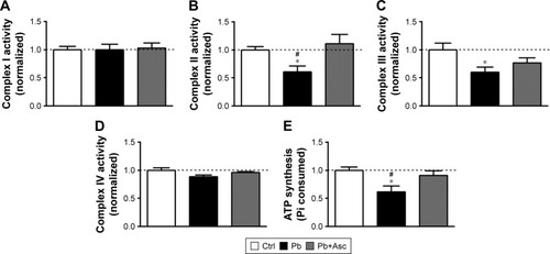
We also observed a decrease in ATP synthesis in presence of substrates malate and glutamate in SM fractions isolated from Pb-exposed rats; however, complete recovery was observed in rats supplemented with ascorbic acid (). On the other hand, ascorbic acid alone did not have any effects on the bioenergetic properties of SM ().
The data suggest that reduction in bioenergetic activity of SM from prepubertal rats developmentally exposed to Pb is rescued with ascorbate supplementation.
Discussion
In the brain, Pb neurotoxicity is prominent in prefrontal cortex, hippocampus and cerebellum,Citation10 where it results in morphological, structural and pathological changes in neuronal cells and their synaptic connections.Citation8,Citation59,Citation60 Mecha nisms of lead neurotoxicity are complex. Many of the toxic mechanisms associated with Pb neurotoxicity are because of its ability to substitute for divalent Ca2+ and Zn2+ cations,Citation6 which critically regulate important cellular processes including cell signaling and gene expression. Among the primary targets of Pb neurotoxicity are synaptic signaling and its maintenance and plasticity.Citation5,Citation9,Citation14 In fact, both electrophysiological and biochemical data point to a reduced efficiency of both pre-synaptic and post-synaptic machinery upon Pb exposure.Citation8,Citation14 While alterations in neurotransmitter (glutamate) release and N-methyl-D-aspartate receptor physiology are the best studied mechanisms of Pb-induced synaptic dysfunction,Citation5,Citation9 an interplay of several factors is thought to influence disruption of synaptic functions in Pb neurotoxicity. These include deleterious effects of Pb on redox and calcium homeostasis; cell signaling and death pathways; and membrane receptor trafficking and gene expression.Citation15,Citation16
Developing brain is particularly vulnerable to Pb neurotoxicity because of its immature blood–brain barrier.Citation16 In fact, chronic early-life exposure to Pb is known to have permanent detrimental effects on development, maintenance and function of the synapses,Citation16 leading to irreparable neurodevelopmental deficits in sensory-motor and cognitive skills.Citation3,Citation4,Citation61 This is because neuronal signaling (both spontaneous and evoked) at the synapse in the developing brain critically affects synapse pruning and proper brain development and function,Citation26,Citation29 and any alterations in synapse functions early in the brain development would lead to permanent cognitive and behavioral effects.
Synapse-specific mitochondria (SM) form a specialized class of mitochondria different from free neuronal mitochondria because of their ability of acting as calcium buffers to prevent excitotoxicity and as energy providers for the compelling process of synaptic signaling.Citation21–Citation23,Citation25 Indeed, SM are smaller in size compared to free non-SM and have an altered enzyme profile as well as a modified proteome and lipidome, and metabolome.Citation21,Citation24 Additionally, SM population has elevated vulnerability to pathological stresses and calcium overload and swelling.Citation21–Citation23 Alterations in proper functioning of cortical SM in prepubertal rats upon Pb exposure as observed in our study could potentially lead to critical disruptions in synaptic signaling, which in turn would affect higher order brain functions. Although, Pb-induced mitochondrial dysfunction has been observed earlier, most of the studies have been limited to in vitro cell culture systemsCitation62–Citation65 or in vitro mitochondrial preparations.Citation66,Citation67 Many in vivo studies using rodent models have observed mitochondrial dysfunction in adult or aged rats.Citation12,Citation68 Few studies have reported the effects of early-life Pb exposure on brain mitochondria in prepubertal rats. For example, ultrastructural morphological changes in hippocampalCitation44 and retinalCitation69 mitochondria have been observed upon exposure of neonatal rats to Pb. Evidence for altered mitochondrial bioenergetic functions in primary cerebellar granular neurons of rats exposed to PbCitation70 and hippocampal and cerebellar neuronsCitation71 has also been provided. Other studies indicate that perinatal Pb exposure increases ROS levels in a calcium-dependent mannerCitation72 and reduces antioxidant system of brain mitochondria.Citation42,Citation73 However, none of these studies have specifically looked at the effects of Pb exposure on synapse-specific mitochondria of brain cerebral cortex, which is an important regulator of synapse function and plasticity. In this study, we sought to assess the deleterious effects of early-life (prenatal and postnatal) exposure to Pb on synapse-localized mitochondrial population in the developing rat brain.
Our results suggest that early-life exposure to Pb leads to disruptions in redox homeostasis of SM quite early during development and consequent compromises in its membrane potential and energy metabolism activity. Given the critical importance of SM in the synapse physiology, this potentially constitutes as a major pathogenic event in synaptic dysfunction leading to detrimental behavioral outcomes observed in events of Pb exposure. Because ascorbic acid (vitamin C) is capable of both reducing oxidative stress and chelating metal ions, it can potentially serve as a detoxifying agent for Pb toxicity. Use of ascorbic acid in amelioration of Pb poisoning has been elucidated previously.Citation12,Citation37–Citation39 We used a dose of 500 mg ascorbic acid per kg body weight because it reduces the number of degenerating neurons upon Pb exposure to levels comparable to control untreated rats.Citation37 In comparison, a dose of 100 mg/kg body weight could only slightly reduce the number of degenerating neurons, which still remained significantly higher than control rats.Citation38,Citation39 While supplementation of ascorbic acid alone did not affect bioenergetic properties of SM, we observed appreciable recovery of Pb-induced oxidative stress and functional compromise of SM by ascorbic acid supplementation. Our results suggest that rescue of Pb-mediated disruptions in SM functions by ascorbate could potentially be used as a therapeutic strategy against early-life Pb exposure.
Acknowledgments
The study was partly funded by Deanship of Scientific Research, Imam Abdulrahman Bin Faisal University, Saudi Arabia (Project No. 2016-087-IRMC). The authors thank Dr Khaldoon Alsamman, Dr Hatem K Herzallah and Dr Sultan T Al-Otaibi for assistance in experiments and in the analysis and review of the results.
Supplementary material
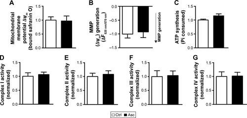
Figure S1 Supplementation of ascorbic acid alone does not affect the bioenergetics of cortical SM.
Notes: MMP at both basal states (A) and in presence of substrates (B) was not altered in SM fraction isolated from rat pups supplemented with ascorbic acid when compared to aged matched controls. Ascorbic acid supplementation did not affect other mitochondrial functions, including ATP synthesis (C) and activities of complexes I–IV (D–G). Data are represented as mean ± SEM (n=5 pups from 2 litters). Difference between the groups were assessed statistically using unpaired two-tailed Student’s t-test.
Abbreviations: SM, synapse-specific mitochondria; MMP, mitochondrial membrane potential; Ctrl, control; Asc, ascorbic acid; SEM, standard error of the mean.
Disclosure
The authors report no conflicts of interest in this work.
References
- MeyerPABrownMJFalkHGlobal approach to reducing lead exposure and poisoningMutat Res20086591–216617518436472
- AssiMAHezmeeMNHaronAWSabriMYRajionMAThe detrimental effects of lead on human and animal healthVet World20169666067127397992
- ChiodoLMJacobsonSWJacobsonJLNeurodevelopmental effects of postnatal lead exposure at very low levelsNeurotoxicol Teratol200426335937115113598
- AllenKAIs prenatal lead exposure a concern in infancy? What is the evidence?Adv Neonatal Care201515641642026372041
- NealAPGuilarteTRMolecular neurobiology of lead (Pb2+): effects on synaptic functionMol Neurobiol201042315116021042954
- GarzaAVegaRSotoECellular mechanisms of lead neurotoxicityMed Sci Monit2006123RA57RA6516501435
- BellingerDCBellingerAMChildhood lead poisoning: the torturous path from science to policyJ Clin Invest2006116485385716585952
- WhiteLDCory-SlechtaDAGilbertMENew and evolving concepts in the neurotoxicology of leadToxicol Appl Pharmacol2007225112717904601
- ToscanoCDGuilarteTRLead neurotoxicity: from exposure to molecular effectsBrain Res Brain Res Rev200549352955416269318
- SharmaPChambialSShuklaKKLead and neurotoxicityIndian J Clin Biochem20153011225646035
- MarchettiCMolecular targets of lead in brain neurotoxicityNeurotox Res20035322123612835126
- FloraGGuptaDTiwariAToxicity of lead: a review with recent updatesInterdiscip Toxicol201252475823118587
- SweattJDNeural plasticity and behavior – sixty years of conceptual advancesJ Neurochem2016139Suppl 2179199
- SadiqSGhazalaZChowdhuryABüsselbergDMetal toxicity at the synapse: presynaptic, postsynaptic, and long-term effectsJ Toxicol2012201213267122287959
- VerstraetenSVAimoLOteizaPIAluminium and lead: molecular mechanisms of brain toxicityArch Toxicol2008821178980218668223
- NealAPGuilarteTRMechanisms of lead and manganese neurotoxicityToxicol Res (Camb)2013229911425722848
- HagbergHMallardCRoussetCIThorntonCMitochondria: hub of injury responses in the developing brainLancet Neurol201413221723224457191
- WinnekeGDevelopmental aspects of environmental neurotoxicology: lessons from lead and polychlorinated biphenylsJ Neurol Sci20113081–291521679971
- GrandjeanPHerzKTTrace elements as paradigms of developmental neurotoxicants: lead, methylmercury and arsenicJ Trace Elem Med Biol20153113013425175507
- HsiangJDíazELead and developmental neurotoxicity of the central nervous systemCurr Neurobiol2011213542
- FedorovichSVWaseemTVPuchkovaLVBiogenetic and morphofunctional heterogeneity of mitochondria: the case of synaptic mitochondriaRev Neurosci201728436337328195557
- VosMLauwersEVerstrekenPSynaptic mitochondria in synaptic transmission and organization of vesicle pools in health and diseaseFront Synaptic Neurosci2010213921423525
- DuHGuoLYanSSosunovAAMcKhannGMYanSSEarly deficits in synaptic mitochondria in an Alzheimer’s disease mouse modelProc Natl Acad Sci U S A201010743186701867520937894
- DubinskyJMHeterogeneity of nervous system mitochondria: location, location, location!Exp Neurol2009218229330719464292
- LyCVVerstrekenPMitochondria at the synapseNeuroscientist200612429129916840705
- RaefskySMMattsonMPAdaptive responses of neuronal mitochondria to bioenergetic challenges: roles in neuroplasticity and disease resistanceFree Radic Biol Med201710220321627908782
- JeanneteauFArango-LievanoMLinking mitochondria to synapses: new insights for stress-related neuropsychiatric disordersNeural Plast20162016398506326885402
- CavallucciVFerrainaCD’AmelioMKey role of mitochondria in Alzheimer’s disease synaptic dysfunctionCurr Pharm Des201319366440645023432718
- LiZOkamotoKHayashiYShengMThe importance of dendritic mitochondria in the morphogenesis and plasticity of spines and synapsesCell2004119687388715607982
- AhmadAShahSABadshahHNeuroprotection by vitamin C against ethanol-induced neuroinflammation associated neurodegeneration in the developing rat brainCNS Neurol Disord Drug Targets201615336037026831257
- KimHJSongWJinEHCombined low-intensity exercise and ascorbic acid attenuates kainic acid-induced seizure and oxidative stress in miceNeurochem Res20164151035104126646003
- de FreitasPZanoniJNAlvesAMde Miranda NetoMHNeuroprotection and neurodegeneration in submucosal VIP-IR neurons in the jejunum of ascorbic acid supplemented aging Wistar ratsNutr Neurosci201215628328822889609
- RiceMEAscorbate regulation and its neuroprotective role in the brainTrends Neurosci200023520921610782126
- MorettiMFragaDBRodriguesALSAscorbic acid to manage psychiatric disordersCNS Drugs201731757158328600627
- PatrickLToxic metals and antioxidants: part II. The role of antioxidants in arsenic and cadmium toxicityAltern Med Rev20038210612812777158
- HsuPCGuoYLAntioxidant nutrients and lead toxicityToxicology20021801334412324198
- SadeghiAEbrahimzadeh BideskanAAlipourFFazelAHaghirHThe effect of ascorbic acid and garlic administration on lead-induced neural damage in rat offspring’s hippocampusIran J Basic Med Sci201316215716424298384
- ChangBJJangBJSonTGAscorbic acid ameliorates oxidative damage induced by maternal low-level lead exposure in the hippocampus of rat pups during gestation and lactationFood Chem Toxicol201250210410822056337
- HanJMChangBJLiTZProtective effects of ascorbic acid against lead-induced apoptotic neurodegeneration in the developing rat hippocampus in vivoBrain Res20071185687417959157
- AcharyaURRathoreRMMishraMRole of vitamin C on lead acetate induced spermatogenesis in swiss miceEnviron Toxicol Pharmacol200313191421782643
- PatraRCSwarupDDwivediSKAntioxidant effects of alpha tocopherol, ascorbic acid and L-methionine on lead induced oxidative stress to the liver, kidney and brain in ratsToxicology20011622818811337108
- GottipoluRRDavuljigariCBPerinatal exposure to lead: reduction in alterations of brain mitochondrial antioxidant system with calcium supplementBiol Trace Elem Res20141621–327027725161091
- HarryGJSchmittTJGongZBrownHZawiaNEvansHLLead-induced alterations of glial fibrillary acidic protein (GFAP) in the developing rat brainToxicol Appl Pharmacol1996139184938685912
- Baranowska-BosiackaIStrużyńskaLGutowskaIPerinatal exposure to lead induces morphological, ultrastructural and molecular alterations in the hippocampusToxicology201330318720023146751
- LaiJCWalshJMDennisSCClarkJBSynaptic and non-synaptic mitochondria from rat brain: isolation and characterizationJ Neurochem197728362563116086
- DuaRGillKDEffect of aluminium phosphide exposure on kinetic properties of cytochrome oxidase and mitochondrial energy metabolism in rat brainBiochim Biophys Acta20041674141115342109
- ValentiDVaccaRAde PintoMCDe GaraLMarraEPassarellaSIn the early phase of programmed cell death in Tobacco Bright Yellow 2 cells the mitochondrial adenine nucleotide translocator, adenylate kinase and nucleoside diphosphate kinase are impaired in a reactive oxygen species-dependent mannerBiochim Biophys Acta200717671667817184729
- DuttaMGhoshAKRangariVSilymarin protects against copper-ascorbate induced injury to goat cardiac mitochondria in vitro: involvement of antioxidant mechanisms(s)Int J Pharm Pharm Sci201468422429
- El-MasryTAEmaraAMEl-ShitanyNAPossible protective effect of propolis against lead-induced neurotoxicity in animal modelJ Evol Biol Res201131411
- BuegeJAAustSDMicrosomal lipid peroxidationMethods Enzymol197852302310672633
- KatalinicVModunDMusicIBobanMGender differences in antioxidant capacity of rat tissues determined by 2,2′-azinobis (3-ethylbenzothiazoline 6-sulfonate; ABTS) and ferric reducing antioxidant power (FRAP) assaysComp Biochem Physiol C Toxicol Pharmacol20051401475215792622
- CherianESudheeshNPJanardhananKKPataniGFree-radical scavenging and mitochondrial antioxidant activities of Reishi-Ganoderma lucidum (Curt: Fr) P. Karst and Arogyapacha-Trichopus zeylanicus Gaertn extractsJ Basic Clin Physiol Pharmacol200920428930720214017
- ValentiDde BariLDe FilippisBRicceriLVaccaRAPreservation of mitochondrial functional integrity in mitochondria isolated from small cryopreserved mouse brain areasAnal Biochem2014444253124018341
- EmausRKGrunwaldRLemastersJJRhodamine 123 as a probe of transmembrane potential in isolated rat-liver mitochondria: spectral and metabolic propertiesBiochim Biophys Acta198685034364482873836
- PowellCSJacksonRMMitochondrial complex I, aconitase, and succinate dehydrogenase during hypoxia-reoxygenation: modulation of enzyme activities by MnSODAm J Physiol Lung Cell Mol Physiol20032851L189L19812665464
- SpinazziMCasarinAPertegatoVSalviatiLAngeliniCAssessment of mitochondrial respiratory chain enzymatic activities on tissues and cultured cellsNat Protoc2012761235124622653162
- KatewaSDKatyareSSA simplified method for inorganic phosphate determination and its application for phosphate analysis in enzyme assaysAnal Biochem2003323218018714656523
- IannettiEFWillemsPHPellegriniMTowards high-content screening of mitochondrial morphology and membrane potential in living cellsInt J Biochem Cell Biol201563667025668473
- GąssowskaMBaranowska-BosiackaIMoczydłowskaJPerinatal exposure to lead (Pb) induces ultrastructural and molecular alterations in synapses of rat offspringToxicology2016373132927974193
- WilsonMAJohnstonMVGoldsteinGWBlueMENeonatal lead exposure impairs development of rodent barrel field cortexProc Natl Acad Sci U S A200097105540554510805810
- HuHTéllez-RojoMMBellingerDFetal lead exposure at each stage of pregnancy as a predictor of infant mental developmentEnviron Health Perspect2006114111730173517107860
- DabrowskaAVeneroJLIwasawaRPGC-1α controls mitochondrial biogenesis and dynamics in lead-induced neurotoxicityAging (Albany NY)20157962964726363853
- GeierDAKingPGGeierMRMitochondrial dysfunction, impaired oxidative-reduction activity, degeneration, and death in human neuronal and fetal cells induced by low-level exposure to thimerosal and other metal compoundsToxicol Environ Chem2009913–473574924532866
- YeFLiXLiFCyclosporin A protects against Lead neurotoxicity through inhibiting mitochondrial permeability transition pore opening in nerve cellsNeurotoxicology20165720321327725305
- LiuGWangZKWangZYYangDBLiuZPWangLMitochondrial permeability transition and its regulatory components are implicated in apoptosis of primary cultures of rat proximal tubular cells exposed to leadArch Toxicol20169051193120926082307
- DeviCBJyotsnaVKumariKKIndravathiGThakurAIn vitro effect of lead on cholinergic and bioenergetic systems in synaptosomal and mitochondrial fractions of rat brain regionsInt J Adv Life Sci201471110
- HoltzmanDShen HsuJMortellPIn vitro effects of inorganic lead on isolated rat brain mitochondrial respirationNeurochem Res197832195206673116
- FloraSJSaxenaGMehtaAReversal of lead-induced neuronal apoptosis by chelation treatment in rats: role of reactive oxygen species and intracellular Ca(2+)J Pharmacol Exp Ther2007322110811617431133
- PerkinsGAScottRPerezAEllismanMHJohnsonJEFoxDABcl-xL-mediated remodeling of rod and cone synaptic mitochondria after postnatal lead exposure: electron microscopy, tomography and oxygen consumptionMol Vis2012183029304823288995
- Baranowska-BosiackaIGutowskaIMarchettiCAltered energy status of primary cerebellar granule neuronal cultures from rats exposed to lead in the pre- and neonatal periodToxicology20112801–2243221108985
- Lalith KumarVMuralidharaAmeliorative effects of ferulic Acid against lead acetate-induced oxidative stress, mitochondrial dysfunctions and toxicity in prepubertal rat brainNeurochem Res201439122501251525322819
- YangXWangBZengHRole of the mitochondrial Ca(2+) uniporter in Pb(2+)-induced oxidative stress in human neuroblastoma cellsBrain Res20141575122124881885
- SannadiSKadeyalaPKGottipoluRRReversal effect of monoisoamyl dimercaptosuccinic acid (MiADMSA) for arsenic and lead induced perturbations in apoptosis and antioxidant enzymes in developing rat brainInt J Dev Neurosci201331758659723906897