
Abstract
Background
Subarachnoid hemorrhage (SAH) can induce apoptosis in many regions of the brain including the cortex and hippocampus. However, few studies have focused on apoptosis in the hypothalamus after SAH. Although some antiapoptotic strategies have been developed for SAH, such as anti-tumor necrosis factor-alpha (TNF-α) antibody, the molecular mechanisms underlying this condition have yet to be elucidated. Therefore, the purpose of this study was to evaluate whether SAH could induce apoptosis in the hypothalamus and identify the potential molecular mechanisms underlying the actions of anti-TNF-α antibody, as a therapeutic regimen, upon apoptosis.
Materials and methods
SAH was induced in a rat model. Thirty minutes prior to SAH, anti-TNF-α antibody or U0126, an extracellular signal-regulated kinase (Erk) inhibitor, was microinjected into the left lateral cerebral ventricle. In addition, phorbol-12-myristate-13-acetate was injected intraperitoneally immediately after the anti-TNF-α antibody microinjection. Then, real-time polymerase chain reaction, Western blotting and immunohistochemistry were used to detect the expression of caspase-3, bax, bcl-2, phosphorylated Erk (p-Erk) and Erk. Finally, anxiety-like behavior was identified by using open field.
Results
Levels of caspase-3, bax and bcl-2, all showed a temporary rise after SAH in the hypothalamus, indicating the induction of apoptosis in this brain region. Interestingly, we found that the microinjection of anti-TNF-α antibody could selectively block the elevated levels of bax, suggesting the potential role of anti-TNF-α antibody in the inhibition of SAH-induced apoptosis in the hypothalamus. Moreover, we found that Erk activation was necessary for apoptosis after SAH and that the microinfusion of anti-TNF-α antibody could inhibit apoptosis by suppressing the increase of p-Erk in the hypothalamus. Finally, our data indicated that the infusion of anti-TNF-α antibody could improve anxiety-like behavior.
Conclusion
Collectively, our data demonstrate that anti-TNF-α antibody attenuates apoptosis in the hypothalamus by inhibiting the activation of Erk, which plays an important role in the treatment of SAH.
Video abstract
Point your SmartPhone at the code above. If you have a QR code reader the video abstract will appear. Or use:
Introduction
Subarachnoid hemorrhage (SAH), a fetal cerebrovascular disease with high morbidity and mortality rates, is usually associated with long-term poor outcome.Citation1 Our current understanding is that early brain injury (EBI) is considered to refer to direct brain damage occurring after SAH.Citation2 Therefore, the elimination of EBI is becoming an important alternative therapy for SAH. Increasing evidence indicates that neural apoptosis, a key process in the pathogenesis of EBI after SAH, has become a key target in the prevention of brain cell death.Citation3,Citation4 Despite the development of some antiapoptotic strategies for SAH, current therapeutic strategies remain unsatisfactory and we still know little about the molecular mechanisms involved.
Tumor necrosis factor-alpha (TNF-α), a cytokine involved in neuronal inflammation, apoptosis and necrosis, is known to be upregulated in both the cerebral cortex and hippocampus after SAH.Citation5 A previous study showed that apoptotic cell death, overlapping with inflammation, could further strengthen the interactions between distinct mediators of brain injury after SAH.Citation6 Inhibition of TNF-α has been approved for therapeutic use in a range of inflammatory disorders, including Crohn’s disease, rheumatoid arthritis, spondyloarthritis and psoriasis.Citation7,Citation8 Our previous study confirmed that the functional blockade of TNF-α could prevent apoptosis-associated gene expression in the hippocampus and prefrontal cortex after SAH, suggesting that TNF-α could represent a potential therapeutic target for acute brain injury in SAH-induced apoptosis in these brain regions.Citation9 This was the first study, to our knowledge, to show the therapeutic effect of anti-TNF-α antibody in SAH. However, the potential molecular mechanisms underlying the inhibition of SAH-induced apoptosis by anti-TNF-α antibody need to be further elucidated.
Treatment with anti-TNF-α agents could modulate the expression levels of a number of genes. Previous experiments have demonstrated that anti-TNF-α-neutralizing antibody reduced apoptosis in podocytes in diabetic nephropathy by inhibiting the activation of mitogen-activated protein kinases.Citation10 Interestingly, studies have shown that extracellular signal-regulated kinase (Erk), the downstream signaling molecule of mitogen-activated protein kinases, is aberrantly upregulated during neuronal apoptosis induced by SAH, cerebral ischemia, stroke and neurodegenerative diseases.Citation11–Citation13 Furthermore, blocking the activation of Erk using an Erk inhibitor, dominant-negative or constitutively active forms of Erk, has been confirmed to prevent the induction of apoptosis.Citation14–Citation16 However, it remains unknown as to whether the effect of anti-TNF-α antibody upon apoptosis induced by SAH is related to Erk activation or not.
Interestingly, numerous reports have demonstrated regional specificity in both the pathology and pharmacology of the central neural system.Citation17,Citation18 In particular, apoptotic changes have been detected in most brain regions in animal models of SAH, especially in the basal cortex and hippocampus.Citation19 Recent studies have begun to highlight the role of the hypothalamus in brain injury and have shown that traumatic brain injury can induce apoptosis in the hypothalamus.Citation20 Furthermore, SAH may lead to dysfunction of the hypothalamus.Citation21 However, thus far, very few studies have investigated whether SAH could lead to apoptosis in the hypothalamus. Since hypothalamus is involved in regulating homeostasis, motivation and emotional behavior,Citation22 elucidating the apoptotic pathway in the hypothalamus and developing appropriate neuroprotective strategies will be highly desirable in the treatment of SAH. Taken together, in this study, we investigated the effect of anti-TNF-α antibody on SAH-induced apoptosis in the hypothalamus of rats, as well as the potential molecule mechanisms involved.
Materials and methods
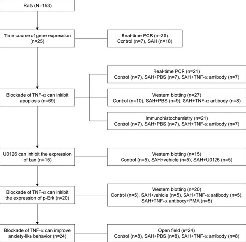
Male Wistar rats (250–350 g, N=153) were used throughout this study. The flow diagram of the study is shown in . All animals were treated humanely in accordance with the guidelines for the Care and Use of laboratory Animals by the National Institute of Health, and the study was approved by the Animal Care and Use Committee of the Second Hospital of Shandong University.
Figure 1 Flow diagram of the study.
An animal model of SAH
The SAH procedure was performed as described in our previous study.Citation9 In brief, male Wistar rats were anesthetized by an intraperitoneal injection of 5% chloral hydrate (0.75 mL/100 g). The left femoral artery was exposed, and 0.3 mL of fresh non-heparinized blood was withdrawn using a 27-gauge needle. Then, the artery was ligated at the proximal area. The head of the rats was then positioned in a stereotaxic apparatus. The arch of the atlas, occipital bone and atlanto-occipital membrane were exposed with a small suboccipital incision. Then, the fresh non-heparinized blood (0.3 mL) from the autologous femoral artery was injected into the cisterna magna at a rate of 0.05 mL/min using a 27-gauge infusion cannula connected to a microsyringe pump. After the induction of SAH, rats were kept in a head-down position until they awoke from the anesthesia, to ensure adequate subarachnoid distribution of the injected blood. The same operation was repeated 48 h later. After surgery, the rats were returned to the cages for recovery.
Drug treatment
For intracerebroventricular microinfusion, animals were placed in a stereotaxic apparatus (RWD Life Science Co., Ltd, 8001, Shenzhen, China) with the bregma used as the zero point. The left lateral cerebral ventricle was entered by using the following coordinates: anteroposterior, −0.8 mm; lateral, −1.5 mm and vertical, −4.0 mm. Anti-mouse/rat TNF-α antibody (dissolved in PBS; 250 ng/μL, 10 μL per rat; eBioscience™, 14-7423-85, San Diego, CA, USA), U0126 (dissolved in PBS/2% dimethyl sulfoxide, 5 μg/μL, 6 μL per rat; ApexBio Technology LLC, A1337, Houston, TX, USA) or the same volume of vehicle was microinfused into the left lateral cerebral ventricle 30 min before SAH. The infusions were performed at a rate of 0.5 μL/min using a microinfusion pump, and injection cannulas were left for an additional 2 min. Phorbol-12-myristate-13-acetate (PMA, dissolved in PBS/1% dimethyl sulfoxide, 100 μg/mL, 500 μg/kg; Sigma, P1585, St Louis, MO, USA) or the same volume of vehicle was injected intraperitoneally immediately after the microinjection of anti-TNF-α antibody.
RNA extraction and real-time polymerase chain reaction (PCR) analysis
To examine the time course of apoptotic gene expression in the hypothalamus after SAH, rats were sacrificed at day 1, day 2 and day 7, respectively. For the animals infused with anti-TNF-α antibody or the same volume of PBS, rats were killed 48 h (day 2) after SAH surgery. As a control, the sham-operated rats were sacrificed from their home cages. The hypothalamus was dissected out from each rat and RNA was extracted using the Ultrapure RNA Kit (CW Biotech, CW0597S, Beijing, China), in accordance with the manufacturer’s instructions. In brief, fresh or frozen samples (−70°C) were first cut into small tissue fragments. Then, 30–50 mg of tissue was added to 1 mL TRIzol reagent homogenized and then incubated for 5 min at room temperature. To each homogenate, a 1/5 volume of chloroform was added and the mixture was vortex mixed and centrifuged at the maximum speed for 10 min at 4°C. The upper aqueous phase was removed and placed into a new tube. The same volume of 70% ethanol (RNase-free) was added and the resulting solution was transferred into a spin column with a collection tube and then centrifuged at 12,000 rpm for ~20 s. The solution in the collection tube was discarded and the spin column was washed three times with wash buffer. After drying out at room temperature, RNA was then dissolved with 30–50 μL of fresh RNase-free water.
Then 0.5 μg of total RNA was reverse transcribed into cDNA using the PrimeScript™ RT Reagent Kit (Takara, RR037A, Otsu, Japan). Real-time PCRs of cDNA samples were performed in a total volume of 20 μL with SYBR (Takara, RR086A), primers at 800 nM and 2 μL of DNA template. The primer sequences used for PCR amplification were as follows: 5′ TGT GGA TGA CTG AGT ACC TGA ACC 3′ and 5′ CAG CCA GGA GAA ATC AAA CAG AGG 3′ forward and reverse primers for bcl-2; 5′ CGG CGA ATT GGA GAT GAA CTG G 3′ and 5′ CTA GCA AAG TAG AAG AGG GCA ACC 3′ forward and reverse primers for bax; 5′ GTG GAA CTG ACG ATG ATA TGG C 3′ and 5′ CGC AAA GTG ACT GGA TGA ACC 3′ forward and reverse primers for caspase-3; 5′ GGA GAT TAC TGC CCT GGC TCC TA 3′ and 5′ GAC TCA TCG TAC TCC TGC TTG CTG 3′ forward and reverse primers for β-actin. Quantitative real-time PCR was performed in a LightCycler2.0 (Roche, Basel, Switzerland). Thermal cycler parameters included 30 s at 95°C, and 40 cycles of denaturation at 95°C for 5 s, annealing at 62°C for 5 s and extension at 72°C for 10 s. The mRNA levels of target genes were normalized for each well to the mRNA levels of the housekeeping gene β-actin using the 2−ΔΔCT method.Citation23
Western blotting
Rats under different treatments were killed 48 h after SAH. The hypothalamus was dissected from each rat and homogenized in RadioImmunoPrecipitation Assay buffer consisting of 10 mM Tris pH 8.0, 150 mM NaCl, 1% NP-40, 1 mM EDTA pH 8.0 and 10% glycerol with protease inhibitors. Protein lysates, mixed with sodium dodecyl sulfate sample buffer, were boiled for 5 min. Protein concentration was then measured using a BCA Protein Assay Kit (Thermo Fisher Scientific, Waltham, MA, USA). Next, 30 μg of protein was loaded in each lane and separated in a 10% sodium dodecyl sulfate-polyacrylamide gel and then transferred to a polyvinylidene difluoride membrane. Blots were blocked with 5% non-fat dry milk in PBS for 2 h at room temperature. Subsequently, the blots were separately incubated overnight at 4°C with the following antibodies: rabbit anti-caspase-3 (1:1,000; Cell Signaling Technology, #9665, Danvers, MA, USA), rabbit anti-bax (1:1,000; Cell Signaling Technology, #2772), rabbit anti-bcl-2 (1:1,000; Cell Signaling Technology, #2870), rabbit anti-phosphorylated Erk (p-Erk, 1:2,000; Cell Signaling Technology, #4370), rabbit anti-Erk (1:2,000; Cell Signaling Technology, #4695), rabbit anti-TNF-α (1:1,000; R&D Systems, AF-510-SP, Minneapolis, MN, USA) or rabbit anti-β-actin (1:1,000, as a control; Cell Signaling Technology, #4970). After the blots were washed with PBS for three times, they were incubated with horse radish peroxidase-conjugated secondary antibodies (1:5,000; Cell Signaling Technology, #7074) for 2 h at room temperature. Finally, Western blot signals were detected using the enhanced chemiluminescence system (EMD Millipore, WBKLS0100, Billerica, MA, USA). Scanned images were visualized with ImageJ software.
Immunohistochemistry
For immunohistochemistry, rats from each group (microinfused with anti-TNF-α antibody, microinfused with PBS or sham-operated rats) were anesthetized by an intraperitoneal injection of 5% chloral hydrate (0.75 mL/100 g) 48 h after SAH. Rats were then perfused transcardially with 0.9% saline followed by 4% paraformaldehyde. Brains were removed and post-fixed in 4% paraformaldehyde overnight, and then equilibrated in 30% sucrose/PBS at 4°C for at least 3 days. Brains were then coronally sectioned at 20 μm thickness with a cryotome. Sections were washed with PBS three times and then blocked in 10% normal goat serum/PBS/0.3% Triton X-100 for 2 h at room temperature. Next, the sections were incubated overnight at 4°C with polyclonal anti-caspase-3 (1:100; Bioworld Technology, Louis Park, MN, USA), anti-bax (1:50, Bioworld Technology) or anti-bcl-2 (1:50, Bioworld Technology). After washing three times in PBS, the sections were then incubated with biotinylated goat anti-rabbit antibody (Elite Kit; Vector Laboratories, Burlingame, CA, USA) for 2 h at room temperature. Staining was detected with a biotin–streptavidin amplification system. Fluorescence images were acquired using a Nikon 80i microscope configured with a charge-coupled device camera interfaced to a personal computer.
Open field
The open field apparatus consisted of a 100×100×50 cm wooden cube with a white floor, which was divided into 5×5 identical squares. Experiments were recorded using a video camera above the open-field arena in a dim room, which was connected to a video recorder. Rats were placed in the center of the open field and allowed to explore for 10 min. The apparatus was cleaned with a 50% ethanol solution prior to the introduction of each animal. The total traveled distance and the time spent in the inner 4×4 squares were recorded to allow us to investigate anxiety-like behavior.
Statistical analysis
Data were analyzed using the Student’s t-test or one-way analysis of variance (ANOVA) followed by least significant difference post hoc comparisons. The significance level was set to 0.05 for all statistical analyses, and data were presented as mean ± standard error of the mean. Data analyses were performed using SPSS statistical program version 17.0.
Results
Time course of apoptotic-related gene expression following SAH in the hypothalamus
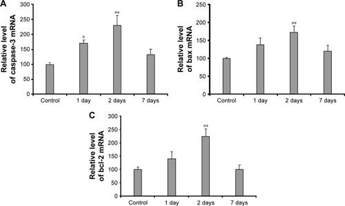
To assess whether SAH could induce apoptosis in hypothalamus, we used real-time PCR to investigate the expression levels of apoptotic-related genes at various time-points, including caspase-3, bax and bcl-2. Gene expression levels were increased after SAH, not only for apoptotic molecules, such as caspase-3 (ANOVA: F(3,21)=8.221, p=0.001; ) and bax (ANOVA: F(3,20)=3.480, p=0.035; ), but also for the antiapoptotic molecule, bcl-2 (ANOVA: F(3,19)=6.928, p=0.002; ). mRNA levels of caspase-3 increased from 24 h after SAH and peaked at 48 h. Compared with the temporal changes in caspase-3 mRNA levels, bcl-2 and bax mRNA showed a delayed increase, with both beginning to show increased mRNA levels 48 h after SAH. The observed temporal changes in these molecules suggest that SAH induced both apoptotic and antiapoptotic pathways in the hypothalamus.
Figure 2 Changes in caspase-3, bax and bcl-2 mRNA at different time points after SAH in the hypothalamus.
Abbreviation: SAH, subarachnoid hemorrhage.
Blockade of TNF-α can inhibit apoptosis induced by SAH in the hypothalamus
Previous studies have shown that when treating brain disorders, the effects of drugs are inconsistent in different brain regions. Our previous study confirmed that anti-TNF-α antibody could inhibit apoptosis in the prefrontal cortex and hippocampus.Citation9 To investigate whether TNF-α is involved in apoptosis in the hypothalamus, rats in this study were divided into three groups: control group, PBS group and TNF-α antibody group. Rats received an infusion of anti-TNF-α antibody or the same volume of PBS and were killed 48 h after SAH surgery. The sham-operated control rats were sacrificed from their home cages. We initially observed changes in TNF-α protein in the hypothalamus by Western blotting. As shows, the expression of TNF-α increased 48 h after SAH, suggesting the induction of TNF-α synthesis by SAH in the hypothalamus. In addition, injection of anti-TNF-α antibody could sufficiently block SAH-induced TNF-α increase (ANOVA: F(2,18)=4.592, p=0.024; ).
Figure 3 The effects of TNF-α antibody on the expression of caspase-3, bax and bcl-2 in the hypothalamus after SAH.
Abbreviations: SAH, subarachnoid hemorrhage; TNF-α, tumor necrosis factor-alpha.
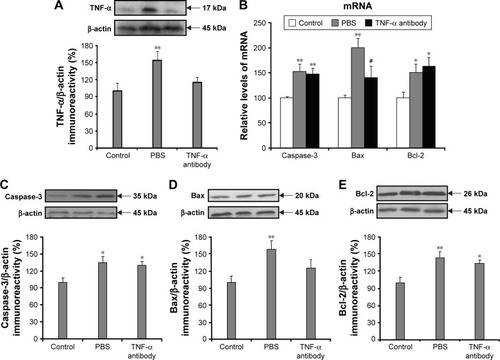
Next, to assess the effect of anti-TNF-α antibody upon SAH-induced apoptosis, we observed changes in the gene expression of caspase-3, bax and bcl-2 at 48 h after SAH. Compared with the PBS group, real-time PCR analysis showed that the microinfusion of anti-TNF-α antibody abolished increased bax mRNA levels in the hypothalamus (ANOVA: F(2,16)=10.406, p=0.001; ), but not the expression levels of caspase-3 (ANOVA: F(2,17)=6.448, p=0.008; ) and bcl-2 (ANOVA: F(2,12)=4.512, p=0.035; ).
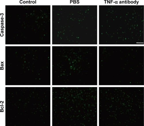
Similar to the changes in mRNA levels, Western blot analysis showed increased expression of caspase-3, bax and bcl-2 protein levels in the hypothalamus in PBS-treated SAH rats relative to sham-operated controls 48 h after SAH. In addition, the infusion of anti-TNF-α antibody effectively suppressed increased levels of bax protein in the hypothalamus (ANOVA: F(2,23)=4.810, p=0.018; ), but not caspase-3 (ANOVA: F(2,18)=4.543, p=0.025; ) or bcl-2 (ANOVA: F(2,21)=6.834, p=0.005; ). Furthermore, immunohistochemistry revealed that the numbers of caspase-3-, bax- and bcl-2-positive cells were significantly increased in the hypothalamus 48 h after SAH, as compared to the sham group (). The elevated expression of bax, but not caspase-3 and bcl-2, was attenuated by blocking TNF-α in the hypothalamus (), which strongly confirmed the Western blotting data. It has been shown that the overall ratio of pro- to antiapoptotic signals determines whether or not cells die.Citation24 The inhibition of bax, but not bcl-2, suggests that anti-TNF-α antibody blocks the SAH-induced apoptosis in the hypothalamus. Together, these results further indicate that anti-TNF-α antibody can block apoptosis induced by SAH in the hypothalamus following selective inhibition of bax expression.
Figure 4 Immunohistochemistry showing the changes of caspase-3, bax and bcl-2 in the hypothalamus 48 h after SAH.
Abbreviations: SAH, subarachnoid hemorrhage; TNF-α, tumor necrosis factor-alpha.
Blockade of TNF-α can inhibit the increased expression of p-Erk in the hypothalamus
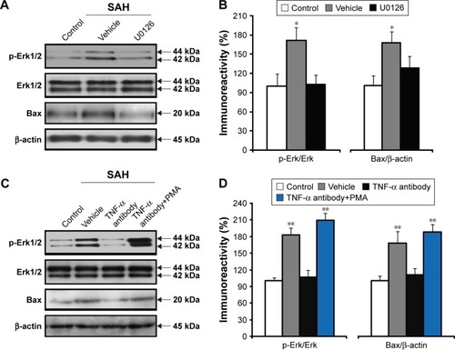
Previous research has shown that inhibition of Erk activation can reduce hippocampal neuronal apoptosis following SAH.Citation13 Next, we wanted to investigate whether the effect of anti-TNF-α antibody on SAH-induced apoptosis was due to regulating p-Erk levels. To address this, we initially examined whether Erk activation was required for apoptosis in the hypothalamus. Rats were divided into three groups: control group, vehicle group and U0126 group. U0126, a specific Erk inhibitor, or the same volume of vehicle was microinjected and then rats were sacrificed 48 h after SAH. As shown in , quantitative densitometric analyses showed elevated p-Erk levels in the hypothalamus of the vehicle group. However, this elevation was significantly reduced by the microinjection of U0126 (ANOVA: F(2,12)=4.815, p=0.029; ). In addition, administration of U0126 could eliminate the SAH-induced upregulation of bax levels (ANOVA: F(2,12)=4.005, p=0.047; ), which suggests that increased Erk activation is necessary for SAH-induced apoptosis. Furthermore, we found that the microinjection of anti-TNF-α antibody could prevent Erk activation 48 h after SAH, as compared with the vehicle group (). This inhibition could be reversed by the co-injection of PMA, a potent agonist of protein kinase C and its downstream effectors in the MEK/ERK-dependent pathwayCitation25 (ANOVA: F(3,16)=25.02, p<0.001; ). Additionally, bax was significantly elevated in response to PMA treatment, as compared with only anti-TNF-α antibody injection (ANOVA: F(3,16)=9.066, p=0.001; ), which further confirmed that p-Erk is not only necessary, but also sufficient for SAH-induced apoptosis. These results suggest that anti-TNF-α antibody can protect the hypothalamus against apoptosis by decreasing Erk phosphorylation.
Figure 5 The effects of anti-TNF-α antibody on the levels of p-Erk in the hypothalamus 48 h after SAH.
Abbreviations: Erk, extracellular signal-regulated kinase; PMA, phorbol-12-myristate-13-acetate; SAH, subarachnoid hemorrhage; TNF-α, tumor necrosis factor-alpha.
Blockade of TNF-α can improve anxiety-like behavior
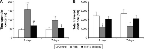
Psychiatric disorders often develop after SAH, the most frequent of which are depression and depression-related disorders, including anxiety. Next, we determined if blockade of TNF-α could relieve anxiety-like behavior as a consequence of SAH. Open field was used to investigate the effect of anti-TNF-α antibody on anxiety-like behavior 2 and 7 days after SAH (). Compared with the control group, SAH rats with PBS injection spent more time in the center compartment of the open field, suggesting that these changes may be associated with SAH surgery. Compared with the PBS group, SAH rats receiving an injection of anti-TNF-α antibody spent a reduced amount of time in the center of the open field both at 2 days (ANOVA: F(2,21)=7.662, p=0.003; ) and 7 days after surgery (ANOVA: F(2,21)=4.353, p=0.026; ). In addition, SAH rats with PBS injection showed decreased total traveled distance as compared with the control group both at 2 and 7 days after SAH. In contrast, no difference was found in the traveled distance between the control group and TNF-α antibody group (2 days, ANOVA: F(2,21)=3.753, p=0.04; 7 days, ANOVA: F(2,21)=4.090, p=0.032; ). As the hypothalamus is involved in emotional behavior, these series of experiments suggest that the effect of anti-TNF-α antibody on improving anxiety-like behavior may be associated with the inhibition of apoptosis in the hypothalamus.
Figure 6 The effects of anti-TNF-α antibody on anxiety-like behavior.
Abbreviations: SAH, subarachnoid hemorrhage; TNF-α, tumor necrosis factor-alpha.
Discussion
The related molecular mechanisms underlying antiapoptotic strategies in the treatment of SAH are still not completely understood. In this study, we found that SAH induced apoptotic and antiapoptotic pathways in the hypothalamus. Neutralizing TNF-α with an anti-TNF-α antibody was shown to inhibit apoptosis induced by SAH in the hypothalamus, which was related to a reduction in Erk activation. In addition, anti-TNF-α antibody ameliorated SAH-induced anxiety-like behavior. Collectively, these results support the notion that TNF-α represents an effective target for the treatment of SAH-induced apoptosis in the hypothalamus.
Apoptosis plays a major role in the pathophysiological process of EBI, a condition that occurs rapidly after SAH. However, whether SAH can induce apoptosis in the hypothalamus is still unknown. Therefore, we considered that it was very important to explore the molecular mechanisms underlying apoptosis in the hypothalamus. Recent research has shown that intracranial hypertension, or global cerebral ischemia/reperfusion injury, can induce apoptosis of hypothalamic cells in rat models.Citation20,Citation26 Consistent with these previous reports, the data described herein showed increased expression levels of caspase-3 and bax in the hypothalamus after SAH, which suggests that SAH can induce apoptosis in the hypothalamus by caspase-3- and bax-dependent pathways. To our knowledge, this is the first study showing the SAH-induced apoptosis in the hypothalamus. Interestingly, our previous report showed that SAH increased the levels of caspase-3 and/or bax, but did not affect bcl-2 expression levels in the hippocampus and prefrontal cortex.Citation9 In a manner which was very different to the hippocampus and prefrontal cortex, the increased expression level of bcl-2 in the hypothalamus suggests that different molecular pathways are induced in selective brain regions during SAH-induced apoptosis. This result further confirms the regional specificity in the pathology of the central neural system after SAH. Because of the functional differences of these brain regions, it is not surprising to figure out that the apoptosis-related molecular pathways in various brain regions play different roles in the outcome of SAH.
Next, we showed that blockade of TNF-α can reverse the apoptosis induced by SAH in the hypothalamus via inhibiting the expression of specific apoptotic molecule. Apoptosis can be regulated by inflammatory cytokines. A previous study has demonstrated that inflammatory injury does, indeed, occur in the hypothalamus in global cerebral ischemia/reperfusion injury by determining TNF-α, IL-1β and IL-6 levels.Citation26 In our study, SAH stimulated the release of TNF-α in the hypothalamus, which suggests that TNF-α may plays a major role in SAH-induced apoptosis and inhibition of TNF-α is a potential therapeutic treatment for it. Although many selective inhibitors, such as caspase inhibitors, have been identified and can effectively protect cells from apoptosis,Citation27 the molecular mechanisms of inhibiting TNF-α are still not completely understood. An earlier study confirmed that TNF-α antibody strategy was useful for the treatment of acute injury to the central nervous system.Citation28 Moreover, our own previous study demonstrated that the use of anti-TNF-α antibody can protect rats from apoptosis by reducing the elevated levels of bax and/or caspase-3 in the hippocampus and prefrontal cortex,Citation9 suggesting a new therapeutic strategy for improving the outcome of SAH. In this study, we further confirmed that anti-TNF-α antibody could specifically suppress increased levels of bax in the hypothalamus after SAH, but not caspase-3. One recent study showed that TNF-α can mediate cell death by ligating its receptor TNF receptor 1 (TNFR1) and uptake of ligand-bound TNFR1 activates caspase-8 and -3 in cord blood monocytes.Citation29 The reduced levels of bax in this study suggest a different apoptosis pathway induced by TNF-α after SAH. Collectively, these results strongly suggest the differential molecular mechanisms of action for anti-TNF-α antibody in different brain regions during the inhibition of apoptosis after SAH.
Next, we showed that anti-TNF-α antibody could block the increased levels of p-Erk in the hypothalamus after SAH. Previous studies have shown that the Erk signaling pathway is a major determinant in controlling diverse cellular processes, including proliferation, migration, differentiation and apoptosis.Citation30–Citation32 In addition, Erk activity has been confirmed to be involved in neurodegenerative diseases and ischemia stroke.Citation33 A recent study also reported that Erk plays a key proapoptotic role in the hippocampus in a rat model of SAH.Citation13 Consistent with these findings, our present data further confirm that levels of p-Erk increase in the hypothalamus following SAH. Interestingly, inhibition of Erk activation could block the increased expression of bax. These results strongly indicate the involvement of the Erk signaling pathway in the induction of apoptosis in the hypothalamus. Moreover, the increased levels of p-Erk could be blocked by neutralizing TNF-α, which further confirms the neuroprotective effect of anti-TNF-α antibody. The reduced ratios of p-Erk/Erk, accompanied by the blockade of bax, suggest that anti-TNF-α antibody protects neurons from apoptosis by inhibiting the phosphorylation of Erk. However, there are conflicting results showing the neuroprotective effect with Erk activation. Intracerebroventricular administration of brain-derived neurotrophic factor protects the neonatal rat brain from hypoxic-ischemic injury via Erk activation.Citation34,Citation35 The possible reason for the discrepancy is that the role of Erk activation may be depending on multiple signaling mechanisms. Transient Erk activation by growth factors is associated with neuroprotection, while prolonged and persistent activation induces cell death.Citation36 Overall, in this study, injection of anti-TNF-α antibody might have inhibited apoptosis by reducing the prolonged and persistent activation of Erk in the hypothalamus, as the levels of p-Erk were elevated 48 h after SAH.
Finally, we found that blockade of TNF-α can improve the anxiety-like behavior. A previous study has shown that increased intracranial pressure, a high-risk factor for the development of SAH, and stress play important roles in the dysfunction of the hypothalamus,Citation20 which is known to be involved in regulating emotional behavior. In addition, SAH is always accompanied by stress, a condition closely linked to anxiety.Citation37 Our open field results further confirmed that SAH could increase the levels of anxiety. Increasing evidence indicates that cytokines play a critical role in the development of neuropsychiatric disorders, including anxiety.Citation38,Citation39 TNF-α, one of the cytokines, is considered to be a key regulator in this process.Citation40 TNF-α expression levels were elevated both in patients with anxiety disorder and in animals with anxiety-like behavior. Consistent with these findings, TNF-α expression levels increased in the hypothalamus after SAH. Interestingly, we also found that anti-TNF-α antibody could reduce SAH-induced anxiety levels, which demonstrated the important role of TNF-α in the hypothalamus in SAH pathology. Furthermore, Karimi et al showed that intraperitoneal injection of 3,4-methylenedioxymethamphetamine increased the anxiety-like behavior and apoptotic cell numbers in the rat hippocampus, thus indicating that neuronal apoptosis may be linked to anxiety levels.Citation41 Due to the significant inhibition of apoptosis in response to treatment with anti-TNF-α antibody, these results further suggest that the improved anxiety-like behavior is attributed to the suppression of apoptosis in the hypothalamus. Additionally, since prolonged Erk activation can aggravate both anxiety-like behavior in post-traumatic stress disordered miceCitation42 and apoptosis, the effective role of anti-TNF-α antibody on SAH-induced anxiety-like behavior is considered to be linked to the inhibition of p-Erk expression levels.
Limitations
Additional studies using inhibitors of apoptosis and Erk in combination with other modulators of neural apoptosis will allow us to directly elucidate their role in the hypothalamus in SAH-induced anxiety-like behavior. Moreover, since TNF-α can bind to two receptor subtypes, TNFR1 (p55-R) and TNF receptor 2 (p75-R), we next wish to examine which receptor is involved in SAH-induced apoptosis.
Conclusion
In summary, our data show that anti-TNF-α antibody blocked SAH-induced apoptosis in the hypothalamus by inhibiting the phosphorylation of Erk. Injection of anti-TNF-α antibody improved the SAH-induced anxiety-like behavior. Our findings will help to improve our understanding of the precise role of anti-TNF-α antibody in SAH-induced apoptosis. In addition, the reduced expression of apoptosis-related molecules in the hypothalamus further suggests that anti-TNF-α antibody acts at the molecular level in a regional-specific manner in the brain during the treatment of SAH.
Acknowledgments
This work was supported by the Foundation for Excellent Young Scientist of Shandong Province (No BS2014SW002), Science and Technology Development Project of Shandong Province (No 2012GSF11802) and Seed Fund of the Second Hospital of Shandong University (No S2015010021).
Disclosure
The authors report no conflicts of interest in this work.
References
- RinkelGJAlgraALong-term outcomes of patients with aneurysmal subarachnoid haemorrhageLancet Neurol201110434935621435599
- CahillJCalvertJWZhangJHMechanisms of early brain injury after subarachnoid hemorrhageJ Cereb Blood Flow Metab200626111341135316482081
- MatzPGFujimuraMLewenAIncreased cytochrome c-mediated DNA fragmentation and cell death in manganese-superoxide dismutase-deficient mice after exposure to subarachnoid hemolysateStroke200132250651511157190
- SunQWuWHuYCEarly release of high-mobility group box 1 (HMGB1) from neurons in experimental subarachnoid hemorrhage in vivo and in vitroJ Neuroinflammation20141110624924349
- ZhangBFSongJNMaXDEtanercept alleviates early brain injury following experimental subarachnoid hemorrhage and the possible role of tumor necrosis factor-alpha and c-Jun N-terminal kinase pathwayNeurochem Res201540359159925542238
- PrunellGFSvendgaardNAAlkassKMathiesenTInflammation in the brain after experimental subarachnoid hemorrhageNeurosurgery20055610821092 discussion 1082.s–1092.s15854258
- YarilinaAPark-MinKHAntonivTHuXIvashkivLBTNF activates an IRF1-dependent autocrine loop leading to sustained expression of chemokines and STAT1-dependent type I interferon-response genesNature Immunol20089437838718345002
- JaniMBartonAWarrenRBGriffithsCEChinoyHThe role of DMARDs in reducing the immunogenicity of TNF inhibitors in chronic inflammatory diseasesRheumatology201453221322223946436
- JiangYLiuDWHanXYNeuroprotective effects of anti-tumor necrosis factor-alpha antibody on apoptosis following subarachnoid hemorrhage in a rat modelJ Clin Neurosci201219686687222516550
- GuoYSongZZhouMInfiltrating macrophages in diabetic nephropathy promote podocytes apoptosis via TNF-alpha-ROS-p38MAPK pathwayOncotarget2017832532765328728881810
- TianHPHuangBSZhaoJHuXHGuoJLiLXNon-receptor tyrosine kinase Src is required for ischemia-stimulated neuronal cell proliferation via Raf/ERK/CREB activation in the dentate gyrusBMC Neurosci20091013919943942
- Takahashi-NikiKKato-OseIMurataHEpidermal growth factor-dependent activation of the extracellular signal-regulated kinase pathway by DJ-1 protein through its direct binding to c-Raf proteinJ Biol Chem201529029178381784726048984
- FengDWangBMaYThe Ras/Raf/Erk pathway mediates the subarachnoid hemorrhage-induced apoptosis of hippocampal neurons through phosphorylation of p53Mol Neurobiol2016535737574826497030
- DrostenMSumEYLechugaCGLoss of p53 induces cell proliferation via Ras-independent activation of the Raf/Mek/Erk signaling pathwayProc Natl Acad Sci U S A201411142151551516025288756
- CagnolSChambardJCERK and cell death: mechanisms of ERK-induced cell death–apoptosis, autophagy and senescenceFEBS J2010277122119843174
- RubinoTViganoDPremoliFChanges in the expression of G protein-coupled receptor kinases and beta-arrestins in mouse brain during cannabinoid tolerance: a role for RAS-ERK cascadeMol Neurobiol200633319921316954596
- MichalskiDHartigWKrugelKRegion-specific expression of vesicular glutamate and GABA transporters under various ischaemic conditions in mouse forebrain and retinaNeuroscience201323132834423219666
- NohHJeonJSeoHSystemic injection of LPS induces region-specific neuroinflammation and mitochondrial dysfunction in normal mouse brainNeurochem Int20146935.s40.s24607701
- ParkSYamaguchiMZhouCNeurovascular protection reduces early brain injury after subarachnoid hemorrhageStroke200435102412241715322302
- TanHYangWWuCAssessment of the role of intracranial hypertension and stress on hippocampal cell apoptosis and hypothalamic-pituitary dysfunction after TBISci Rep20177380528630478
- KaramouzisIPaganoLProdamFClinical and diagnostic approach to patients with hypopituitarism due to traumatic brain injury (TBI), subarachnoid hemorrhage (SAH), and ischemic stroke (IS)Endocrine2016523441.s450.s26573924
- NunnNWomackMDartCBarrett-JolleyRFunction and pharmacology of spinally-projecting sympathetic pre-autonomic neurones in the paraventricular nucleus of the hypothalamusCurr Neuropharmacol2011926227722131936
- LivakKJSchmittgenTDAnalysis of relative gene expression data using real-time quantitative PCR and the 2(-Delta Delta C(T)) methodMethods200125402.s408.s11846609
- PhilchenkovACaspases: potential targets for regulating cell deathJ Cell Mol Med20048443244415601572
- MiyazakiMTakemasaTTSC2/Rheb signaling mediates ERK-dependent regulation of mTORC1 activity in C2C12 myoblastsFEBS Open Bio201773424433
- YuBRuanMZhangZNChengHBShenXCSynergic effect of borneol and ligustrazine on the neuroprotection in global cerebral ischemia/reperfusion injury: a region-specificity studyEvid Based Complement Alternat Med20162016407280927547227
- MeguroTChenBParentADZhangJHCaspase inhibitors attenuate oxyhemoglobin-induced apoptosis in endothelial cellsStroke200132256156611157197
- GenoveseTMazzonECrisafulliCImmunomodulatory effects of etanercept in an experimental model of spinal cord injuryJ Pharmacol Exp Thera2006316310061016
- DreschersSGilleCHaasMSeubertFPlatenCOrlikowskyTWReduced internalization of TNF-a/TNFR1 down-regulates caspase dependent phagocytosis induced cell death (PICD) in neonatal monocytesPLoS One2017128e018241528793310
- WagnerEFNebredaARSignal integration by JNK and p38 MAPK pathways in cancer developmentNat Rev Cancer20099853754919629069
- FreminCMelocheSFrom basic research to clinical development of MEK1/2 inhibitors for cancer therapyJ Hematol Oncol20103820149254
- McCainJThe MAPK (ERK) pathway: investigational combinations for the treatment of BRAF-mutated metastatic melanomaP T20133829610823599677
- ReinhardtPSchmidBBurbullaLFGenetic correction of a LRRK2 mutation in human iPSCs links parkinsonian neurodegeneration to ERK-dependent changes in gene expressionCell Stem Cell201312335436723472874
- HanBHHoltzmanDMBDNF protects the neonatal brain from hypoxic-ischemic injury in vivo via the ERK pathwayJ Neurosci2000205775578110908618
- KilicEKilicUSolizJBrain-derived erythropoietin protects from focal cerebral ischemia by dual activation of ERK-1/-2 and Akt pathwaysFASEB J200519142026202816207820
- JonesNMBergeronMHypoxia-induced ischemic tolerance in neonatal rat brain involves enhanced ERK1/2 signalingJ Neurochem20048915716715030400
- LeeSRheeDKEffects of ginseng on stress-related depression, anxiety, and the hypothalamic-pituitary-adrenal axisJ Ginseng Res201741458959429021708
- MaesMLeonardBFernandezA(Neuro)inflammation and neuroprogression as new pathways and drug targets in depression: from antioxidants to kinase inhibitorsProg Neuropsychopharmacol Biol Psychiatry20113565966321376099
- HouRBaldwinDSA neuroimmunological perspective on anxiety disordersHum Psychopharmacol20122761422213434
- ChenJSongYYangJThe contribution of TNF-alpha in the amygdala to anxiety in mice with persistent inflammatory painNeurosci Lett201354127528023415758
- KarimiSJahanshahiMGolalipourMJThe effect of MDMA-induced anxiety on neuronal apoptosis in adult male rats’ hippocampusFolia Biol (Praha)201460418719125152052
- XiangMJiangYHuZYangYBotchwayBOAFangMStimulation of anxiety-like behavior via ERK pathway by competitive serotonin receptors 2A and 1A in post-traumatic stress disordered miceNeurosignals201725395328977803