
Abstract
This case discusses the course of a woman with a history of epilepsy, alcohol use disorder, herpes simplex virus (HSV) encephalitis, and Wernicke encephalopathy (WE) who presented with altered mental status following approximately 48 hours of vomiting. After experiencing a tonic–clonic seizure in the emergency department, she developed a fluent aphasia. Aphasias are ordinarily attributed to structural changes in the brain parenchyma, often from stroke, neoplasm, or infection. When the magnetic resonance imaging of brain failed to show changes that could explain her fluent aphasia, the neurology team consulted psychiatry to workup psychogenic aphasia. During an admission 9 months earlier, she had been diagnosed with HSV encephalitis and possible WE. There was a high degree of suspicion for recurrent HSV infection, intermittent focal seizure activity, postictal psychosis, pseudobulbar affect, or a vascular cause of her fluent aphasia. After 3 days of treatment with levetiracetam, high-dose intravenous thiamine, and aripiprazole, the patient’s fluent aphasia reversed. The authors conclude that the patient’s reversible fluent aphasia was not psychiatric in etiology but likely caused by her seizures, the result of subtherapeutic phenytoin levels; her electroencephalogram showed focal seizure activity in the temporal lobes, possibly affecting her language centers. Language-related neurological conditions, or aphasias, can mimic psychiatric conditions such as conversion disorder or psychosis. In patients with substance use disorders, the line between psychiatric and neurological conditions becomes even more difficult to distinguish. The paper also discusses how unique aspects of her medications – levetiracetam conferring neuron membrane fluidity; aripiprazole, a drug shown to halt brain atrophy in mouse models; and parenteral thiamine to address her deficiency and WE – have aided in the reversal of the fluent aphasia. Levetiracetam should be considered in WE and the rare occurrence of aphasia after seizures.
Introduction
Language-related neurological conditions, or aphasias, can mimic psychiatric conditions such as conversion disorder or psychosis. In patients with substance use disorders, the line between psychiatric and neurological conditions becomes even more difficult to distinguish. Patients in alcohol withdrawal could manifest a wide range of presentation starting with initial withdrawal symptoms during the first 2–3 days and then leading to complications including delirium tremens, Wernicke’s encephalopathy (WE), or hepatic encephalopathy (HE). Hallucinations are more prominent than confusion in delirium tremens, while confusion is prominent in HE. Fluent aphasia typically is not present in these three complications. A feared end-stage presentation for chronic alcoholics is Korsakoff syndrome, described by Victor et alCitation1 as “an abnormal mental state in which memory and learning are affected out of all proportion to other cognitive functions in an otherwise alert and responsive patient.” Prior to this irreversible state however, is the acute and reversible presentation of WE caused by thiamine deficiency. It should be noted that WE, while more easily recognized in alcohol abuse, can also be caused by anorexia nervosaCitation2 and bariatric surgery.Citation3 Classically, WE is characterized as ataxia, confusion, and nystagmus. An alternative clinical definition considers WE if two of the following conditions are met: dietary deficiencies, oculomotor dysfunction, and either altered mental status or mild memory dysfunction.Citation4 In this case, we will examine the course of a patient with a long history of alcohol abuse and seizure disorder who presented with WE symptoms in addition to a fluent aphasia. She experienced a reversal of her acute aphasia under our care; to our knowledge, there is only one other case report detailing such a reversal.Citation5
Case report
A 52-year-old right-handed woman, who we will call RR, presented to an academic medical center with multiple seizures. She had a history of alcohol abuse, liver cirrhosis, and epilepsy for which she took phenytoin; she had not had any seizures for several years. Additionally, she was treated for herpes simplex virus (HSV) encephalitis 9 months prior. Initially, RR presented with nausea and vomiting for 48 hours, likely from alcohol withdrawal. Her boyfriend found her at home, appearing confused, and witnessed her have a generalized tonic–clonic seizure. She was then brought by ambulance to the emergency department (ED) where she had another seizure (tonic–clonic movements, right > left, with eyes deviated to the left and not responding). Her medications included aspirin, hydrocodone-acetaminophen, folic acid, thiamine, pyridoxine, and phenytoin 100 mg daily. She typically drank 2–6 beers a day. Prior to the admission for encephalitis, she was seen in the ER 1 year before for alcohol abuse and fall. After the admission for encephalitis and prior to this current episode, she was seen in ER four times, all for falls and related fractures. As the patient has been lost to follow-up, we are unable to confirm the patient’s consent for publication of this report. All data has been sufficiently anonymized not to cause harm to the patient or her family.
Vital signs at arrival-blood pressure 113/72, heart rate 106, temperature 102°F (38.9°C), Respiratory rate 16, body mass index (BMI) 23.77 kg/m2. In the ED, RR was given 4 mg of lorazepam, 2 mg of intravenous (IV) diazepam and fosphenytoin 15 PE units/kg. She remained unresponsive to verbal prompts with rhythmic beating of her left foot, prompting a neurology consult. Lacosamide was added and one dose of IV acyclovir was administered for empiric recurrent HSV encephalitis. Initially unresponsive on neurological examination, she slowly started following commands but became fluently aphasic. The differential diagnoses at this point included seizures with concern for status epilepticus, subtherapeutic medication, alcohol withdrawal, electrolyte derangement, and infection.
Laboratory results
RR’s laboratory results were reported as follows: 1. cerebrospinal fluid: negative for infection or inflammation, PCR negative. 2. complete blood count: increased red blood cell distribution width 21.2 (normal 11%–15.0%), decreased platelets 101 (150–400 K/mL). 3. complete metabolic panel: decreased potassium 2.8 (3.3–5.1), elevated anion gap 25 (4–16) decreased blood urea nitrogen 4 (7–22 mg/dL), increased glucose 228 (70–100 mg/dL), alkaline phosphatase 167 (30–110 IU/L), AST 125 (10–40 IU/L). 4. blood: lactate 3.4 (0.9–1.7 mM/L). 5. urine/drug: urinalysis was negative for urinary tract infection. Urine drug screen and blood alcohol were negative, and phenytoin levels subtherapeutic 4.2. 6. electrocardiogram: sinus tachycardia. 7. imaging: chest X-ray and computed tomography-head were both negative. The magnetic resonance imaging (MRI) was inconclusive, showing less prominent/no definite asymmetric flair abnormality in the left medial temporal lobe compared to her admission for HSV encephalitis 9 months prior.
RR was transferred to the neuro intensive care unit and placed on alcohol withdrawal protocol (Clinical Institute Withdrawal Assessment for alcohol; CIWA). video electroencephalogram (vEEG) did not reveal spikes or epileptiform activity (although this was after medication loading and gaze deviation resolution) and she had continued fluent aphasia. On day 2, the cranial nerve exam showed diminished blink to threat on the right, bilateral nystagmus on horizontal gaze with a left gaze preference, and gait that was found to be impaired, requiring assistance.
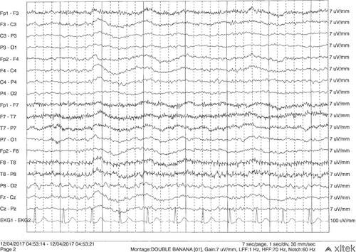
Phenytoin and lacosamide were replaced with levetiracetam 750 mg twice a day (BID) (). RR became agitated and was given haloperidol 2 mg IV. Minutes later, she was found to be awake, with an unremarkable cranial nerve exam and without nystagmus, but intermittent fluent aphasia continued. Continuous 17-hour video EEG monitoring was performed. Video EEG () while aphasic showed focal slowing over the left temporoparietal region, consistent with an underlying structural or functional lesion, but no clear epileptiform activity.
Table 1 Medication administration and CIWA
Figure 1 Continuous 17-hour video EEG monitoring.
Abbreviation: EEG, electroencephalogram.
At this point, psychiatry was consulted for possible psychogenic aphasia (conversion disorder). On exam, the patient was neither able to sustain attention nor follow commands and presented with fluent aphasia, repeating the phrase “It was as big as your nose, from your chin to your ear… as big as your nose.” Psychiatry recommended increasing thiamine from 100 per os (PO) to 500 mg q8h IV and adding aripiprazole 2.5 mg PO for delirium. On day 4, mental status exam showed intermittent fluent aphasia with perseveration, as well as signs of disinhibition with episodes of inappropriate laughter. By the end of the day the patient could say, “I feel much better, I want to go home tomorrow.” Transient ischemic attack workup, including an electrocardiogram with Doppler, computed tomography angiogram, and fasting lipid panel, came back negative. On day 5, the patient sustained a 30-minute conversation with few paraphasic errors, but frequently giggled, continuing to show signs of disinhibition.
She scored a 27 out of 30 on mini mental status exam, losing two points in recall and one point in the three-stage command. She was started on acamprosate for alcohol cravings and discharged to a subacute rehabilitation facility.
Discussion
Upon admission to the neurology intensive care unit, the medical team was managing a patient with a history of alcohol use disorder and epilepsy who was presenting at subtherapeutic phenytoin levels with altered mental status after throwing up for nearly 48 hours. During an admission 9 months earlier, she had been diagnosed with HSV encephalitis and possible WE syndrome. There was a high degree of suspicion for recurrent HSV infection, intermittent focal seizure activity, postictal psychosis, or pseudobulbar affect or a vascular cause of her fluent aphasia. There were no concerns for HE because the following: total bilirubin was 1.5, patient was not jaundiced, no asterixis was present, and patient’s confusion got better without any administration of lactulose. The absence of corresponding findings on MRI or EEG raised a concern for psychogenic etiology of aphasia, resulting in a psychiatric consult.
Postictal psychosis occurs almost exclusively in adults with chronic and treatment-refractory epilepsy, usually 15–20 years after their initial diagnosis affecting 25%–50% of this population.Citation6 It presents with hallucinations and disorders of thought. Even though RR was confused, no hallucinations or delusions were present, reducing suspicion for this diagnosis.
Pseudobulbar affect commonly presents as emotional incontinence, or uncontrollable outbursts of laughter or crying at inappropriate times, and is found in patients with a history of brain injury or neurodegenerative disease.Citation7 While RR demonstrated some disinhibition and laughed excessively, she had no history of neurodegenerative disease or brain damage and imaging was negative for corticobulbar tract degeneration.
Psychogenic aphasia, or conversion disorder, can be found in 4%–5% of adult patients with neurologic disorders.Citation8 It is characterized by inconsistent mispronunciations and frequent stuttering. Unlike true aphasias, psychogenic aphasia worsens with time. RR’s abnormal speech began suddenly, was fluent, and without stuttering.
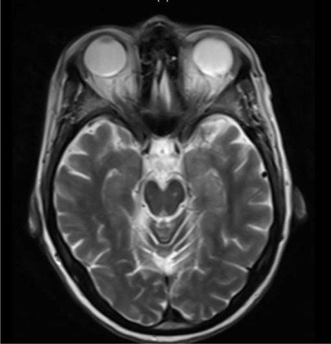
Suspicion for recurrent HSV encephalitis was low, and our literature search of language changes in HSV encephalitis did not show evidence of fluent aphasias. A 2012 Brazilian case report details anomic aphasia, or the misidentification of objects using related words (saying “comb” instead of “hair”) in a 13-year-old patient with herpes simplex encephalitis (HSE).Citation9 Another case report from Italy notes a similar difficulty in naming objects in patients with HSE, particularly living objects (fruits, vegetables, and animals).Citation10 None of the elements of our patient’s exam are consistent with anomic aphasia. RR’s most recent MRI () failed to show a temporal FLAIR abnormality; such a finding on a previous MRI () resulted in her initial HSV encephalitis diagnosis. It is possible however that the residual effects of her previous infection resulted in the temporal structural/functional lesion found in the vEEG ().
Figure 2 Current admission shows less pronounced left mesial temporal abnormality.
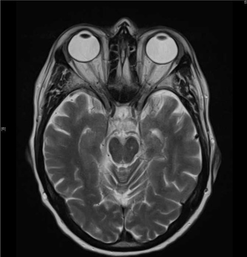
Figure 3 Admission 9 months prior showing nonspecific FLAIR abnormality in the left mesial temporal lobe.
The focus now shifted to the patient’s extensive alcohol abuse history. Fluent aphasia may be confused for confabulation, an unintentional fabrication of ideas and circumstances inconsistent with the truth.Citation11 Confabulation is associated with permanent memory damage and can be seen in WK syndrome. Momentary or provoked confabulation is commonly seen in Korsakoff syndrome and is irreversible.Citation12 Her exam during hospitalization for suspected HSV encephalitis 9 months prior revealed spontaneous confabulation, causing the team at the time to believe she had WE. Spontaneous confabulation subsides in WE after clouding of consciousness subside.Citation12 The patient’s reversible language abnormality did not include the creation of narratives or stories but, rather, was a slew of disconnected words and phrases, more consistent with aphasia. Therefore, we do not believe that her presentation could be accounted for solely by her alcohol-induced WE.
Aphasias are commonly due to stroke, though trauma, infection, and malignancies have been known to produce them as well.Citation13 While less common, postictal aphasias have been documented. Several case reports describe postictal motor and sensory aphasias, affecting language output and verbal memorization ability.Citation14,Citation15 A review of the literature revealed only one other case of a fluent aphasia in the postictal state: a 2012 British case report details a 60-year-old patient with sudden-onset fluent aphasia after prolonged status epilepticus, like our patient RR.Citation5 This patient’s aphasia was reversed in 3 days with levetiracetam 750 mg BID and 500 mg of intravenous thiamine every 8 hours; our patient’s regimen varied only with the addition of aripiprazole 2.5 mg. The near-complete reversal of RR’s aphasia prompted the question of what in her treatment regimen produced the return to baseline.
A partial dopamine agonist and antipsychotic, aripiprazole, has documented neuroprotective effects on animal models. A 2018 study redemonstrated previous studies’ findings on aripiprazole, it is able to reduce cortical atrophy and restore motor function following ischemic attacks in mouse models.Citation16 However, RR was on a low dose (2.5 mg). It was therefore the antiepileptic levetiracetam and its previously unexplored effect on aphasias and neuroprotection that gained the focus. The effect of the antiepileptic levetiracetam on reducing the severity of withdrawal-related GABA and glycine receptor hyperactivity is well summarized in a 2008 report.Citation17
Levetiracetam belongs to the racetam class of drugs, molecules with a pyrrolidone backbone, that also includes piracetam; the two are racemic structures and levetiracetam has an additional ethyl group.Citation18 The neurorestorative properties of piracetam in patients with stroke-induced aphasia revealed a mild increase in “any language measure” over patients who did not take piracetam.Citation19 A 2006 meta-analysis shed more light on its neurorestorative effects. A derivative of GABA, the molecule is believed to improve neuron membrane fluidity and therefore neurotransmission, thus acting as a restorative agent.Citation20 This analysis also looked at a study that examined alcohol-related damage to phospholipid bilayers. Piracetam can confer protection in alcohol-induced stress by shielding the membrane from alcohol and increasing its fluidity, inducing individual molecules in the neuronal bilayer to reorganize.Citation21 Given levetiracetam’s similarity in structure to piracetam, its effects may have been to strengthen neuronal membranes in the temporal lobes, the areas hit by her seizures and a common location for alcohol-related brain changes.Citation22 The patient was monitored on the CIWA protocol, receiving lorazepam 2 mg once totally after CIWA was initiated. Levetiracetam’s effect on alcohol withdrawal could explain the CIWA score decreasing with minimal benzodiazepine use.
RR’s extensive history of alcohol dependence made us strongly consider withdrawal as the source of her seizures. Thiamine deficiency and alcohol withdrawal may have contributed to patient’s vomiting. Seizures and delirium are complications of alcohol withdrawal, which also presents with language abnormalities such as slurred speech. Fluent aphasia has not been reported to be associated with acute withdrawal, and her vEEG changes suggested that an underlying lesion was the etiology of her seizures. Prior to the admission for encephalitis, she was seen in ER 1 year before for alcohol abuse and fall. After the admission for encephalitis and prior to the episode described in our manuscript, she was seen in ER four times, all for falls and related fractures. While many factors may have contributed to fall, WE is a strong possibility. Inconclusive MRI is a characteristic of WE.Citation23 Aphasia was not identified during the previous visits. During the current admission, fluent aphasia was noted. It is possible the patient had WE at home, was confused, and was not able to care for herself and did not take Dilantin and then seized, followed by fluent aphasia which was observed in ER and hospital admission. We concluded that her seizures were likely the inciting factor of the patient’s aphasia.
Conclusion
This patient’s course was complicated by her history of epilepsy, alcohol abuse (with likely WE), and HSV encephalitis. We believe that her fluent aphasia was caused by her seizures. Her WE may have contributed to her acute altered mental status, and it almost certainly caused her occasional documented confabulation. The unique features of the following combination likely resulted in a reversal of RR’s fluent aphasia: 1) high doses of levetiracetam, possibly conferring neuron membrane fluidity, 2) aripiprazole, a drug shown to halt brain atrophy in mouse models, and 3) a high dose (1,500 mg) of parenteral thiamine to address her deficiency and likely WE.Citation24 The administration of parenteral thiamine along with levetiracetam’s effectiveness in reducing neuronal hyperactivity may have been instrumental in her improvement as an alcoholic patient. Levetiracetam should be considered in WE and the rare occurrence of aphasia after seizures.
Disclosure
The authors report no conflicts of interest in this work.
References
- VictorMAdamsRDCollinsGHThe Wernicke–Korsakoff SyndromePhiladelphia, PAF. A. Davis1971
- SaadLSilvaLFBanzatoCEDantasCRGarciaCAnorexia nervosa and Wernicke-Korsakoff syndrome: a case reportJ Med Case Rep2010421720646296
- OudmanEvan der StigchelSPostmaAWijniaJWNijboerTCA Case of Chronic Wernicke’s Encephalopathy: A Neuropsychological StudyFront Psychiatry201455924904442
- CaineDHallidayGMKrilJJHarperCGOperational criteria for the classification of chronic alcoholics: identification of Wernicke’s encephalopathyJ Neurol Neurosurg Psychiatry199762151609010400
- PatilBOwareADe-novo simple partial status epilepticus presenting as Wernicke’s aphasiaSeizure201221321922222115817
- DevinskyOCommon “postictal Psychosis: Dangerous, and Treatable”Epilepsy Currents200882313418330462
- AhmedASimmonsZPseudobulbar affect: prevalence and managementTher Clin Risk Manag2013948348924348042
- BinderLMSpectorJYoungjohnJRPsychogenic stuttering and other acquired nonorganic speech and language abnormalitiesArch Clin Neuropsychol201227555756822789718
- Soares-IshigakiECCeraMLPieriAOrtizKZSiqueiraECAphasia and herpes virus encephalitis: a case studySao Paulo Med J2012130533634123174874
- BarbarottoRCapitaniELaiaconaMNaming deficit in herpes simplex encephalitisActa Neurol Scand2009934272280
- TrzepaczPTBakerRWThe Psychiatric Mental Status ExaminationOxfordOxford University Press1993
- KopelmanMDThomsonADGuerriniIMarshallEJThe Korsakoff syndrome: clinical aspects, psychology and treatmentAlcohol Alcohol200944214815419151162
- GoodglassHUnderstanding AphasiaSan DiegoAcademic Press1993104
- KudoTFunakoshiATanakaMPostictal aphasia and its generating mechanism in 3 patients with localization-related epilepsySeishin Shinkeigaku Zasshi19939521251507685919
- TrebuchonALambertIGuisianoBThe different patterns of seizure-induced aphasia in temporal lobe epilepsiesEpilepsy Behav20187825626429128469
- GilCHKimYRLeeHJAripiprazole exerts a neuroprotective effect in mouse focal cerebral ischemiaExp Ther Med201815174575029399080
- Sarid-SegalOPiechniczek-BuczekJKnappCThe effects of levetiracetam on alcohol consumption in alcohol-dependent subjects: an open label studyAm J Drug Alcohol Abuse200834444144718584574
- LöscherWRichterAPiracetam and levetiracetam, two pyrrolidone derivatives, exert antidystonic activity in a hamster model of paroxysmal dystoniaEur J Pharmacol2000391325125410729365
- GreenerJEnderbyPWhurrRPharmacological treatment for aphasia following strokeCochrane Database Syst Rev20014CD000424
- WinbladBPiracetam: A Review of Pharmacological Properties and Clinical UsesCNS Drug Rev2006112169182
- PeuvotJSchanckADeleersMBrasseurRPiracetam-induced changes to membrane physical properties. A combined approach by 31P nuclear magnetic resonance and conformational analysisBiochem Pharmacol1995508112911347488225
- TuXWangJLiuXZhengJAberrant regional brain activities in alcohol dependence: a functional magnetic resonance imaging studyNeuropsychiatr Dis Treat20181484785329606878
- AntunezEEstruchRCardenalCUsefulness of CT and MR imaging in the diagnosis of acute Wernicke’s encephalopathyAJR Am J Roentgenol19981714113111379763009
- ThomsonADGuerriniIMarshallEJThe evolution and treatment of Korsakoff’s syndrome: out of sight, out of mind?Neuropsychol Rev2012222819222569770