
Abstract
Background
The association between circulating magnesium (Mg) and Parkinson’s disease (PD) remains ambiguous and controversial. Thus, a meta-analysis was conducted to evaluate the circulating Mg levels in PD patients and to clarify whether high circulating Mg levels should be considered as a potential risk factor for PD.
Methods
In this study, 17 case–control published studies were selected in our meta-analysis by searching the electronic databases of Web of Science, PubMed, and China National Knowledge Infrastructure (CNKI) before June 1, 2018. Overall, 848 PD cases and 784 healthy controls (HC), 1,023 PD cases and 911 HC, and 180 PD cases and 144 HC met the inclusion criteria for this study Mg levels in serum, peripheral blood, and cerebrospinal fluid (CSF), respectively. Standardized mean difference (SMD) in random-effects model and 95% CI were used to assess the correlation strength through the comparison of the two groups.
Results
Meta-analysis showed that the serum Mg levels in PD cases were significantly higher than those in HC individuals (SMD =1.09, 95% CI =0.52, 1.66). Furthermore, this result was further confirmed by the combined analysis of serum and whole blood studies together (SMD =0.64, 95% CI =0.10, 1.19). In addition, the higher CSF Mg levels in patients of PD were observed in comparison with normal range (SMD =0.55, 95% CI =0.21, 0.88). However, this data did not further discuss and analyze because of the smaller sample size of CSF studies.
Conclusion
Our findings supported the notion that the increase of circulating Mg levels appears in the patients with PD.
Introduction
Parkinson’s disease (PD), the second most frequent neurodegenerative disorder of aging, is characterized by the dopamine reduction in striatal and progressive and selective loss of dopaminergic neurons in substantia nigra caused by unknown etiopathogenesis.Citation1,Citation2 It affects typical motor function including muscle rigidity, abnormal posture, bradykinesia, resting tremor, and other cognitive impairments, not only affecting the life quality of patients but also increasing the society burden.Citation3 As we know, PD is a multifactorial disorder, many risk factors, including lifestyle, environmental factors, and micronutrients disturbances are involved in the occurrence and development of PD.Citation4,Citation5 Compelling evidences suggest that genetic mutations in α-synuclein, Parkin, PINK, LRRK2, and other genes are also key factors to induce the PD, accounting for almost 10% of PD cases.Citation6–Citation8 In addition, the rest of PD patients caused by unexplained reason are usually rare and sporadic.
Metal homeostasis plays an important role in biological processes involved in signal transduction, cell respiration, metalloproteinase activity, and other physiological processions because of their ability to accept or donate electrons.Citation9,Citation10 However, the imbalance of metal homeostasis in brain, such as zinc, copper, manganese, or iron, also had potentially dangers for the onset and progression of neurodegenerative diseases, including PD.Citation11,Citation12 For instance, metals could catalyze the formation of reactive oxygen species, which caused oxidative stress leading to the death of dopaminergic neurons.Citation13 In addition, metals even at low concentrations also could readily foster the oligomerization and aggregation of α-synuclein associated with the pathophysiology of PD.Citation14,Citation15 Therefore, it is meaningful to understand the relation with metal homeostasis and PD for developing an effective preventive targeting strategy for PD.
Magnesium (Mg) is the most abundant divalent cation in human cells.Citation16 In central nervous system (CNS), as a cofactor for various key enzymes, Mg is considered indispensable for a wide variety of neural functions, including energy production, neuromuscular conduction, and neurotransmitter release.Citation17,Citation18 Disturbances in Mg homeostasis have been implicated in a broad spectrum of neurodegenerative disorders of aging, including PD and Alzheimer’s disease.Citation19,Citation20 Numerous studies pointed out the possible role of Mg levels in patients with PD, but the conclusions were ambiguous and inconsistent. Several studies have shown that systemic Mg levels were reduced in PD patients compared with healthy controls (HC).Citation21–Citation23 In contrast, other articles reported a considerably higher Mg levels in PD patients.Citation24–Citation27 Also, a few studies probably tended no statistical difference in Mg levels between PD patients and healthy populations.Citation28,Citation29
Thus, we conducted the meta-analysis to comprehensively assess the serum, peripheral blood, and cerebrospinal fluid (CSF) levels of Mg variations in patients with PD compared with healthy cases and to provide additional insights into the maintenance for a healthy nutritional status and the prevention of PD.
Methods
Search strategy
This meta-analysis fulfilled the Preferred Reporting Items for Systematic reviews and Meta-Analyses (PRISMA) guidelines and the Cochrane Collaboration definition of both terms.Citation30,Citation31 We primarily searched relevant literature from Web of Science, PubMed, and China National Knowledge Infrastructure (CNKI) before June 1, 2018. The subject terms included a combination of key words, such as “magnesium” and “Parkinson’s disease”, “Parkinson”, “serum”, “blood”, “plasma”, “CSF”, “metals”, or “trace element” without any language limits. Also, other potentially missing relevant studies were further supplemented by handsearching the references of included articles.
Inclusion criteria
Eligible publication for inclusion in the current meta-analysis simultaneously followed the following criteria: 1) case– control study design; 2) human study with PD and HC groups; and 3) studies with sample size and Mg concentration in serum, blood, or CSF in cases and controls. Exclusion criteria are as follows: 1) abstract, letter, review, editorials, or case reports; 2) repeated data; 3) animal studies; 4) studies without numerical data of Mg concentration; and 5) studies without Mg concentration for HC.
Data extraction
Both authors (XJ and D-F Z) independently completed the data extraction from text, tables, and figures. Data information includes first author, publication year, country, Mg concentration, case number, mean age, sample source, percentage of female, analysis method, duration of PD, and other study characteristics. If available, Mg concentration were expressed with mean ± SD, otherwise, estimated data from the median and range. All underlying units of Mg concentration varied with different studies were converted to mg/L.
Statistical analyses
The standardized mean difference (SMD) and 95% CI were invoked as the strength of the association between Mg concentrations and PD risk. The value of I2 was used to estimate the proportion of total variation attributable across study, with values >75% indicative of significant heterogeneity between studies.Citation32 The random-effects model was selected if I2 value >50%, otherwise, the fixed-effects model was adopted.Citation33 Subgroup analyses stratified by the locations and analytic methods were conducted to clarify the possible sources of between-study heterogeneity. Meta-regression analysis was employed to investigate the potentially important covariates that might affect between-study heterogeneity.Citation34 Sensitivity analysis was used to examine whether the pooled SMD was markedly influenced by the significant difference of individual studies.Citation35 Egger’s test was used to assess publication bias comparing the effect sizes with their standard error.Citation36 Cumulative meta-analysis was established to evaluate the temporal effect. All statistical analyses were performed using STATA 12.0 (StataCorp LP, College Station, TX, USA). Two-tailed P-value <0.05 was considered as statistically significant.
Results
Characteristics of eligible studies
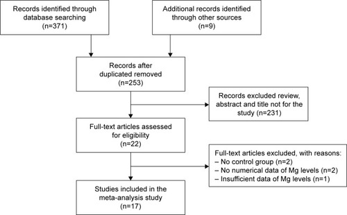
A total of 380 potential articles matched the inclusion after preliminary search from the Web of Science, PubMed, CNKI, and other relevant databases. After further screening, 17 final epidemiological studies were finally enrolled in current study (a total of 1,203 PD cases and 1,055 HC). The strategy of study search was provided in .
Figure 1 Meta-analysis flow diagram of the study search strategy.
The sample sizes of PD patients ranged from 19 to 250. The average age of PD cases ranged from 57.6 to 65.7 years old. The percentage of female patients ranged from 7.7% to 55.0%. Methods of Mg concentration analysis consisted of the atomic absorption spectrometry, inductively coupled plasma-atomic emission spectrometry, inductively coupled plasma-mass spectrometry (ICP-MS), inductively coupled plasma optical emission spectrometry (ICP-OES), and colorimetry. The geographic populations were European, American, Asian, and African, respectively. The detailed characteristics were summarized in .
Table 1 Characteristics of the included studies in this meta-analysis (arranged by publication time)
Meta-analysis of serum Mg levels between PD patients and HC subjects
The overall data from 12 articles compared the serum Mg levels between PD patients and HC subjects in a random-effects model because significant heterogeneity (I2=95.6%, P=0.000) was discovered across these studies. The pooled sample size encompassed a total of 1,632 subjects including 848 PD cases and 784 HC subjects. The results demonstrated that serum Mg levels in PD patients were significantly higher than those in HC subjects (SMD =1.09, 95% CI =0.52, 1.66; ). Subgroup analysis accounting for location disclosed that the Mg levels increased in PD patients in Asian populations (SMD =2.22, 95% CI =0.82, 3.63; P for Z=0.002) and African populations (SMD =1.18, 95% CI =0.80, 1.55; P for Z=0.000). In addition, the subgroup analysis stratified by analysis methods showed that a similar pattern in ICP-MS/ICP-AES (SMD =1.55, 95% CI =0.54, 2.57; P for Z=0.003). However, neither the location nor the analysis method contributed to the source of heterogeneity ().
Table 2 The subgroup analysis of studies reporting serum Mg levels in PD
Figure 2 Forest plot of serum Mg levels between PD patients and HC subjects.
Abbreviations: HC, healthy control; PD, Parkinson’s disease; SMD, standardized mean difference.
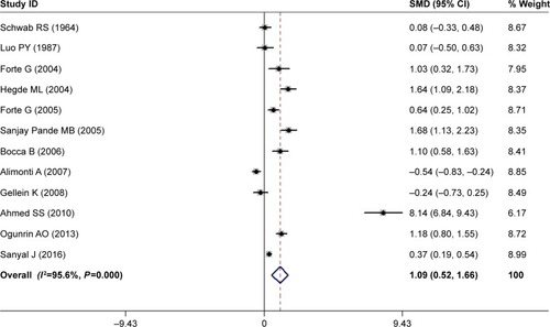
Meta-regression found that the duration, average age, and gender of PD patients had no influences on between-study heterogeneity (). Sensitivity analysis showed that Ahmed and Santosh’s study has an excessive influence on the outcome;Citation53 however, after the exclusion of this study, the statistical significance of the combined SMD was not unduly changed, indicating the robustness of the results. Cumulative meta-analysis revealed that no temporal effect affected the overall analysis results. Moreover, Egger’s test detected no publication bias in all included studies (P=0.054) and visual observation of the funnel plot also revealed symmetrical approximately.
Table 3 The meta-regression of studies reporting serum Mg levels in PD
Meta-analysis of peripheral blood Mg levels between PD patients and HC subjects
We further constructed a combined analysis of Mg serum and blood levels to evaluate Mg levels in peripheral blood. Fourteen articles were collected with a pooled sample size of 1,934 subjects consisting of 1,023 cases and 911 controls (). Meta-analysis results showed that the peripheral blood Mg levels of PD patients had a significant rising trend in random-effects model (SMD =0.64, 95% CI =0.10, 1.19; ). However, significant heterogeneity was found among these studies (I2=96.2%, P=0.000). In addition, results of subgroup analysis stratified by locations showed that compared with HC subjects, the pooled SMD was 1.78 (95% CI =0.70, 2.86; P for Z=0.001) for Asian populations, and 1.18 (95% CI =0.80, 1.55; P for Z=0.000) for African populations. In addition, in stratified analysis by HC subjects matched by analysis methods, the pooled SMD was 1.13 (95% CI =0.17, 2.09; P for Z=0.021). However, the problem of high heterogeneity was not solved by the location or analysis method ().
Figure 3 Forest plot of peripheral blood Mg levels between PD patients and HC subjects.
Abbreviations: HC, healthy control; PD, Parkinson’s disease; SMD, standardized mean difference.
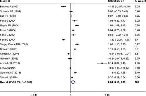
Table 4 The subgroup analysis of studies reporting peripheral blood Mg levels in PD
Furthermore, meta-regression analyses with the covariates of duration, average age, and gender of PD patients showed that no above-mentioned covariates conferred significant impact on heterogeneity (). Sensitivity analyses indicated that none of these studies significantly reverse the result. Cumulative meta-analysis showed no temporal effect affected on the overall results. Finally, no publication bias was observed in our study, according to the Egger’s tests (P=0.324).
Table 5 The meta-regression of studies reporting peripheral blood Mg levels in PD
Meta-analysis of CSF Mg levels between PD patients and HC subjects
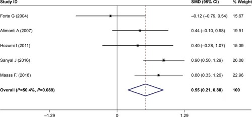
Currently, the research literature on CSF Mg levels in patients with PD is relatively less. Finally, only five articles were searched with less pooled population encompassing 324 subjects composed of 180 PD cases and 144 HC subjects (). Our investigation found that PD patients had higher Mg levels in CSF in comparison with HC subjects using a random-effects model (SMD =0.55, 95% CI =0.21, 0.88; ) with moderate heterogeneity among these studies (I2=50.4%, P=0.089). Limited by fewer number of studies, no further in-depth analysis was carried out.
Figure 4 Forest plot of CSF Mg levels between PD patients and HC subjects.
Abbreviations: CSF, cerebrospinal fluid; HC, healthy control; PD, Parkinson’s disease; SMD, standardized mean difference.
Discussion
To date, available literature on circulating Mg levels in PD etiology remains controversial. Thus, we aimed the previous literature comparing Mg concentrations in serum, peripheral blood, and CSF between PD patients and HC individuals to uncover their differences in PD. As we know, this is the first meta-analysis to quantify and report the association of circulating Mg levels with PD. Herein, we found that PD patients had a higher serum Mg levels than the HC subjects (SMD =1.09, 95% CI =0.52, 1.66). Furthermore, this result accorded with the combination of its serum and blood levels analysis (SMD =0.64, 95% CI =0.10, 1.19). The random-effects model was applied because of the existence of significant heterogeneity (I2.50%) among the studies. Next, we investigated the sources of heterogeneity by means of subgroup analyses and meta-regression analysis. However, above-mentioned covariates had no significant contribution to the between-study heterogeneity. The study of Ahmed and Santosh has shown a highly heterogeneous result.Citation53 However, excluding the study, the recalculated values of I2 in above two meta-analyses were attenuated to 91.4% and 94.4%, respectively, the significant heterogeneity still existed. This result indicated that the study of Ahmed and Santosh was not the main reason related to significant heterogeneity. A sensitivity analysis demonstrated the effect of leaving-out any individual study on the overall effect estimate each time was minimal, indicating that our findings were relatively certain and stable. The cumulative meta-analysis results were steady, suggesting the analytical instruments used for measuring serum Mg have become more advanced and precise in the later years. Our study suggested that the patients with PD had a tendency toward elevated circulating Mg levels, despite the high heterogeneity among studies.
Mg homeostasis is regulated mainly by Mg transporter proteins, which are located on the plasma membrane, such as TRPM6, 7 (intake) and SLC41A1 (efflux).Citation37,Citation38 Under normal conditions, extracellular Mg ions are actively transported by Mg transporter proteins into the cell and accumulated in mitochondria.Citation39 Thus, intracellular Mg levels are higher than those in extracellular fluid. However, many degenerative diseases including PD, often cause protein expression alterations or gene variant of these transporter proteins, thus finally lead to deranged Mg homeostasis.Citation40 For example, in substantia nigra area PD patients, the mRNA and protein expression levels of TRPM7 significantly decreased, suggesting that the Mg influx was reduced during the progress of PD.Citation41 Moreover, SLC41A1 is a coding region of the novel PD susceptibility locus PARK16. Its coding variant p.A350V that only occurred in the PD patients enhanced Mg efflux and then decreased intracellular Mg levels.Citation42,Citation43 Furthermore, previous clinical study also showed that due to mitochondrial cytopathies, intracellular Mg concentration in brain tissues of patients with neurologic disorders was lowered.Citation44 Although the reason is unknown, we hypothesized that this phenomenon might be related to Mg efflux from impaired mitochondria. Finally, the joint action of above-mentioned various mechanisms leads to the higher Mg levels in PD patients. Consequently, we further performed meta-analysis to investigate the CSF Mg levels in PD patients. The results showed that Mg levels significantly increased in PD patients compared with healthy population (SMD =0.55, 95% CI =0.21, 0.88). However, the limited number of studies (only five publications) and the small sample size (180 PD patients and 144 healthy individuals) conducted on CSF perhaps lead to an unreliable meta-analysis result. In CNS, Mg transporters across the blood–brain barrier transport Mg ion from blood into CSF, which create the phenomena that Mg levels in CSF are high like that in serum.Citation45 Finally, peripheral high concentration of Mg gets into the CNS, causing CSF Mg levels rise, which could partly explain the origin of higher Mg CSF levels. In general, CSF Mg levels keep relatively steadily state even if their serum levels has been changed, thus estimating CSF Mg levels may be more precise than its blood levels.Citation46 It was worth noting that high Mg CSF concentrations, in turn, accelerated the formation of α-synuclein oligomers, both from reaction rate and from formation size, that exacerbated dopamine neurons damage and contributed to the progression of PD.Citation47–Citation49 For the future, continuous investigations are necessary to assess the CSF Mg levels of PD patients and expound its impact on the progression of PD.
Nevertheless, this meta-analysis still included several limitations and shortcoming despite the strengths. First, we cited the most recent and comprehensive papers, but, the number of included studies was still limited, especially those for blood and CSF Mg levels. Future studies are necessary to confirm our conclusion with larger sample sizes. Second, we limited the search range to articles written in Chinese and English, and therefore several important papers published in other languages were not inevitably included. However, these cited articles probably met our inclusion criteria. Third, confounding factors, including the severity of PD, time of taking L-dopa, dietary or drug-induced Mg intake, socioeconomic status, and risky lifestyles (eg, alcohol, tobacco, and obesity) were difficult to collect from most of the included studies. Fourth, we only studied Mg levels in blood, serum, and CSF in the meta-analysis. However, most of total body Mg is intracellular, which makes examining Mg levels in erythrocytes of PD patients to comprehensively evaluate Mg possible implication in the pathology.
Conclusion
To our knowledge, this is the first comprehensive and systematic meta-analysis that evaluated the association between circulating Mg levels and PD patients to deeply understand the role of Mg homeostasis in PD. This meta-analysis emphasized that Mg serum, peripheral blood, and CSF levels in PD patients were obviously higher than those in healthy individuals. Due to the high heterogeneity, further investigations are needed to verify the results with large-scale cohort studies.
Author contributions
All authors contributed to data analysis, drafting or revising the article, gave final approval of the version to be published, and agree to be accountable for all aspects of the work.
Acknowledgments
This work was funded by the National High Technology Research and Development Program of China (2018ZX09711001-008-006), Scientific Research Fund of Liaoning Provincial Education Department (Grant Number: LK201608), National Natural Science Foundation of China (Grant Number: 81501098, 81603112, and 81803751), Program for Liaoning Innovation Research Team in University (Grant Number: LT2014016), Key Laboratory Foundation from Shenyang S&T Projects (Grant Number: F16-094-1-00), Double Hundred Program for Shenyang Scientific and Technological Innovation Projects (Grant Number: 100040), Liaoning Province Scientific Research Foundation (Grant Number: 2014226033), and National Science and Technology Major Projects for Significant New Drugs Development (Grant Number: 2017ZX09101003-008-006).
Disclosure
The authors report no conflicts of interest in this work.
References
- LeesAJHardyJReveszTParkinson’s diseaseLancet200937396802055206619524782
- de LauLMGiesbergenPCde RijkMCHofmanAKoudstaalPJBretelerMMIncidence of parkinsonism and Parkinson disease in a general population: the Rotterdam StudyNeurology20046371240124415477545
- SveinbjornsdottirSThe clinical symptoms of Parkinson’s diseaseJ Neurochem2016139Suppl 131832427401947
- PowersKMSmith-WellerTFranklinGMLongstrethWTSwansonPDCheckowayHParkinson’s disease risks associated with dietary iron, manganese, and other nutrient intakesNeurology200360111761176612796527
- BettiolSSRoseTCHughesCJSmithLAAlcohol consumption and Parkinson’s disease risk: a review of recent findingsJ Parkinsons Dis20155342544226406123
- OhCKSultanAPlatzerJS-Nitrosylation of PINK1 attenuates PINK1/Parkin-dependent mitophagy in hiPSC-based Parkinson’s disease modelsCell Rep20172182171218229166608
- GuoYBChenJZhangXDXuSBLiuHYMolecular dynamics simulations to understand LRKK2 mutations in ParkinsonMol Simul20164216470
- PapagiannakisNKorosCStamelouMAlpha-synuclein dimerization in erythrocytes of patients with genetic and non-genetic forms of Parkinson’s DiseaseNeurosci Lett20186721314514929129675
- SinglaNDhawanDKInfluence of zinc on calcium-dependent signal transduction pathways during aluminium-induced neurodegenerationMol Neurobiol201450261362524500000
- WenHQinYZhongWLiCLiuXShenYTrivalent metal ions based on inorganic compounds with in vitro inhibitory activity of matrix metalloproteinase 13Enzyme Microb Technol20169291727542739
- AizenmanEMastroberardinoPGMetals and neurodegenerationNeurobiol Dis2015811326542885
- LangleyMRGhaisasSAyMManganese exposure exacerbates progressive motor deficits and neurodegeneration in the MitoPark mouse model of Parkinson’s disease: relevance to gene and environment interactions in metal neurotoxicityNeurotoxicology20186424025528595911
- MedeirosMSSchumacher-SchuhACardosoAMIron and oxidative stress in Parkinson’s disease: an observational study of injury biomarkersPLoS One2016111e014612926751079
- WangXMouallaDWrightJABrownDRCopper binding regulates intracellular alpha-synuclein localisation, aggregation and toxicityJ Neurochem2010113370471420141569
- LiYSunLCaiTAlpha-Synuclein overexpression during manganese-induced apoptosis in SH-SY5Y neuroblastoma cellsBrain Res Bull2010814–542843319932157
- GrubbsRDMaguireMEMagnesium as a regulatory cation: criteria and evaluationMagnesium1987631131273306178
- LairesMJMonteiroCPBichoMRole of cellular magnesium in health and human diseaseFront Biosci2004926227614766364
- SwaminathanRMagnesium metabolism and its disordersClin Biochem Rev2003242476618568054
- ShindoYYamanakaRSuzukiKHottaKOkaKIntracellular magnesium level determines cell viability in the MPP(+) model of Parkinson’s diseaseBiochim Biophys Acta20151853123182319126319097
- OyanagiKKawakamiEKikuchi-HorieKMagnesium deficiency over generations in rats with special references to the pathogenesis of the Parkinsonism-dementia complex and amyotrophic lateral sclerosis of GuamNeuropathology200626211512816708544
- BarbeauAJasminGDuchastelYBiochemistry of Parkinson’s diseaseNeurology196313565813966496
- GelleinKSyversenTSteinnesETrace elements in serum from patients with Parkinson’s disease – a prospective case-control study: the Nord-Trøndelag Health Study (HUNT)Brain Res2008121911111518538747
- AlimontiARistoriGGiubileiFSerum chemical elements and oxidative status in Alzheimer’s disease, Parkinson disease and multiple sclerosisNeurotoxicology200728345045617267042
- ForteGBoccaBSenofonteOTrace and major elements in whole blood, serum, cerebrospinal fluid and urine of patients with Parkinson’s diseaseJ Neural Transm200411181031104015254791
- ForteGAlimontiAPinoAMetals and oxidative stress in patients with Parkinson’s diseaseAnn Ist Super Sanita200541218919516244392
- BoccaBAlimontiASenofonteOMetal changes in CSF and peripheral compartments of parkinsonian patientsJ Neurol Sci20062481–2233016765382
- MaassFMichalkeBLehaAElemental fingerprint as a cerebrospinal fluid biomarker for the diagnosis of Parkinson’s diseaseJ Neurochem2018145434235129388213
- SchwabRSPoryaliAAmesANormal serum magnesium levels in Parkinson’s diseaseNeurology19641485585614215602
- SanyalJAhmedSSNgHKMetallomic biomarkers in cerebrospinal fluid and serum in patients with Parkinson’s disease in Indian populationSci Rep201663509727752066
- MoherDLiberatiATetzlaffJAltmanDGPRISMA GroupPreferred reporting items for systematic reviews and meta-analyses: the PRISMA statementInt J Surg20108533634120171303
- GreenSMcDonaldSCochrane Collaboration: more than systematic reviews?Intern Med J20053513515667459
- HigginsJPThompsonSGDeeksJJAltmanDGMeasuring inconsistency in meta-analysesBMJ2003327741455756012958120
- SongFGilbodySBias in meta-analysis detected by a simple, graphical test. Increase in studies of publication bias coincided with increasing use of meta-analysisBMJ19983167129471
- HigginsJPThompsonSGControlling the risk of spurious findings from meta-regressionStat Med200423111663168215160401
- TobiasAAssessing the in fluence of a single study in the meta-analysis estimateStata Tech Bull19998471517
- DuvalSTweedieRTrim and fill: a simple funnel-plot-based method of testing and adjusting for publication bias in meta-analysisBiometrics200056245546310877304
- SunYSukumaranPSchaarASinghBBTRPM7 and its role in neurodegenerative diseasesChannels20159525326126218331
- KolisekMNestlerAVormannJSchweigel-RöntgenMHuman gene SLC41A1 encodes for the Na+/Mg2+ exchangerAm J Physiol Cell Physiol20123021C318C32622031603
- KolisekMZsurkaGSamajJWeghuberJSchweyenRJSchweigelMMrs2p is an essential component of the major electrophoreticMg2+ influx system in mitochondriaEMBO J20032261235124412628916
- KolisekMMontezanoACSponderGPARK7/DJ-1 dysregulation by oxidative stress leads to magnesium deficiency: implications in degenerative and chronic diseasesClin Sci2015129121143115026453619
- CookNLvan den HeuvelCVinkRCharacterisation of TRPM channel mRNA levels in Parkinson diseaseMagnes Res2009223188189
- TucciANallsMAHouldenHGenetic variability at the PARK16 locusEur J Hum Genet201018121356135920683486
- KolisekMSponderGMastrototaroLSubstitution p.A350V in Na+/Mg2+ exchanger SLC41A1, potentially associated with Parkinson’s disease, is a gain-of-function mutationPLoS One201388e7109623976986
- BarbiroliBIottiSCortelliPLow brain intracellular free magnesium in mitochondrial cytopathiesJ Cereb Blood Flow Metab199919552853210326720
- JimersonDCPostRMCarmanJSCSF calcium: clinical correlates in affective illness and schizophreniaBiol Psychiatry19791413751420907
- LevineJSteinDRapoportAKurtzmanLHigh serum and cerebrospinal fluid Ca/Mg ratio in recently hospitalized acutely depressed patientsNeuropsychobiology1999392637010072661
- HoyerWAntonyTChernyDHeimGJovinTMSubramaniamVDependence of alpha-synuclein aggregate morphology on solution conditionsJ Mol Biol2002322238339312217698
- LoweRPountneyDLJensenPHGaiWPVoelckerNHCalcium(II) selectively induces alpha-synuclein annular oligomers via interaction with the C-terminal domainProtein Sci200413123245325215537754
- ZhangQSHengYYuanYHChenNHPathological α-synuclein exacerbates the progression of Parkinson’s disease through microglial activationToxicol Lett2017265303727865851
- LuoPYZhuXZhangYPDetermination of serum trace elements levels and CT scans of brain in Parkinson’s diseaseActa Univ Med Second Shanghai198774332334
- HegdeMLShanmugaveluPVengammaBSerum trace element levels and the complexity of inter-element relations in patients with Parkinson’s diseaseJ Trace Elem Med Biol200418216317115646263
- Sanjay PandeMBNagabhushanPHegdeMLRaoTSRaoKSAn algorithmic approach to understand trace elemental homeostasis in serum samples of Parkinson diseaseComput Biol Med200535647549315780860
- AhmedSSSantoshWMetallomic profiling and linkage map analysis of early Parkinson’s disease: a new insight to aluminum marker for the possible diagnosisPLoS One201056e1125220582167
- OgunrinAOKomolafeMASanyaEOTrace metals in patients with Parkinson’s disease: a multi-center case-control study of Nigerian patientsJ Neurol Epidemiol2013113138
- FengJYangXLMaQPWangYLYaoYNThe case-control study on relationship between blood metal elements and Parkinson disease in Uygur of Hetian region in XinjiangJ Xinjiang Med Univ2012353342345
- AlimontiABoccaBPinoARuggieriFForteGSancesarioGElemental profile of cerebrospinal fluid in patients with Parkinson’s diseaseJ Trace Elem Med Biol200721423424117980814
- HozumiIHasegawaTHondaAPatterns of levels of biological metals in CSF differ among neurodegenerative diseasesJ Neurol Sci20113031–2959921292280