Abstract
Background
The purpose of this study was to describe and evaluate a surgical approach, known as internal limiting membrane (ILM) peeling, as an adjunct to repair of recurrent retinal detachment due to proliferative vitreoretinopathy (PVR).
Methods
This was a retrospective case series. All eyes underwent repair of recurrent PVR-related rhegmatogenous retinal detachment incorporating macular indocyanine green-assisted ILM peeling. Patients with primary detachments, diabetes, staphyloma, or macular holes were excluded. The main outcome measure was the anatomic success of single surgery. The characteristics of the group were studied, including the number and types of previous detachment repair attempts, as well as the subsequent surgeries.
Results
Fourteen eyes from 14 patients were included. Anatomic success with single surgery was achieved in 11 of 14 (79%) of the operated eyes using this technique, and eventual success was achieved in all eyes (100%). Among the failed repairs prior to ILM peeling, 8/14 eyes had scleral buckles, 7/14 had silicone oil tamponade, and two had inferior retinectomies. There was no subsequent development of epiretinal membranes after ILM peeling.
Conclusion
ILM peeling in conjunction with vitrectomy and peeling of peripheral membranes is an effective technique with a high anatomic success rate in the challenging scenario of PVR-related recurrent detachments. We describe the technique as an alternative to the traditional retinectomy.
Introduction
Proliferative vitreoretinopathy (PVR) is the leading cause of failure of retinal detachment surgery.Citation1–Citation3 Its recurrent nature can lead to redetachment, multiple surgeries, and even total loss of vision and phthsis.Citation1 Many approaches are employed as a means of reducing retinal traction caused by preretinal and subretinal proliferative membranes. The approaches range from membrane peeling to removal of tense retinal tissue.Citation1,Citation4 In extreme situations, a retinectomy or circumferential excision of the retina is performed to allow reapposition of the neurosensory retina.Citation5–Citation11 Recent studies report single surgery success rates ranging from 60% to 87% with traditional retinectomy and relaxing retinotomy in PVR cases.Citation12–Citation14 Because of the sometimes problematic after effects associated with retinectomy, such as the frequent necessity for long-term silicone oil tamponade, recurrent PVR, hypotony, or corneal decompensation, this surgical strategy should be viewed as an option of last resort.Citation5–Citation11,Citation15
Because of its rigidity, the internal limiting membrane (ILM) is considered a significant source of retinal stiffness, especially in posterior pole disease.Citation16,Citation17 A minimally destructive form of retinal dissection, ILM peeling, is commonly and successfully employed for release of retinal tension in a variety of posterior pole diseases. Intraoperative tissue staining is widely used for optimum visualization in ILM peeling when complete and thorough posterior pole ILM removal is thought to be critical to the surgery’s success.Citation18–Citation20 Indocyanine green-assisted ILM peel has been described in many ocular disease states, including macular hole repair, repair of staphylomatous posterior pole retinal detachments, macular hole-related retinal detachments, myopic traction maculopathy, and epiretinal membrane prevention in retinal detachments.Citation21–Citation28 Further, it is now frequently employed in diabetic macular edema which is recalcitrant to focal laser photocoagulation and intravitreal antivascular endothelial growth factor or anti-inflammatory therapy, and is thought to have a mechanical effect.Citation29–Citation32 A recent study focusing on optical coherence tomography findings in patients who underwent ILM peeling in conjunction with PVR-retinal detachment repair demonstrates that the technique is now also being adapted for difficult retinal detachment cases.Citation33
ILM peeling of the posterior pole is a non-standard approach to PVR detachments, and is not represented well in the literature. The rationale for its use is based on other well studied surgical applications in posterior pole diseases, such as macular pucker and macular hole. Theoretically, ILM peeling reduces retinal tension transmitted to the posterior pole. Removal of this tensile layer offers the mechanical advantage of relaxing the surface tractional forces (). It increases retinal compliance by the creation of a central “soft spot”, which allows adjacent areas to relax better. ILM removal can also create a plane by which to undermine and dissect PVR,Citation34 especially if it is posteriorly located. Additionally, it reduces the likelihood of recurrence of posterior epiretinal membrane/PVR formationCitation28 and subsequent redetachments, while maximizing macular visual function.
Figure 1 A schematic showing the theoretical mechanism of action. (A) posterior pole after staining, and (B) after ILM (internal limiting membrane)-rhexis, with vectors (arrows) of retinal relaxation after removal of ILM layer.

The primary objective of this study was to describe and evaluate a surgical approach using indocyanine green-assisted ILM peeling as an adjunct in vitrectomy for repair of recurrent retinal detachment due to PVR. The primary outcome measure was the anatomic success with single surgery. The utility of this technique as a means to relieve retinal tension is examined in terms of the surgical success of detachment repair. The authors hypothesize that posterior pole ILM removal in some cases helps counteract the pathologic tractional sequelae of PVR, and improves the chance of stable long-term retinal reattachment.
Materials and methods
This is a retrospective case series examining 14 consecutive cases in which indocyanine green-assisted ILM peeling was used as an adjunct in pars plana vitrectomy for recurrent PVR-related rhegmatogenous retinal detachment. All cases were performed by a single surgeon (MAV) from April 2001 to November 2007. The main outcome measure was the anatomic success rate using single surgery. This was defined as stable total retinal reattachment for a minimum follow-up of 6 months after surgery. The characteristics of the group were studied, including the visual outcomes, the number of previous retinal detachment repair attempts, the use of scleral buckles, silicone oil, and traditional extensive peripheral retinectomies. The numbers and types of surgeries after the ILM peel, such as subsequent retinal detachments repair attempts, if applicable, were also studied. There was a minimum follow-up length of 6 months, and a mean follow-up of 37 (range 6–101) months.
Inclusion/exclusion criteria
Only recurrent rhegmatogenous retinal detachments which were PVR-related were considered for the review. Primary retinal detachments, eyes with proliferative or tractional diabetic disease, staphylomatous detachments, and detachments due to macular holes were excluded from the series. While not an inclusion criterion, all of the patients had macula-off detachments. Fourteen eyes of 14 patients were identified that met the inclusion criteria. The group had an average number of prior surgical repair attempts of 2.5 (range 1–5) before the vitrectomy featuring indocyanine green-assisted ILM peeling. The overall surgical chronology was complex, indicating a problematic disease course in many of the patients (). Most of the cases (11/14) had advanced (grade C) PVR.Citation35 Also, the large majority (12/14) of the cases had at least some PVR located posterior to the equator. The classification of the PVR and the general location of the retinal detachments are given in .
Table 1 Chronology of surgical procedures for each of the 14 cases
Table 2 Features of retinal detachment at time of surgery with ILM-peeling
Surgical technique
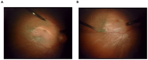
Either 25-gauge (10/14) or 20-gauge (4/14) instrumentation was used. While all eyes had undergone previous vitrectomy, close vitreous base shaving with external scleral depression was again performed. Peeling and removal of any visible PVR membranes and bands was also performed in all cases prior to ILM removal. Indocyanine green staining of the posterior pole was accomplished with several brief directed pulses of dye around the macula with a soft tip catheter under balanced salt solution with avoidance of the fovea. The staining was followed by prompt and complete removal of the dye with the cutter on aspiration mode. After effective staining, ILM dissection was initiated in the posterior pole under a wide-angle viewing system visualization with end-grasping forceps, and carried out as far as possible, to the arcades or beyond, if necessary (). ILM peeling over the mobile detached retina offers uncertain countertraction compared with dissection in the attached retina, so good staining greatly facilitates the grasping and removal. In cases of posterior PVR, ILM removal was extended to the areas of PVR formation as a means to undermine the pathologic membranes. After subretinal fluid drainage was performed, tamponade was accomplished with either SF6, C3F8, or silicone oil (). Scleral buckling was not performed concurrently with this technique.
Figure 2 ILM peeling over detached macula using asymmetric 25-gauge end-grasping forceps. View the surgical video using this link: http://youtu.be/7KWk2Jyngrs.
Results
In our series using this ILM peeling technique, anatomic success using single surgery was achieved in 11 of 14 eyes (79%). Eventual success, if not achieved directly after the vitrectomy with ILM peel, was achieved in all eyes (100%). The group had an average number of prior surgical repair attempts of 2.5 (range 1–5) before indocyanine green-assisted ILM peeling. The group had an average number of subsequent reoperations for detachment repair of 0.7 (range 0–6). Among the failed detachment repairs prior to ILM peeling, all had at least one prior PPV, 8/14 eyes had prior scleral buckling procedures, 7/14 had prior silicone oil tamponade, and 2/14 had prior inferior retinectomies (). The mean final best-corrected visual acuity was logMAR 0.9206, or 20/167 (range 20/30 to hand motion). There was no subsequent development of macular epiretinal membranes in any of the cases after ILM peeling within the duration of each patient follow-up.
Table 3 Surgical features and clinical outcomes of study group
Discussion
Using indocyanine green-assisted ILM peeling in conjunction with vitrectomy and peeling of peripheral membranes is an effective technique with a high anatomic success rate (79%) in the challenging scenario of PVR-related recurrent retinal detachments. We describe the technique as a surgeon’s tool in PVR-related redetachments, and offer it as a tissue-sparing alternative to the more extensive traditional retinectomy in select cases. The anatomic success of single surgery using this technique was comparable with other studies in which relaxing retinotomies and traditional retinectomies were featured. In our data set, there are two examples of achieving anatomic success with ILM removal, even after traditional retinectomies done by different surgeons had failed.
The authors acknowledge the limitations of a retrospective case series, in which certain cases with severe PVR may have been appropriately selected for more aggressive tissue removal, and therefore were not included in the study group. The process of selecting which cases are most appropriate for ILM peel is entirely based on the surgeon’s experience and individual assessment of retinal compliance, and it is therefore difficult to compare this directly with other more aggressive surgical techniques. While a control group for the study would have been desirable, it is difficult to assign control cases in a retrospective case series without selection bias, especially given the diverse spectrum of PVR severity, and the uniqueness of the individual cases. Therefore, comparison of surgical success with other modern PVR studies featuring traditional methods is probably the best reference.Citation12–Citation14 There is still clearly a role for traditional retinectomy in the most advanced cases, which the authors favor doing under silicone oil. Nevertheless, we believe the “virtual” retinectomy is an excellent conservative compromise in salvageable PVR cases, offering improved retinal compliance, prevention of epiretinal membrane and macular pucker formation, and subsequent posterior surface PVR. As such, we propose that it can be considered as an additional measure to minimize the chance of recurrent detachment.
The applicability of ILM peeling in detachment surgery may not be limited to PVR. Retinal detachment caused by other pathologies, such as diabetic tractional disease, staphyloma, or macular holes, were not included in this study. However, it is worth noting that the same technique has been employed in our clinical practice for these different varieties of rhegmatogenous retinal detachment with good effect.
The category of patients examined in this review represents one of the most challenging subsets of retinal pathology with which vitreoretinal surgeons are faced. This particular group of patients had somewhat limited visual outcomes, in general, logMAR 0.9206, or 20/167, on average, ranging from 20/30 to hand motion. However, if the cases with severe vision loss were excluded in this small retrospective study, the visual outcomes would be skewed towards a much more satisfactory level. The visual acuity outcomes are influenced by multiple factors, such as numerous macular redetachments, corneal decompensation, other ocular disease, and post-surgical sequelae (). Despite the somewhat guarded visual prognosis in eyes with multiple detachments due to PVR, maintaining anatomic success long term is a tenable goal. Beyond the restoration and preservation of vision, postoperative stability and absence of phthisis is a major quality of life issue with patients. For these reasons, we describe our results using this alternative technique which frees the tangential traction from the posterior pole in the management or recurrent PVR rhegmatogenous retinal detachments.
Disclosures
This work was presented at the American Society of Retina Specialists 28th annual meeting, August 28–September 1, 2010, Vancouver, Canada. The authors report no conflicts of interest in this work.
References
- MachemerRMassive periretinal proliferation: a logical approach to therapyTrans Am Ophthalmol Soc197775556586418548
- LewisHAabergTMAbramsGWCauses of failure after initial vitreoretinal surgery for severe proliferative vitreoretinopathyAm J Ophthalmol199111118141985496
- LewisHAabergTMCauses of failure after repeat vitreoretinal surgery for recurrent proliferative vitreoretinopathyAm J Ophthalmol1991111115191985484
- CharlesSVitrectomy for retinal detachmentTrans Ophthalmol Soc U K198010045425496947605
- FedermanJLEagleRCJrExtensive peripheral retinectomy combined with posterior 360 degrees retinotomy for retinal reattachment in advanced proliferative vitreoretinopathy casesOphthalmology19909710130513202243681
- ShalabyKARelaxing retinotomies and retinectomies in the management of retinal detachment with severe proliferative vitreoretinopathy (PVR)Clin Ophthalmol201041107111420957056
- HanDPLewisMTKuhnEMRelaxing retinotomies and retinectomies. Surgical results and predictors of visual outcomeArch Ophthalmol199010856946972334327
- MorseLSMcCuenBW2ndMachemerRRelaxing retinotomies. Analysis of anatomic and visual resultsOphthalmology19909756426472342810
- BoveyEHDe AncosEGonversMRetinotomies of 180 degrees or moreRetina19951553943988594631
- MetgeFMassinPGaudricARetinectomies in the treatment of retinal detachments with vitreoretinal proliferationJ Fr Ophtalmol1997205345349 French9238471
- IversonDAWardTGBlumenkranzMSIndications and results of relaxing retinotomyOphthalmology19909710129813042243680
- TsengJJBarileGRSchiffWMInfluence of relaxing retinotomy on surgical outcomes in proliferative vitreoretinopathyAm J Ophthalmol2005140462863616226515
- QuiramPAGonzalesCRHuWOutcomes of vitrectomy with inferior retinectomy in patients with recurrent rhegmatogenous retinal detachments and proliferative vitreoretinopathyOphthalmology2006113112041204716952397
- TanHSMuraMObersteinSYde SmetMDPrimary retinectomy in proliferative vitreo-retinopathyAm J Ophthalmol2010149344745220172071
- BlumenkranzMSAzenSPAabergTRelaxing retinotomy with silicone oil or long-acting gas in eyes with severe proliferative vitreoretinopathy. Silicone Study Report 5. The Silicone Study GroupAm J Ophthalmol199311655575648238214
- KuhnFInternal limiting membrane removal for macular detachment in highly myopic eyesAm J Ophthalmol2003135454754912654379
- SayanagiKIkunoYTanoYTractional internal limiting membrane detachment in highly myopic eyesAm J Ophthalmol2006142585085217056366
- KadonosonoKItohNUchioENakamuraSOhnoSStaining of internal limiting membrane in macular hole surgeryArch Ophthalmol200011881116111810922208
- KwokAKYeungYSLeeVYWongTHIndocyanine green assisted peeling of the retinal ILMOphthalmology20021096104012045036
- BurkSEDa MataAPSnyderMERosaRHJrFosterREIndocyanine green-assisted peeling of the retinal internal limiting membraneOphthalmology2000107112010201411054324
- Da MataAPBurkSERiemannCDIndocyanine green-assisted peeling of the retinal internal limiting membrane during vitrectomy surgery for macular hole repairOphthalmology200110871187119211425673
- KwokAKLaiTYMan-ChanWWooDCIndocyanine green assisted retinal internal limiting membrane removal in stage 3 or 4 macular hole surgeryBr J Ophthalmol2003871717412488266
- Da MataAPBurkSEFosterRELong-term follow-up of indocyanine green-assisted peeling of the retinal internal limiting membrane during vitrectomy surgery for idiopathic macular hole repairOphthalmology2004111122246225315582081
- OieYEmiKTakaokaGIkedaTEffect of indocyanine green staining in peeling of internal limiting membrane for retinal detachment resulting from macular hole in myopic eyesOphthalmology2007114230330617194478
- UemotoRYamamotoSTsukaharaITakeuchiSEfficacy of internal limiting membrane removal for retinal detachments resulting from a myopic macular holeRetina200424456056615300077
- FutagamiSInoueMHirakataARemoval of internal limiting membrane for recurrent myopic traction maculopathyClin Experiment Ophthalmol200836878278519128386
- KwokAKLaiTYInternal limiting membrane removal in macular hole surgery for severely myopic eyes: a case-control studyOphthalmology2003877885889
- ArasCAriciCAkarSPeeling of internal limiting membrane during vitrectomy for complicated retinal detachment prevents epimacular membrane formationGraefes Arch Clin Exp Ophthalmol2009247561962319107502
- HartleyKLSmiddyWEFlynnHWJrMurrayTGPars plana vitrectomy with internal limiting membrane peeling for diabetic macular edemaRetina200828341041918327132
- YanyaliAHorozogluFCelikENohutcuAFLong-term outcomes of pars plana vitrectomy with internal limiting membrane removal in diabetic macular edemaRetina200727555756617558316
- GandorferAMessmerEMUlbigMWKampikAResolution of diabetic macular edema after surgical removal of the posterior hyaloid and the inner limiting membraneRetina200020212613310783944
- HallerJAQinHApteRSVitrectomy outcomes in eyes with diabetic macular edema and vitreomacular traction. Diabetic Retinopathy Clinical Research Network Writing CommitteeOphthalmology2010117610871093e320299105
- OdrobinaDCMichalewskaZMichalewskiJNawrockiJHigh-speed, high-resolution spectral optical coherence tomography in patients after vitrectomy with internal limiting membrane peeling for proliferative vitreoretinopathy retinal detachmentRetina201030688188620182404
- SakamotoHYamanakaIKubotaTIshibashiTIndocyanine green-assisted peeling of the epiretinal membrane in proliferative vitreoretinopathyGraefes Arch Clin Exp Ophthalmol2003241320420712644944
- MachemerRAabergTMFreemanHMIrvineARLeanJSMichelsRMAn updated classification of retinal detachment with proliferative vitreoretinopathyAm J Ophthalmol199111221591651867299