Abstract
Background
The purpose of this study was to compare the outcomes of trabeculectomy using single sutures or releasable sutures.
Methods
This retrospective study analyzed the medical records of 61 patients who had undergone trabeculectomy using single sutures (n = 33, 54.1%) or releasable sutures (n = 28, 45.9%). The scleral flap was secured with a mean 3.9 (range 3–5) single sutures in 33 patients and with three releasable sutures in 28 patients. Primary outcomes were the success rate, based on intraocular pressure and medication usage, and the frequency of complications and post-surgical interventions. The criteria used to determine complete success were, first, intraocular pressure < 18 mmHg and, second, ≤21 mmHg and ≥20% intraocular pressure reduction without glaucoma medication.
Results
All patients had an intraocular pressure ≤ 21 mmHg; 87.5% in the single suture group and 92.6% in the releasable suture group had an intraocular pressure < 18 mmHg at 24 months. There was a highly significant reduction in intraocular pressure to baseline values in both groups at the last visit. Applying the first criterion, complete success was achieved in 57.6% of patients with single sutures and 71.4% with releasable sutures, and based on the second criterion, 66.7% and 71.4%, respectively. No significant difference was found between the groups with regard to intraocular pressure, or success or complication rates.
Conclusion
The results of trabeculectomy using single sutures or releasable sutures are equivalent. Therefore, the choice of suture technique should be based on individual patient requirements and surgeon experience.
Introduction
At present, trabeculectomy is the most commonly performed and effective surgical method for medically uncontrolled glaucoma.Citation1 However, suture adjustment and bleb interventions are frequently necessary after trabeculectomy.Citation1–Citation5 In particular, the use of mitomycin C requires tight suturing of the scleral flap to prevent postoperative hypotony.Citation5 Therefore, several modifications of trabeculectomy have been described, suggesting variation in shapes and sizes of the scleral flap, use of antimetabolites, and use of laserable, releasable, or adjustable sutures.Citation6–Citation11 The aim of this study was to compare the reduction in intraocular pressure achieved using either laser lysis of standard single sutures or removal of releasable sutures after trabeculectomy, with consideration of their advantages and disadvantages. In addition, we assessed the incidence of complications and further surgical interventions for outflow adjustment at the scleral flap site.
Materials and methods
Patients
We retrospectively analyzed the medical records of 61 consecutive patients who underwent trabeculectomy with mitomycin C using either standard single sutures or releasable sutures between January 2008 and January 2009 at the University Eye Hospital, Wuerzburg, Germany. Baseline information was reviewed for each patient, including age, gender, localization of eye undergoing surgery, type of glaucoma, and ocular medication. Preoperatively, all patients underwent a standard ophthalmic examination to obtain best corrected visual acuity, which was converted to the logarithm of the minimum angle of resolution (logMAR), intraocular pressure using Goldmann applanation tonometry, angle grading by gonioscopy, slit-lamp biomicroscopy for anterior segment examination, and indirect ophthalmoscopy for assessment of the optic nerve head and peripheral retina. Inclusion criteria were medically uncontrolled primary or secondary open-angle or angle-closure glaucoma treated with trabeculectomy and mitomycin C with single sutures or releasable sutures between January 2008 and January 2009.
Exclusion criteria included congenital glaucoma or secondary glaucoma, such as neovascular glaucoma or glaucoma associated with iridocorneal endothelial syndrome, second trabeculectomy, and more than two cyclodestructive procedures or laser trabeculoplasties prior to trabeculectomy, and use of both single and releasable sutures to secure the scleral flap.
Surgical technique
Details of the surgical procedure used have been described previously.Citation9–Citation12 Trabeculectomy with laserable sutures was performed by four experienced glaucoma surgeons and with releasable sutures by a single glaucoma surgeon. A corneal traction suture was placed at 6 o’clock, and a fornix-based conjunctival flap was created.Citation7 Episcleral blood vessels were then cauterized. Four sponges (2 × 8 mm) soaked in different concentrations of mitomycin C depending on the individual risk of scarring were placed under the conjunctival flap for 3 minutes. The majority of patients in each group received mitomycin C 0.02%, ie, 27 patients (81.8%) in the single suture group and 22 patients (78.6%) in the adjustable suture group. Mitomycin C 0.01% was used in four eyes (14.3%) with releasable sutures and in three eyes (9.1%) with single sutures. Sponges soaked in mitomycin C 0.05% were placed in two eyes (7.1%) in the releasable suture group and in three eyes (9.1%) in the single suture group. There was no statistically significant difference in the amount of mitomycin C used between the groups (P = 0.531). The area of application was then washed with 30 mL of balanced saline solution. Thereafter, a rectangular scleral flap measuring 4 × 3 mm was dissected and a trabeculectomy of 0.8 × 2 mm was performed followed by an iridectomy. The scleral flap was fixed with single sutures or releasable sutures. Single sutures were placed at both corners of the scleral flap and towards the center of one or two sides. Additional sutures were placed if needed. The mean number of single sutures used was 3.9 (range 3–5) in 33 eyes from 61 patients (54.1%). The scleral flap was secured with three releasable sutures to allow suture removal in 28 patients (45.9%), as shown in the video http://dvpr.es/JAPzcw. Finally, the conjunctiva was closed with a 10.0 nylon running mattress suture.Citation11
Postoperative management
All patients received topical prednisolone acetate every 1–2 hours for a week tapering over 6–8 weeks, antibiotics, such as gentamicin three times daily for one week or as needed, and a cycloplegic agent, such as atropine twice per day for 1–2 weeks. All antiglaucomatous medication was discontinued after surgery. Patients were followed up at day 1, weeks 1 and 4, months 3, 6, and 12, and at the final follow-up visit (at a mean 24.9 ± 5.5 months for the single suture group and 23.9 ± 5.3 months for the releasable suture group), with additional visits whenever necessary, for documentation of intraocular pressure, number of intraocular pressure-lowering drugs, best corrected visual acuity, results of anterior and posterior segment examination, frequency of complications and postsurgical interventions, and filtering bleb score, ie, the Wuerzburg Bleb Classification Score (WBCS).Citation13 The WBCS is an objective grading system for assessment of the appearance of a filtering bleb and is used to decide whether to treat an imminent bleb scarring earlier. It contains several parameters, ie, vascularization, corkscrew vessels, encapsulation, and microcysts, each scored 0 to 3. The total bleb score is calculated as the sum of each parameter. Patients with a higher bleb score have better clinical morphology of the filtering bleb and a lower postoperative intraocular pressure. Citation13 Laser suture lysis of single suturesCitation10–Citation12,Citation14 or removal of releasable suturesCitation10,Citation19–Citation21 was undertaken in patients with increased intraocular pressure and flat filtering blebs which inflated after ocular massage, and in patients who did not reach their target intraocular pressure. Ocular massage immediately posterior to the scleral flap was performed to produce aqueous flow through the fistula under the conjunctiva.
Further intensive postoperative care to control wound healing included increased application of topical steroids, early bleb injections of 5-fluorouracil at the beginning of bleb scarring, and needling for encapsulated blebs, as proposed by Marquardt et al in 2004.Citation16 Subconjunctival injections of 5-fluorouracil were repeatedly applied according to the WBCS.Citation13
Two criteria for complete success were defined according to the World Glaucoma Association guidelines:
intraocular pressure ≤ 21 mmHg and an intraocular pressure reduction ≥ 20% versus baseline values without antiglaucomatous medication or
intraocular pressure < 18 mmHg without glaucoma medication.
Qualified success was determined, fulfilling the above-mentioned criteria with or without intraocular pressure-lowering medication.
Statistical analysis
SPSS© version 19.0 for Windows (SPSS Inc, Chicago, IL) was used for statistical analyses and to create the accompanying figures. Analyses of variance for repeated measurements were used to examine intraocular pressure. The Student’s t-test was used for continuous variables with normal distribution. Otherwise, differences in single discrete variables between groups were tested for significance using the Mann-Whitney U-test. Categorical variables were evaluated using the χ2 (Fisher’s Exact) test. A maximum two-tailed P value of <0.05 was considered to indicate statistical significance.
Results
The approach of using single sutures for scleral flap tying was performed in 33 eyes (54.1%). Releasable sutures were placed in 28 eyes (45.9%). Both groups were comparable with regard to sample size, gender, age, preoperative intraocular pressure, number of preoperative medications, and type of glaucoma (). The last follow-up visit occurred on average at 24.9 ± 5.5 months and at 23.9 ± 5.3 months for patients with single sutures and releasable sutures, respectively. Baseline data and surgical outcomes at 12 months and at the last follow-up visit are summarized in .
Table 1 Group characteristics
Table 2 Data preoperatively and at 12 and 24 months
Intraocular pressure
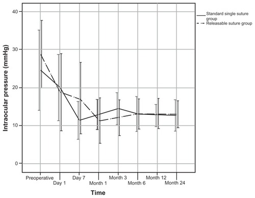
shows changes in intraocular pressure during follow-up for both groups. The mean preoperative intraocular pressure was 25.9 ± 10.7 mmHg in the single suture group and 28.8 ± 8.9 mmHg in the releasable suture group (P = 0.269). We observed a highly significant reduction of intraocular pressure at all times in both groups compared with the preoperative intraocular pressure (P < 0.0001). Intraocular pressure was 12.8 ± 4.0 mmHg in the single suture group and 13.0 ± 3.6 mmHg in patients with releasable sutures at the last follow-up visit. Moreover, there was no significant difference in intraocular pressure between the single suture group and the releasable suture group during follow-up (P = 0.776, ).
Figure 1 Intraocular pressure development during follow-up for both groups.
Removal of releasable sutures or laser suture lysis of single sutures was performed in the event of inadequate filtration and unacceptable intraocular pressure. Twelve patients (42.9%) in the releasable suture group needed release of sutures, most often during the first week after trabeculectomy (range 1–27 days). Removal of one releasable suture was performed under slit-lamp with topical anesthesia in nine patients after 5.8 ± 7.4 days, and a second suture was released in three patients after 25.7 ± 21.1 days. Laser suture lysis of the first single suture was necessary in 18 patients (54.6%) with single sutures, mostly within the first 7 (range 1–79) days. Additionally, nine patients (27.3%) underwent laser suture lysis of a second suture and four patients (12.1%) had laser suture lysis of a third suture.
Medication
Preoperatively, 63.6% of patients with single sutures and 78.6% with releasable sutures received 3.0 ± 1.2 and 3.0 ± 1.0 topical glaucoma medications, respectively. Prior to scheduled surgery, 12 patients with single sutures and six patients with releasable sutures were on systemic glaucoma medication (acetazolamide) after topical medication was discontinued. Seven patients (21.2%) with single sutures needed 0.3 ± 0.7 (range 1–2) topical antiglaucomatous medications at the 12-month visit and eight patients (24.2%) received 0.4 ± 0.8 (range 1–3) medications at the last visit. In contrast, six patients (21.4%) in the releasable suture group were on 0.6 ± 1.3 (range 1–4) topical medications after 12 months and eight patients (28.6%) on 0.7 ± 1.4 (range 1–4) topical medications at the last follow-up visit. No statistically significant difference in postoperative need for glaucoma medication was found between the two groups during follow-up ().
Qualified success
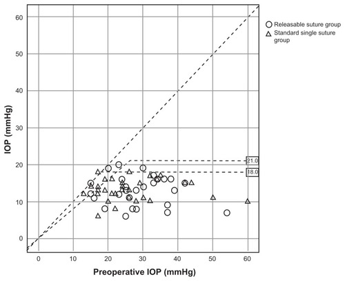
According to the criteria for qualified success, 26 patients (78.8%) with single sutures had an intraocular pressure of 21 mmHg or less and at least a 20% reduction in intraocular pressure to baseline measures and 29 patients (87.9%) had an intraocular pressure of less than 18 mmHg at the last follow-up visit. In the releasable suture group, 26 patients (92.9%) fulfilled both criteria for a qualified success at the last visit. Statistical analysis showed no statistically significant differences between the groups (, ).
Figure 2 Scatter plot of intraocular pressure results at 12 months postoperatively compared with baseline for each eye.
Abbreviations: IOP, intraocular pressure.
Complete success
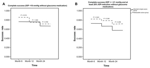
Complete success was achieved by 19 patients (57.6%) with single sutures, by having an intraocular pressure of 21 mmHg or less and at least a 20% reduction of intraocular pressure, whereas 22 patients (66.7%) had an intraocular pressure of less than 18 mmHg at the last visit. Overall, 20 patients (71.4%) with releasable sutures had complete success, fulfilling both criteria (). Although there was a slight tendency for a higher success rate using releasable sutures, no statistically significant difference in complete success was found between the groups at any time point ().
Figure 3 Probability of complete success at months 6 and 12 and at the last visit. (A) Intraocular pressure < 18 mmHg without glaucoma medication. (B) Intraocular pressure ≤ 21 mmHg and at least 20% intraocular pressure reduction without glaucoma medication.
Visual acuity
Mean best corrected visual acuity at the last visit was 0.39 ± 0.42 logMAR in the single suture group and 0.50 ± 0.46 logMAR in the releasable suture group. Visual acuity was not significantly decreased compared with baseline or any of the follow-up visits (P = 0.958, one-way analysis of variance). Moreover, no statistical significance was found between the groups at any time point ().
Suture and bleb interventions
Suture and bleb manipulations following trabeculectomy are shown in . Bleb needling was performed in 10 eyes (30.3%) in the single suture group and in four eyes (14.3%) in the releasable suture group (P = 0.089). Nearly all patients in both groups needed injections of 5-fluorouracil to prevent bleb scarring, ie, 29 patients (92.9%) in the single suture group and 24 patients (87.9%) in the releasable suture group. The mean number of 5-fluorouracil injections was 5.8 ± 3.6 (range 0–15) in patients with standard single sutures and 6.6 ± 5.1 (range 0–18) in patients with releasable sutures (P = 0.133).
Table 3 Suture and bleb interventions
Complications and postsurgical interventions
The incidence of complications and postoperative interventions following trabeculectomy are shown in and . Postoperative ophthalmic examination revealed a flat anterior chamber in three patients (9.1%) in the single suture group and in one patient (3.6%) in the releasable suture group. Treatment of a shallow anterior chamber using sodium hyaluronate injection (Healon©, Abbott Laboratories Inc., Abbott Park, Illinois, USA) was required by two patients (6.1%) in the single suture group and by none in the releasable suture group. Conjunctival wound leakage needing suturing of the conjunctiva was identified in four eyes (12.1%) from patients in the single suture group and in two eyes (7.1%) from patients in the releasable suture group. Seven of 33 patients (21.2%) with single sutures had transient hypotony (intraocular pressure ≤ 5 mmHg) for a mean duration of 5.3 ± 4.2 days. Postoperative hypotony lasting longer than 2.8 ± 2.1 days was found in eight patients (28.6%) with releasable sutures. There was no statistically significant difference in duration of hypotony between the groups (P = 0.178). Scleral flap revision with additional sutures for persistent hypotony and to reduce the risk of choroidal detachment was performed in three patients (9.1%) in the single suture group and in three patients (10.7%) in the releasable suture group. No statistically significant difference was found between the two groups with regard to complications and postsurgical interventions.
Table 4 Incidence of postoperative complications
Table 5 Postsurgical interventions
Discussion
Trabeculectomy is still considered the gold standard for medically uncontrolled glaucoma.Citation9,Citation17 Therefore, several modifications and variations have been developed to maximize the benefits of treatment while minimizing adverse events.Citation6–Citation11 In trabeculectomy, aqueous outflow is determined by the size, shape, and position of the scleral flapCitation8 use of antimetabolites,Citation2,Citation18 suture position, tension, and application of laserable, adjustable, or releasable sutures,Citation9–Citation12,Citation14,Citation15 thereby having an impact on success of treatment based on intraocular pressure, medication required, occurrence of complications, and postoperative bleb and suture management. The technique of flap closure with different types of sutures using either single sutures cut with laser lysis,Citation14 sutures released by corneal incision,Citation19,Citation20 or sutures that can be transconjunctivally adjusted,Citation6,Citation9,Citation15 should be loose enough to permit aqueous outflow but tight enough to reduce complications related to excessive filtration.Citation10,Citation21 Previous studies have evaluated the relative merits of laserable, releasable, or adjustable sutures.Citation10,Citation12 The releasable suture technique was shown to be an effective and safe method of providing sufficient aqueous humor outflow by easily removing the sutures with forceps.Citation19–Citation21 However, complications have been described, including foreign body irritation and keratopathy due to exposed sutures, conjunctival leakage after suture removal, or endophthalmitis caused by externalized sutures that are contaminated with bacteria.Citation6,Citation10,Citation15,Citation19–Citation21 In our study, we investigated the advantages and disadvantages of releasable sutures compared with laser suture lysis of single sutures in trabeculectomy. Our results are based on two comparable groups with a follow-up of 2 years. The data illustrate that both surgical techniques are equally effective, with low complication rates and adequate reduction of intraocular pressure.
A study by Aykan et al,Citation10 in which the surgical outcomes of laser suture lysis of single sutures and removal of releasable sutures were compared, revealed no differences in efficacy or complication rates between the groups. While single sutures were used in both groups, the flap of one group was additionally secured by two releasable sutures. Aykan et al used 2–4 single sutures in addition to two releasable sutures on two sides of the scleral flap (releasable suture group). This is in contrast with our study comparing outcomes of patients with either single sutures or releasable sutures. Moreover, we found highly significant intraocular pressure reductions in both groups during follow-up. In addition, evaluation of success revealed no significant differences between the groups. This is consistent with the findings reported by Aykan et al.Citation10 In our series, complications and revisions were few, relatively minor for both methods, and compare favorably with the findings of other studies.Citation10,Citation12,Citation19,Citation20 Furthermore, we found comparable complication rates between the single suture and releasable suture groups. This is in line with previously published data comparing different types of sutures in trabeculectomy, ie, laser suture lysis of single sutures, removal of releasable sutures, or adjustment of sutures.Citation10,Citation12 The endophthalmitis and blebitis described in early studies after filtration surgery were not encountered in our cases.Citation2,Citation4,Citation5
In practice, focal massage immediately posterior to the filtering bleb allows adjustment of the scleral flap and increases filtration through the fistula. Accordingly, suture adjustment for controlling intraocular pressure in the early postoperative period is indicated after successful bleb massage. While single sutures require laser suture lysis that may lead to subsequent inflammation of the conjunctiva, Tenon’s capsule, and the sclera, as well as conjunctival leak and sudden collapse of the anterior chamber, the adjustment of sutures as well as removal of releasable sutures have been reported to allow controlled reduction of intraocular pressure.Citation12,Citation15 However, we found no significant difference regarding complications induced by laser suture lysis or removal of sutures. Previous studies have favored tying of the scleral flap with adjustable sutures, allowing safer control of intraocular pressure and minimizing the risk of postoperative hypotony.Citation9,Citation12 There has been one report of a longer duration of ocular hypotony in patients with single sutures,Citation10 but there was no statistically significant difference in duration of ocular hypotony between groups in our study.
Our study has some limitations, first related to the fact that our data were collected retrospectively, so is more likely than prospective studies to have more sources of error due to confounding and bias. However, although trabeculectomy was performed by more than one surgeon, postoperative care was identical for all patients included in the present study. Despite the limitations imposed by inclusion of surgical data recorded by different surgeons, the groups of patients were comparable with regard to sample size, gender, age, preoperative intraocular pressure, medication usage, and diagnosis of glaucoma. Many questions remain regarding possible parameters influencing the long-term outcome of both techniques. Therefore, additional prospective studies with greater statistical power to detect small differences will be needed to establish and improve the methods used at the scleral flap site.
In summary, we have shown that trabeculectomy with either single sutures or releasable sutures is an efficient surgical procedure for reduction of intraocular pressure in the medium term. Reduction of intraocular pressure, and success and complication rates, were similar in this study, regardless of whether eyes received single sutures or releasable sutures.
Disclosure
The authors report no conflicts of interest in this work.
References
- CairnsJETrabeculectomy. Preliminary report of a new methodAm J Ophthalmol19686646736794891876
- DeBryPWPerkinsTWHeatleyGKaufmanPBrumbackLCIncidence of late-onset bleb-related complications following trabeculectomy with mitomycinArch Ophthalmol2002120329730011879132
- EdmundsBThompsonJRSalmonJFWormaldRPThe national survey of trabeculectomy. III. Early and late complicationsEye (Lond)200216329730312032721
- MillsKBTrabeculectomy: A retrospective long-term follow-up of 444 casesBr J Ophthalmol198165117907957326225
- WellsAPCordeiroMFBunceCKhawPTCystic bleb formation and related complications in limbus- versus fornix-based conjunctival flaps in pediatric and young adult trabeculectomy with mitomycin cOphthalmology2003110112192219714597529
- JonesEClarkeJKhawPTRecent advances in trabeculectomy techniqueCurr Opin Ophthalmol200516210711315744141
- GrehnFKlinkTA new 6 o’clock traction suture technique for glaucoma filtration surgeryJ Glaucoma2011201282920440218
- BirchallWWakelyLWellsAPThe influence of scleral flap position and dimensions on intraocular pressure control in experimental trabeculectomyJ Glaucoma200615428629016865004
- WellsAPBunceCKhawPTFlap and suture manipulation after trabeculectomy with adjustable sutures: titration of flow and intraocular pressure in guarded filtration surgeryJ Glaucoma200413540040615354079
- AykanUBilgeAHAkinTCertelIBayerALaser suture lysis or releasable sutures after trabeculectomyJ Glaucoma200716224024517473738
- KlinkTGrehnFSuture management after trabeculectomyOphthalmologe20091064364367 German19306003
- KobayashiHKobayashiKA comparison of the intraocular pressure lowering effect of adjustable suture versus laser suture lysis for trabeculectomyJ Glaucoma201120422823320577111
- KlinkTKannGEllingerPKlinkJGrehnFGuthoffRThe prognostic value of the wuerzburg bleb classification score for the outcome of trabeculectomyOphthalmologica20112251556020714183
- KapetanskyFMLaser suture lysis after trabeculectomyJ Glaucoma200312431632012897576
- AshraffNNWellsAPTransconjunctival suture adjustment for initial intraocular pressure control after trabeculectomyJ Glaucoma200514643544016276274
- MarquardtDLiebWEGrehnFIntensified postoperative care versus conventional follow-up: A retrospective long-term analysis of 177 trabeculectomiesGraefes Arch Clin Exp Ophthalmol2004242210611314648140
- GrehnFSurgery of primary open angle glaucomaKlin Monbl Augenheilkd200822513038 German18236367
- GeorgoulasSDahlmann-NoorABrocchiniSKhawPTModulation of wound healing during and after glaucoma surgeryProg Brain Res200817323725418929113
- RainaUKTuliDTrabeculectomy with releasable sutures: A prospective, randomized pilot studyArch Ophthalmol199811610128812939790625
- KolkerAEKassMARaitJLTrabeculectomy with releasable suturesArch Ophthalmol1994112162668285895
- de BarrosDSGheithMESiamGAKatzLJReleasable suture techniqueJ Glaucoma200817541442118703954