Abstract
An aqueous root extract from Nardostachys jatamansi was investigated for its antioxidant and anticataleptic effects in the haloperidol-induced catalepsy rat model of the disease by measuring various behavioral and biochemical parameters. Catalepsy was induced by administration of haloperidol (1 mg/kg, ip) in male albino rats. A significant (P < 0.01) reduction in the cataleptic scores were observed in all the drug-treated groups as compared to the haloperidol-treated group; with maximum reduction observed in the Nardostachys jatamansi (250 and 500 mg/kg body weight) administered group. To estimate biochemical parameters: the generation of thiobarbituric acid reactive substances (TBARS); reduced glutathione (GSH) content and glutathione-dependent enzymes; catalase; and superoxide dismutase (SOD), in the brain were assessed. Haloperidol administration increased generation of TBARS and significantly reduced GSH, which were restored to near normal level with the Nardostachys jatamansi treatment. Catalase and SOD levels were also increased to normal levels, having been reduced significantly by haloperidol administration. Our findings of behavioral studies and biochemical estimations show that Nardostachys jatamansi reversed the haloperidol-induced catalepsy in rats. We conclude that the antioxidant potential has contributed to the reduction in the oxidative stress and catalepsy induced by haloperidol administration.
Introduction
The brain is deficient in oxidative defense mechanisms and hence is at a greater risk of damage mediated by reactive oxygen species (ROS), resulting in molecular and cellular dysfunction.Citation1 ROS can damage virtually any biological molecule in its vicinity including; DNA, essential proteins, and membrane lipids.Citation2 Parkinson’s disease is a neurodegenerative disease characterized by the selective loss of dopamine neurons of the substantia nigra pars compacta. The events, which trigger and/or mediate the loss of nigral dopamine neurons, remains unclear. Neuroleptic-induced catalepsy has long been used as an animal model for screening drugs for parkinsonism.Citation3 Catalepsy is defined as the failure to correct an externally imposed posture. A condition characterized by inactivity, decreased responsiveness to stimuli, and a tendency to maintain an immobile posture. The limbs tend to remain in whatever position they are placed (waxy flexibility). Catalepsy may be associated with psychotic disorders (eg, schizophrenia, catatonia), nervous system drug toxicity, and other conditions. The catalepsy test is widely used to evaluate motor effects of drugs that act on the extrapyramidal system.Citation4 Evidence suggests that immense oxidative stress, free radical formation,Citation5 genetic susceptibility,Citation6 and programmed cell deathCitation7 are the main causes for neurodegeneration associated with Parkinson’s and other related diseases. The neuropathology of the disease is based on depigmentation and cell loss in the dopaminergic nigrostraital tract of the brain, with the corresponding decrease in the striatal dopamine (DA) concentration.Citation8 Besides, dopamine receptor blockade and catecholamine depletion, other neurochemical hypotheses have been proposed for the development of catalepsy such as striatonigral GABAergic (gamma-aminobutyric acid), cholinergic, glutamate, and serotonoergic.Citation9–Citation11 Haloperidol is an antipsychotic drug which is used in the treatment of schizophrenia and other affective disorders. It blocks dopaminergic action in the nigrostraital pathway leading to a high frequency of extrapyramidal motor side effects.Citation12 In animal models, haloperidol induces a behavioral state known as catalepsy in which the animals are unable to correct externally imposed postures.Citation13 The use of haloperidol has been associated with an increased level of oxidative stress in the brain.Citation14 This evidence suggests a possible role for antioxidants in the treatment of haloperidol-induced catalepsy. The brain is made up of 70% lipid and any kind of stress is usually manifested by lipid peroxidative damage.Citation15 The extent of this damage can be used to evaluate the degree of cellular harm. Stress-induced lipid peroxidative damage in the brain can be quantified by either determining the amount of peroxidative products or the rates of enzyme-catalyzed reactions neutralizing free radical intermediates such as superoxide dismutase (SOD). SOD is a primary, natural, and free radical scavenging and antioxidant enzyme in the body. The estimation of the activity of such antioxidant enzymes such as SOD, catalase, or glutathione peroxidase, can be used to assess the therapeutic effects of different antioxidant agents.Citation16
Nardostachys jatamansi (N. jatamansi) (Valirenaceae) is indigenous to the Himalayan regions of India. In Ayurveda, roots and rhizomes of N. jatamansi are used to treat hysteria, epilepsy, and convulsions.Citation17 The decoction of the drug is also used in neurological disorders, insomnia and disorders of cardiovascular system.Citation18 The sesquiterpenes (Jatamansic acid, Jatamansone), lignans and neolignans are reported to be present in the roots of this plant.Citation19,Citation20 Rhizomes are reported to contain a terpenoid ester, Nardostachysin I.Citation21 To date much research has been undertaken to evaluate the drug to treat various neurological and cardiovascular disorders in various animal models and is widely used in ayurvedic formulations. It is reported to possess antidepressant activity, Citation22 anticonvulsant activity,Citation23 antiarrhythmic activity,Citation24 and possess antioxidant, and lipid peroxidation activity in doxorubicin-induced cardiac damage in rats.Citation25 It is also reported to improve learning and memory in miceCitation26 and to enhance biogenic amine activity.Citation27 An acetone extract of N. jatamansi has shown significant inhibition of benzoyl peroxide-induced cutaneous oxidative stress, toxicity, and ear edema in mice.Citation28 It has also been reported to possess protective activity in 6-hydroxydopamine-induced, parkinsonism in rats.Citation29 These pharmacological properties of N. jatamansi prompted us to evaluate its efficacy in haloperidol-induced parkinsonism.
Materials and methods
Plant material
Dried roots of N. jatamansi were purchased from a herbal market (Chennai, Tamil Nadu, India) and authenticated by Dr P Jayaraman, Director of the Plant Anatomy Research Centre, Chennai, India. A specimen voucher was deposited at the department of Pharmacology, C L Baid Metha College of Pharmacy, Chennai, India.
Preparation of aqueous extract of N. jatamansi
The dried roots were coarsely powdered and extracted with distilled water for 48 hours by cold maceration at room temperature before being filtered. The filtrate was concentrated in rotary vacuum evaporator. Percentage yield was calculated with respect to the dried material and screened for its phytoconstituents. The extract obtained was suspended in 1% v/v Tween 80 for oral administration.
HPTLC profile
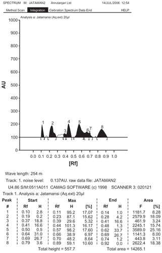
Chromatography was performed on high performance thin layer chromatography (HPTLC) plates coated with 0.25 mm layer of silica gel 60 F254 (Merck, München, Germany). Before using, the plates were washed with methanol and activated at 110°C for 5 minutes Samples were applied as 4 mm wide bands and 6 mm apart by using a Linomat IV sample applicator (Camag Muttenz, Basel, Switzerland) equipped with 100 μL syringe. A constant application rate of 5 μL/second was used. The mobile phase was petroleum ether-acetone (3:1v/v) and chromatograms were monitored at 254 nm ().
Figure 1 HPTLC chromatogram of aqueous extract of N. jatamansi.
Experimental animals
Inbred adult Wistar rats of either sex, weighing 150–200 gm were obtained from the animal house of the C L Baid Metha College of Pharmacy, India. The animals were maintained in a well-ventilated room with a 12-hour light/dark cycle in standard polypropylene cages under controled temperature (26 ± 1°C) and humidity (30%–40%). They were fed with a standard pellet diet obtained from Poultry Research Station, Nandanam, Chennai, India. Water was supplied to the animals ad libitum. Experimental protocols were approved by Institutional Animal Ethics Committee (IAEC) of C.P.C.S.E.A with ref no. IAEC/08/14/CLBMCP/2005–2006, dated 12–20–2005.
Toxicity studies
Acute toxicity studies
Rats selected by a random sampling technique were used in the study. Acute oral toxicity was performed as per Organization for Economic Co-operation and Development (OECD)-423 guidelines.Citation30 Three male Wistar rats weighing between 150–200 g were used for each dose. The dose levels of 5 mg, 50 mg, 500 mg, 1000 mg, 2000 mg, and 5000 mg/kg/body weight, per os were selected. The lethal dose (LD)-50 value of the extract was determined. The drug was administered orally to rats, which were fasted overnight with water ad libitum before the administration of the drug. The body weight of the rat was noted before and after treatment. The animals were observed for toxic symptoms, such behavioral changes, locomotion, convulsions, and mortality for 72 hours.
Repeated oral toxicity studies
In repeated oral toxicity studies drug extracts are administered to the animal for a period covering approximately 10% of the expected life of the animal. Usually, the dose levels are lower than for acute studies and allow chemicals to accumulate in the body before lethality occurs, if the chemical possesses this ability.Citation31 Wistar rats of both sexes, 150–200 g were used for the study and kept in a temperature-controlled environment (26°C ± 1°C) with a 12-hour light/dark cycle. Food and water were freely available and were recorded each at 3-day intervals. The animals were divided into one control group and one treated group, each group consisting of six animals. The control group received Tween 80 and each treated group received the aqueous extract (1000 mg/kg body weight), by gavage once a day for 28 days. The animals were each weighed every 3 days. At the end of the experiment, blood was collected from the orbital sinus, under ether anesthesia, for biochemical and hematological analysis. After the blood collection, the animals were sacrificed by cervical displacement and selected organs (brain, lungs, liver, heart, pancreas, kidney, and testis) were removed and collected in 10% formalin solution in readiness for preparation of sections. The histopathological studies were carried following the method of Mukherjee.Citation32
Experimental design
Adult male Wistar rats (180–250 g) were divided into six groups each containing six animals. Group I received the vehicle 1% Tween 80 solution and served as the control. Group II received haloperidol alone and served as the cataleptic control without any drug treatment. Group III received a combination of l-dopa and carbidopa (100 mg + 25 mg/kg by intraperitoneal administration) and served as positive control. Groups IV, V, and VI received N. jatamansi at a dose of 100, 250, 500 mg/kg body weight, respectively. Thirty minutes after the administration of these drugs, catalepsy was induced by the intraperitonial administration of haloperidol at a dose of 1 mg/kg body weight in normal saline. This procedure was repeated for 15 days. All the behavioral studies were performed at room temperature in a calm room without any external interference. The severity of catalepsy was measured every 30 minutes thereafter for a total duration for 3 hours. Catalepsy of an individual rat was measured in a stepwise manner using a scoring method described below. After 15 days of behavioral studies, animals were sacrificed by cervical dislocation and the whole brain was immediately dissected out and washed in ice-cold saline to remove all traces of blood. The brains were weighed and a 10% tissue homogenate was prepared in 0.025 M Tris–HCl buffer at pH 7.5 and used to measure the activities of thiobarbituric acid reactive substances (TBARS). Enzyme activity was assayed in 10% brain homogenates prepared in 0.2 M phosphate buffer, pH 8.0.
Behavioral studies
Measurement of catalepsy by block methodCitation33
This scoring method followed is in three steps. Step 1: The rat was taken out of the home cage and placed on a table. If the rat failed to move when touched or pushed gently on the back a score of 0.5 was assigned. Step II: The front paws of the rats were placed alternately on a 3-cm high block. If the rat failed to correct the posture within 15 seconds, a score of 0.5 for each paw was added to the score of step 1. Step III: The front paws of the rat were placed alternately on a 9-cm high block, if the rat failed to correct the posture within 15 seconds a score of 1 for each paw was added to the scores of steps I and II. Thus, the highest score for any animal was 3.5 (cut off score) and that reflects total catalepsy ().
Table 3 Effect of aqueous extract of N. jatamansi on haloperidol-induced catalepsy by block method
Behavioral assessment by metal bar test
Behavioral assessment in haloperidol-induced cataleptic rats was studied by the method of Kulkarni.Citation34 Cataleptic behavior was measured with a high bar test method. Catalepsy score was measured for 4 hours at one-hour intervals after haloperidol administration by gently placing both forepaws of the rat over a metal bar (diameter 2–5 mm suspended 6 cm above the table top). The intensity of catalepsy assessed by counting the time in seconds until the rat brought both forepaws down to the tabletop, with a maximum cutoff time of 3 minutes. Finally, scores at different time points (0, 60,120,180 and 240 minutes after haloperidol injection) were added and expressed as a cumulative catalepsy score for comparison purposes ().
Table 4 Effect of aqueous extract of N. jatamansi on Haloperidol induced catalepsy by metal bar test
Biochemical studies
Estimation of lipid peroxidation products
Lipid peroxidation was estimated colorimetrically in brain tissue by quantifying TBARS according to the method of Niehaus and Samuelson.Citation35 In brief; for the estimation of TBARS the supernatant of the tissue homogenate was treated with tertiary butanol–trichloroacetic acid–hydrochloric acid, (TBA–TCA–HCl) reagent and mixed thoroughly. The mixture was kept in boiling water bath for 15 minutes. After cooling, the tubes were centrifuged for 10 minutes and the supernatant taken for measurement. The developed color was read at 535 nm using a UV spectrophotometer (Hitachi 912) against a reagent blank and expressed as mM per 100 g tissue.
Estimation of antioxidants
Catalase (CAT) was assayed colorimetrically at 620 nm and was expressed as micromoles of H2O2 consumed per minute per mg of protein; using the method described by Sinha.Citation36 The reaction mixture (1.5 mL, volume) contained 1.0 mL of 0.01 M pH 7 phosphate buffer, 0.1 mL of tissue homogenate and 0.4 mL of 2 M H2O2. The reaction was stopped by the addition of 2 mL of dichromate-acetic acid reagent (5% potassium dichromate and glacial acetic acid mixed in the ratio of 1:3). The assay for SOD was based on SOD mediated inhibition of the reduction of nitroblue tetrazolium to blue formazan by superoxide anions as described by Beauchamp and Fridovich.Citation37 The total protein present in the homogenate was estimated following the method described by Lowry.Citation38 Units of SOD activity determined were expressed in terms of milligrams of total protein (TP). Reduced glutathione (GSH) was determined by the method of Ellman.Citation39 One mL of supernatant was treated with 0.5 mL of Ellman’s reagent and 3 mL of phosphate buffer (0.2 M, pH 8.0). The absorbance was read at 412 nm. The activity of GSH was expressed as nM GSH formed/g tissue.
Statistical analysis
Each group of rats assigned to a specific drug treatment each group consisted of 6–9 animals. All the values are expressed as mean ± standard error of mean (SEM). The data were analyzed by analysis of variance (anova) followed by Tukey test. The criterion for statistical significance was P < 0.05.
Results
The phytochemical analysis of an aqueous root extract of N. Jatamansi revealed the presence of sterols, carbohydrates, terpenes, phenols, gums and mucilage. The HPTLC finger-print of the aqueous extract showed the presence of eight spots (Rf values 0.11, 0.23, 0.39, 0.44, 0.57, 0.66, 0.70 and 0.84) () at a wavelength of 254 nm. The standard reference compound, Jatamansone, had the Rf value of 0.39 (), hence it is confirmed that the aqueous extract contains terpenoids. The acute oral toxicity was undertaken according to the OECD guidelines 423 (acute toxicity class method). There was no considerable change in body weight either before or after experimental treatment and no signs of toxicity were observed (). The LD50 test of the aqueous extract was found to be greater than 5000 mg/kg body weight after oral administration. Repeated oral toxicity was carried out by administration of the extract at a dose of 1000 mg/kg body weight per os for 28 days. The extract treated rats did not show any significant changes in hematological parameters (hemoglobin red blood corpuscles [RBC], white blood corpuscles [WBC], neutrophils, monocytes, eosinophils and lymphocytes) when compared with the normal control animals (). Histopathological examinations of the internal organs, ie, liver, kidney and brain, did not shown any changes in their normal architecture, suggesting the safety of the drug () as the oral administration of the drug to normal rats had no significant effect. Based on these findings a dose of 100, 250, and 500 mg/kg bodyweight were selected for the study. The cataleptic scores of the present study are given in and , assessed by block method and metal bar test, respectively. Haloperidol induced catalepsy significantly (P < 0.01) at a dose of 1 mg/kg (intraperitoneal administration). Significant reversal in haloperidol-induced catalepsy was observed with the administration of N. jatamansi aqueous extract and combination of l-dopa and carbidopa. The maximal decrease (P < 0.01) in catalepsy was observed in the group receiving aqueous extract of N. Jatamansi at a dose of 250 mg/kg. However, there was no pronounced reduction in the cataleptic scores at a dose of 500 mg/kg, but almost similar results were observed as observed at a dose of 250 mg/kg body weight.
Figure 2 HPTLC chromatogram of standard (Reference compound) Jatamansone.
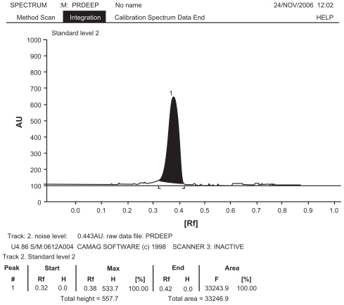
Figure 3 Histopathological slides of Brain (A control, A1 test), Liver (B control, B1 test) and Kidney (C control, C1 test), Repeated oral toxicity study of Nardostachys jatamansi treatment for 28 days in rats showed no pathological changes.
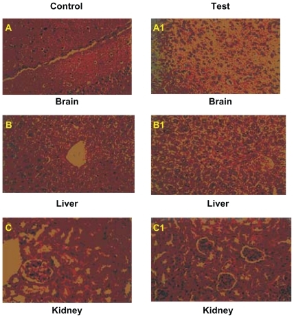
Table 1 Acute toxicity study
Table 2 Repeated oral toxicity studies
The levels of lipid peroxidation products and antioxidants in the brains of haloperidol, drug-treated and control groups are shown in . The haloperidol-treated rats showed a significant increase (P < 0.01) in TBARS and there was also a significant reduction (P < 0.01) in SOD, CAT, and GSH in the brain tissue. Oral administration of the extract along with haloperidol administration significantly restored (P < 0.01) the peroxides and antioxidant levels to near normal in the brains of the test animals. For all the parameters studied, N. jatamansi extract administered at doses of 250 and 500 mg/kg bodyweight showed significant effects. l-dopa and carbidopa also showed a significant effect in all the parameters studied in rats.
Table 5 Effect of N. jatamansi on TBARS, SOD, CAT, and GSH levels in normal and catalepsy-induced rat brain
Discussion
The central nervous system is especially vulnerable to free radical damage because of the brain’s high oxygen consumption, its abundant lipid content, and the relative paucity of antioxidant enzymes as compared with other tissues.Citation40 Evidence also indicates that ROS may stimulate extracellular release of excitatory amino acids.Citation41 Glutamate is the major excitatory amino acid in the brain which acts through various types of ionotropic receptors, the most significant being N-methyl D-aspartate (NMDA) receptors. There seems to be a bidirectional relationship between the ROS production and the release of excitatory aminoacids.Citation42 Free radicals generated in the brain are also reported to influence gene expression, subsequently effecting apoptosis and neuronal death.Citation43 In the brain, an array of cellular defense systems exists to counterbalance the ROS. These include enzymatic and nonenzymatic antioxidants that lower the concentration of free radical species and repair oxidative cellular damage. The brain is known to synthesize molecules like glutathione and NADPH. Glutathione functions as a major antioxidant in tissue defense against free radicals in the brain. However, the concentration of glutathione is, relatively, in lesser quantities in the brain as compared to the other organs of the body.Citation40 The natural antioxidant system present in brain can be in form of enzymes like catalase, peroxidase, superoxide dismutase or low molecular weight antioxidants (ascorbic and lipoic acids, carotenoids or indirectly acting chelating agents).Citation43 Free radical scavengers or antioxidants function as biological bodyguards for essential molecules by either neutralizing reactive species before they mutilate a molecule or they repair damage that has been inflicted.
The present study demonstrates the antioxidant effects of an aqueous root extract of N. Jatamansi in haloperidol-induced, cataleptic oxidative stress in rats. The induction of free radicals in mammals by haloperidol is well established. Previous studies have shown that dopamine receptors in the striatum are involved in neuroleptic-induced catalepsy. It has been demonstrated that the cataleptic effects of haloperidol are apparently mediated by dopamine receptors localized postsynaptically on strial neurons.Citation13 It is also well established that the administration of haloperidol leads to an increase in the oxidative stress in the brain tissue.Citation14 The increase in SOD observed in the present study supports the this concept. Superoxide formation is a major factor in oxygen toxicity and the superoxide dismutase enzyme constitutes an essential defense against it. Under normal conditions, decreased activity of antioxidant enzymes, such as SOD, glutathione peroxidase and catalase, in the brain leads to the accumulation of oxidative free radicals resulting in degenerative effects.Citation44 An increase in these enzymes under normal conditions would represent increased antioxidant activity and a protective mechanism in neuronal tissue, thus, constituting the first line of defense against oxidative stress in our body. However, in the presence of a free radical-quenching agent, the induction of the antioxidant enzymes is minimized. So, any overall decrease in cataleptic scores and SOD activity in the drug treated groups indicates the ability of the drug extract to combat oxidative stress in brain tissue and reduce the severity of haloperidol-induced catalepsy. The altered balance of the antioxidant enzymes caused by the decrease in CAT, SOD, GSH activities may be responsible for the inadequacy of the antioxidant defenses in combating ROS mediated damage. The decreased activities of CAT and SOD may be a response to increased production of H2O2 and O2 by the autooxidation.Citation45 It has been suggested that these enzymes play an important role in maintaining physiological levels of oxygen and hydrogen peroxide by hastening the dismutation of oxygen radicals and eliminating organic peroxides and hydroperoxides. Treatment with N. jatamansi extract increased the activity of these enzymes by quenching the free radicals. Previously N. jatamansi has been reported to be a well known antioxidant and the ethanol extract of N. jatamansi is reported to possess potent antioxidant activity that scavenges free radicals generated after the induction of catalepsy.Citation27 Significantly lower levels of lipid peroxides in the brains of the drug-treated group and increased activities of enzymatic and nonenzymatic antioxidants in the brain suggests that the extract reduces oxidative stress. In previous studies N. jatamansi has been reported to have antilipid peroxidative and protective effect in rat cerebral ischemia.Citation46,Citation47 N. jatamansi has also been reported to enhance biogenic amine activity,Citation48 decrease the level of dopamine and its metabolites and increasing the number of dopaminergic D2 receptors in striatum.Citation29 Such evidence supports our study and indicates that the extract of N. jatamansi inhibits the symptoms of haloperidol-induced catalepsy in rats. The action by which the amelioration takes place may be attributed to one (or) more pharmacological/biochemical mechanisms. To conclude, the brain exhibits numerous morphological and functional alterations during oxidative stress, a factor implicated in the pathogenesis of many CNS disorders. Treatment of such neuronal disorders with N. jatamansi plant extract significantly decreases lipid peroxidation and significantly increases the antioxidants in the brain. The findings of this study suggest the possible antioxidant role of N. jatamansi in overcoming behavioral and neurochemical changes during oxidative stress. Since the catalepsy test has predictive value regarding extrapyramidal effects, the possibility of pharmacological interactions between haloperidol and N. jatamansi extract should be further investigated in clinical studies.
Disclosures
The authors report no conflicts of interest relevant to this research.
References
- GuptaYKMadhur GuptaKohliKNeuroprotective role of melatonin in oxidative stress vulnerable brainIndian J Physiol Pharmacol200347437338615266948
- WolffSPGarnerADeanRTFree radicals lipids and protein degradationTrends Biol Sci1986112731
- Anil kumarKulkarniSKEffect of BR-16A(Mentat), a polyherbal formulation on drug induced catalepsy in miceIndian J Exper Biol200644454816430090
- SanbergPRBunseyMDGiordanoMThe catalepsy test: its up and downsBehav Neurosci19881027487592904271
- JennerPOxidative stress and the pathogenesis of Parkinson’s diseaseNeurology199647161170
- BandmannOMarsdenDCWoodNWGenetic aspects of Parkinson’s diseaseMov Disord (Review)199813203211
- ZivIMelamedENardiNRole of apoptosis in the pathogenesis of Parkinson’s disease: a noval therapeutic opportunityMov Disord1998138658709827608
- Von BohlenHalbachOSchoberAKrieglsteinKGenes, proteins, and neurotoxins involved in Parkinson’s diseaseProg Neurobiol20047315117715236834
- BazianASDivergent and convergent mechanism of integrative activity of mammalian brainZh Vyssh Nerv Deiat Im I P Pavlova20015151452811605433
- OkuyamaSAtypical antipsychotic profile of sigma receptor ligandsNippon Yakurigaku Zasshi1999114132310562961
- Neal BelveauBSJoyceJNLuckiISerotonergic involvement in haloperidol-induced catalepsyJ Pharmacol Exp Ther19932652078386235
- FardeLNordstromALWieselFAPauliSHalldinCSedvallGPositron emission tomographic analysis of central D1 and D2 dopamine receptor occupancy in patients treated with classical neuroleptics and clozapine:Relation to extra-pyramidal side effectsArch Gen Psychiatry1992495385441352677
- SanbergPRHaloperidol-induced catalepsy is mediated by postsynaptic dopamine receptorsNature19802844724737189016
- SagaraYInduction of reactive oxygen species in neurons by haloperidolJour Neurochem199871100210129721725
- KedarNPCan we prevent Parkinson’s and Alzheimer’s disease?J Postgrad Med20034923624514597787
- Albina ArjumanVinod NairGopalkrishnaHNNandiniMEvaluation of the antioxidant potential of NR-ANX-C (a polyherbal formulation) and its individual constituents in reversing haloperidol-induced catalepsy in miceIndian J Pharmacol2007393151154
- BagchiAOshimaYHikinoHNeoligans and lignans of Nardostachys Jatamansi RootsPlanta Med199157969717226134
- UniyalMRIssarRKCommercially and traditionally important medicinal plants of Mandakini valley of Uttarkhand HimalayasJ Res Indian Med1969418396
- ChatterjiAPrakashiSCThe treatise on Indian medicinal plantsNational Institute of Science Communication (Publication and Information Directorate)5New Delhi1997
- AroraRBNardostachys jatamansi, a chemical, pharmacological and clinical appraisalNew Delhi, IndiaMonograph Special Series, Indian Council of Medical Research511965
- ChatterjeeABasakBSahaMStructure and Stereo-chemistry of Nardostachysin, A New Terpenoid ester constituent of the Rhizomes of Nardostachys JatamansiJ Nat Prod200063111531153311087600
- Bharat metkar PalSCVeena KastureSanjay KastureAntidepressant activity of Nardostachys Jatamansi DCInd J Nat Prod19991521013
- RaoVSRaoAKaranthKSAnticonvulsant and neurotoxicity profile of Nardostachys Jatamansi in ratsJ Ethanopharmacol20051023351356
- AroraRBMadanBRAntiarrythmics-Antiarrythmic activity of Nardostachys Jatamansi (an indigenous drug)Ind J Med Res1956442259269
- SubashiniRYogeetaSGnanapragasamADevakiTProtective effect of Nardostachys Jatamansi on oxidative injury and cellular abnormalities during doxorubicin-induced cardiac damage in ratsJ Pharma Pharmacol2006582257262
- JoshiHParleMNardostachys jatamansi improves learning and memory in miceJ Med Food20069111311816579738
- AhamadMSaleemSAhamadASNeuroprotective effect of Withania sominifera on 6-hydroxydopamine induced Parkinsonism in ratsHum Exp Toxicol200524313714715901053
- AliADuaYSiddiquiAWSultanaSRafiullahMRMInhibition of benzoyl peroxide-induced cutaneous oxidative stress, toxicity and ear edema in mice by Nardostachys JatamansiPharmaceutical Biol2005436533539
- AhmadMYousufSKhanBMAttenuation by Nardostachys jatamansi of 6-hydroxydopamine-induced parkinsonism in rats: behavioral, neurochemical, and immunohistochemical studiesPharmacol Biochem Behav200683115016016500697
- EcobichnonDJThe Basis of Toxicity Testing2nd EdNew York, NYCRC Press1997
- WilliamsPDPergamanEComprehensive Toxicology2nd EdOxford, UKPergamon Press1984
- MukherjeeKLMedical Laboratory Technology1st EdNew Delhi, IndiaTata McGraw Hill1989
- ChopdeCTKhistiRTMandaneSNHaloperidol-induced catalepsy: A model for screening antidepressants effective in treatment of depression with Parkinson’s diseasesInd J Exp Biol19973512971301
- KulkarniSKAnil KumarSEffect of BR-16A (Mentat®), a poly herbal formulation on drug induced catalepsy in miceInd J Exp Biol2006444548
- NiehausWGSamuelsonBFormation of malondialdehyde from phospholipid arachidonate during microsomal lipid peroxidationEur J Biochem196861261304387188
- SinhaKAColorimetric assay of catalaseAnal Biochem1972473893944556490
- BeauchampCFridovichISuperoxide dismutase:Improved assays and an assay applicable to acrylamide gelsAnal Biochem197144276874943714
- PetersonGLReview of the folin phenol protein quantification method of Lowry, Rosenbrough, Farr and RandallAnal Biochem1979100201220393128
- EllmanGLTissue sulfhydryl groupsArch Biochem Biophys195982707713650640
- SkaperSDFloreaniMCecconMFacciLGiustiPExcitotoxicity, oxidative stress, and the neuroprotective potential of melatoninAnn NY Acad Sci199989010711810668417
- GilmanSCBonnerMJPellmarTCEffect of oxidative stress on excitatory aminoacid release by cerebral cortical synaptosomesFree Rad Med Biol199315671675
- CoyleJTPuttfarckenPOxidative stress, glutamate, and neurodegenerative disordersScience19932626896957901908
- Gilgun-SherkiYRosenbaumZMelamedEOffenDAntioxidant therapy in acute central nervous system injury: the current statePharmacol Rev20025427128412037143
- NaiduPSSinghAKulkarniSKEffect of Withania somnifera root extract on haloperidol induced orofacial dyskinesia: Possible mechanism of actionJ Med Food2003610711412935321
- AragnoMBrignardelloETamagnoOBoccuzziGDehydroeppiandrosterone administration prevents the oxidative damage induced by acute hyperglycemia in ratsJ Endocrinol19971552332409415057
- TripathiYBEktaTAnilUAntilipid peroxidative property of Nardostachys JatamansiInd J Exp Biol19963411501151
- SalimSAhmadMZafarKSAhmadASIslamFProtective effect of Nardostachys jatamansi in rat cerebral ischemiaPharmacol Biochem Behav200374248148612479970
- PrabhuVKaranthKSRaoAEffects of Nardostachys jatamansi on biogenic amines and inhibitory amino acids in the rat brainPlanta Med19946021141178202559