Abstract
Background: The teaching of anatomy to medical undergraduates continues to develop. Medical imaging can accurately demonstrate anatomy. ‘disect’ is a computer program which manipulates and reconstructs real CT images in 3-D.
Aim: To implement and assess a novel computer-based imaging resource.
Methods: Third-year undergraduate medical students at the University of East Anglia were randomised to different methods of delivering the program – either self-directed use or guided use with worksheets. Knowledge of gastro-intestinal anatomy was assessed using a 20-item test. Attitudes to using ‘disect’ were evaluated using Likert scales.
Results: Most students reported the program was easy to use and a valuable resource for learning anatomy. There was no difference in scores between guided use and self-directed use (10.7 marks versus 10.6 marks, p = 0.52). Students who undertook the anatomy special study module, which involved dissection of the digestive system, performed best (12.8 marks versus 9.9 marks, p = 0.005).
Conclusion: Students can adequately use a computer program to see major anatomical structures derived from CT scans. Students reported that learning anatomy can be aided by the imaging-based resource. Learning anatomy is a multi-modal activity and packages like ‘disect’ can enhance learning by supplementing current teaching methods.
Introduction
The teaching of undergraduate and post-graduate anatomy is being redefined and methods used to deliver this teaching are also changing, including increasing use of medical imaging and computer-based resources (Mitchell Citation2002; McLachlan Citation2004; Miles Citation2005). The evidence for computer assisted learning (CAL) in medical education in general has been recently reviewed (Greenhalgh Citation2001). This report identified 200 studies, of which only 12 were randomised-controlled trials. Many of these investigations had methodological problems, including a lack of statistical power and potential contamination between the intervention and control groups. Our own recent search identified eight quantitative studies (Tam Citation2009) which provided some evidence to support the use of computer-assisted learning specifically in the setting of undergraduate medical anatomy. Identified trials included computer-based teaching for learning anatomy of the inner ear (Nicholson Citation2006), the carpus (Garg Citation1999, Citation2001, Citation2002), surface anatomy of the abdomen (Hallgren Citation2002; Qayumi Citation2004) and anatomy and physiology of the biliary tree (Devitt Citation1999), and in general reported an improvement in knowledge. However, these trials were conducted over short teaching periods and in limited areas of anatomy. One larger retrospective study compared two cohorts of students (Elizondo-Omana Citation2004): one group had traditional teaching involving lectures and dissection, and the second group the same plus access to a multi-media laboratory. The latter scored an average mark of 68% compared to 58% (p < 0.05) in the gross anatomy examination. This suggested that CAL is useful in supporting or consolidating knowledge. Further randomised-controlled trials are needed which assess CAL in more comprehensive areas of anatomy over longer periods of time.
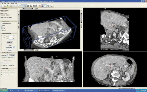
The aim of this randomised-controlled trial was to test different modes of delivery of the ‘disect’ package and secondarily to measure students’ attitudes regarding using a computer-based resource to study anatomy. ‘disect’ (www.disectsystems.com) is a computer-based DiCOM viewer that can be run on PCs and laptops and allows cross-sectional scans, such as CT or MRI to be incorporated and viewed (). DiCOM is the major format of digital radiological images, and many hospitals have changed from films to digital images. ‘disect’ allows interaction with pictures in a similar manner to the dedicated silicon graphics workstations that radiologists use to manipulate and reconstruct images. Real scans are loaded onto ‘disect’ and the user can interact with the images by scrolling through the three orthogonal planes. The program allows a 3-D exploration of the total data provided by a real CT scan ().
Methods
A total of 128 undergraduate medical students in their third year at the School of Medicine, Health Policy and Practice, University of East Anglia were invited to participate in the trial. Students were given an introductory talk and supplied with a participant information sheet. Ethical approval was gained by the Faculty of Health Ethics Committee at the University and written informed consent was obtained from students. All who agreed to participate attended a 1 hour training session in groups of 20, where they learned how to use ‘disect’. The class was interactive, with each student having access to a computer loaded with the ‘disect’ software. They were taught to perform functions that were being simultaneously demonstrated on a screen. Students learned how to scroll through the axial, coronal and sagittal planes, to cross-reference one structure in three different planes, and also to perform basic image reconstructions, for example, of the arterial tree. Further teaching was available if requested by the students.
The 128 students in Year 3 were allocated across 14 problem-based learning (PBL) groups which studied one of three modules taught that year (‘Hormones and Homeostasis’, ‘The Senses’ and the ‘Gastrointestinal System’ module). All students attended lectures, but only two-thirds of the students (those studying ‘Hormones and Homeostasis’ and the ‘Gastrointestinal System’) attended sessions with prosections of the anatomy of the Gastro-intestinal (GI) tract in the dissecting room. In addition, within each PBL group, two students undertook a Selected Special Study (SSS) module in anatomy which involved dissection classes. Therefore, there were several different groups of students – dissectors, those who were on the GI module who had access to prosections, those on the endocrine module who also had prosection-based teaching of the digestive tract and those students studying ‘The Senses’ with no particular incentive to learn GI anatomy at that stage and had no access to prosections.
The study design was a cluster randomised trial of worksheet-guided access (intervention group) compared to self-directed access (control group) to the program. The randomisation was performed by a third party in the Clinical Trials Unit at the University using a Visual Basic randomisation program. The unit of randomisation was the PBL seminar group, each of which was composed of up to 10 students. PBL groups were randomised to either self-directed access to ‘disect’ or worksheet-guided access.
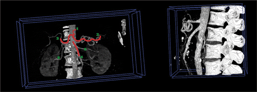
Access to the ‘disect’ program was initiated in week 3. All third-year medical students then had access to the program in the undergraduate medical suite for the remaining six of the eight weeks dedicated to academic learning. The worksheets which were designed to be used with the program, labelled major anatomical structures in the abdomen and guided the student to interact with the CT scan. An example included making a 3-D reconstruction of the blood supply to the gut (). Students who were randomised to not having worksheet support still had access to the program but were advised to use the program in conjunction with their textbooks, lecture notes or an anatomy atlas. The software was accessible by user name, and the worksheets were installed by security group and active directories to ensure that only the appropriate individuals had access to the worksheets.
Figure 2. A labelled reconstruction of the blood supply to the gut. ‘Disect’ allowed zooming and rotation to gain an appreciation of the anatomy of the coeliac axis and superior mesenteric artery.

Participants attended a 1 hour session, where a 20-item written test was given. There were 10 questions testing factual knowledge and a further 10 structures to label on CT scans. The marks could range from 0 to 20. The test was designed with reference to local learning objectives and also to a recent recommended national curriculum (Hanwell Citation2007). Questionnaires were completed at the examination to assess students’ opinions of ‘disect’. Opinion questions were framed as 5-point Likert scales and the frequency distribution of responses was reported. Free text responses as well as any verbal feedback received after the testing session were noted. Mean test scores for the factual components were calculated for the two groups and differences assessed using an unpaired t-test. At 80% power with 5% significance, 14 students were required in each group to detect a difference of 2 marks (assuming a mean score of 12 with a standard deviation of 2). A linear regression model was used to assess if there was an effect on the scores for special study students, prosection and students doing the gastro-intestinal module and the individual PBL groups assuming that these are independent variables.
Results
Of the 128 medical students in Year 3, 85 (66.4%) agreed to participate and attended the initial training sessions. Of these, 34 students (40% of the participants, with 11 in the worksheet group and 23 in the self-directed group) attended the examination and returned the questionnaire. Of the participants, nine students undertook the anatomy special study module, whilst 25 did not. Twenty-seven students had access to prosection, seven did not, and 11 students were enrolled on the GI module whereas 23 were not. Comparison of these variables by group is shown in . The number of dissecting students and the number of students exposed to prosections appeared reasonably well matched but the groups proved not to be particularly evenly matched in terms of their current educational placement, with more of the worksheet group currently studying the GI system (45% worksheet versus 26% self-directed) and more of the self-directed group studying hormones and homeostasis (27% worksheet group versus 57% self-directed group) – but this was adjusted for in the linear regression analysis. A total of 37 questionnaires were returned with three students electing not to sit the test. One stated that this was because they studied at home and did not use the University medical suite computers and therefore could not easily access the program. The other two students stated that they had not used the program much and did not want to do the test.
Table 1. Number and percentage of students in the intervention and control groups
The results showed positive feedback (), including students being strongly in favour of using this program in the future to learn other areas of anatomy. They also reported that the program was easy to use and that structures could be easily identified using the program.
Table 2. Questionnaire results
In the quantitative test of anatomical knowledge, there was no difference between the worksheet and self-directed groups (10.7/20 marks versus 10.6/20 marks, mean difference 0.1, 95% CI −2.2 to 2.4, p = 0.95). The students who did the anatomy special study module performed better than those who did not (12.8 marks versus 9.9 marks, mean difference 2.9, 95% CI 0.6–5.1, p = 0.014). The students who had access to prosected specimens also performed better than those who did not (11.1 marks versus 9.0 marks, mean difference 2.1 marks, 95% CI −0.5 to 4.7, p = 0.10). Students who were doing the GI module scored higher than those who did not, although the difference was not statistically significant (11.5 marks versus 10.2 marks, mean difference 1.3, 95% CI −1.0 to 3.6, p = 0.26). The linear regression model demonstrated no significant effect of the type of access to the program (p = 0.52), but did show strong statistical significance for taking the anatomy special study module (p = 0.005). The effect of prosection did not quite reach statistical significance (p = 0.07) and dedicated GI study was not statistically significant (p = 0.47) (). The individual PBL groups did not have a statistically significant effect on the test scores.
Table 3. Test scores
Discussion
Students gave positive feedback on ‘disect’, but the addition of the worksheet did not affect performance in the test. This suggests that the program was equally well used by both groups of students and that such programs should be considered as a method of complementing existing anatomy teaching techniques. Most participants found ‘disect’ easy to use, that it was good at demonstrating anatomy in 3-D, and useful for understanding CT scans.
The worksheets did not improve knowledge over self-directed use. However, it should be noted that this study was relatively small with limited statistical power, and our confidence limits cannot rule out either a moderate (on our test up to a 2-point) improvement in learning, or a moderate deficit due to the addition of a worksheet guide. The worksheets were only accessible on computer to those randomised to the worksheet group, but it is possible that contamination occurred, i.e. sharing of the worksheets with the self-directed group. This would have diminished our ability to show a between group difference. If we assume that little contamination occurred then this study suggests that self-directed use of the program supported by anatomy textbooks and atlases may well be as effective as the worksheets. It should be noted that those taking part in the study may be the more motivated students who more readily appreciate and understand anatomy. Previous studies have found that CAL may have had a greater impact on the knowledge of the weaker students (Qayumi Citation2004). These reasons may account for why no statistically significant difference was noted for including the worksheet.
Students who undertook the special study module in anatomy scored higher than those who did not. The effect of the special study module, which involved anatomical dissection, may produce an increase in knowledge for several reasons. These students were dissecting and exposed to more anatomy teaching than those who did not. Also, such undergraduates may be more motivated to learn anatomy, possibly due to interest or recognition of its importance. The special study students also demonstrated prosections to fellow students, and so a peer-to-peer effect may have enhanced this group's performance.
Students who were exposed to prosected specimens of the GI tract also scored better than those who did not, but it was of questionable statistical significance potentially due to the small numbers included in this study. Unsurprisingly, the students studying the GI system scored better than those in other modules as they had more gastroenterology teaching but again this did not reach statistical significance.
The strengths of this trial include that the assessment of knowledge was tested over a large area of anatomy and over a longer period of time than previous studies. The main weakness was the limited participation in the test which limited our statistical power to detect between group differences. This may have been due to timetabling as the optional test was offered during the clinical examination week which may have deterred attendance due to students concentrating on preparation for their summative assessments. Also, the views of students who agreed to participate but did not sit the test, and the views of the students who declined to participate are unknown. The latter, for example, may not value computer resources. However, at least one quarter of all medical students reported a positive experience in using the program and suggests that medical schools should continue to develop its use. Further studies are required to determine how best to use these resources in conjunction with current methods for teaching anatomy.
Conclusion
Students were positive about using the computer program to help consolidate their anatomical knowledge. They found it a useful adjunct for studying, whether using it in a directed fashion through the provision of worksheets, or in a self-directed manner. Students undertaking the special study module in anatomy, which involved dissection performed better than those who did not. Students who were also exposed to prosection also performed better. The views of the participating students were that anatomy learning can be enhanced by novel computer, or image-based resources and their use should be considered by medical schools.
Declaration of interest: The authors report no conflicts of interest. The authors alone are responsible for the content and writing of the article.
Additional information
Notes on contributors
M.D.B.S. Tam
M. TAM MA (Oxon), MRCS, FRCR, is Specialist Registrar at the Radiology Academy at the Norfolk and Norwich Radiology Academy with an interest in medical education and is pursuing a Masters in Clinical Education at the School of Allied Health Professionals at the University of East Anglia.
A.R. Hart
A.R. HART FRCP is an Honorary Senior Lecturer in Gastroenterology at The University of East Anglia, Norwich with an interest in the design and conduct of aetiological and clinical studies in digestive diseases.
S.M. Williams
S. WILLIAMS MA (Oxon), MRCS, FRCR, is Consultant Radiologist and is currently Director of the Radiology Department at the Norfolk and Norwich University Hospital. He oversaw the implementation of the Radiology Academy, a purpose built unit designed for post-graduate medical education of Radiology Registrars.
R. Holland
R. HOLLAND BMBCh, DPH, PhD is Reader in Public Health Medicine. He is Health Services Researcher with a particular interest in clinical trials and systematic reviews. He is Course Director of the MBBS degree at the University of East Anglia.
D. Heylings
D. HEYLINGS MBBCh, ILTM is Senior Lecturer in Anatomy at The University of East Anglia, responsible for the anatomy curriculum. In a wider context he has a particular interest in trends in medical education, curriculum design and delivery especially concerning anatomy within the modern educational environment.
S. Leinster
S. LEINSTER FRCS (Ed), FRCS (Eng), MD, SFHEA is Professor of Medical Education and Dean of the School of Medicine, Health Policy and Practice at the University of East Anglia. His clinical discipline is Surgical Oncology.
References
- Devitt P. Computer-aided learning: An overvalued educational resource?. Med Educ 1999; 33: 136–139
- Elizondo-Omana R. Traditional teaching supported by computer-assisted learning for macroscopic anatomy. Anat Rec 2004; 278B: 18–22
- Garg A. Learning anatomy: Do computer models improve spatial understanding?. Med Teach 1999; 21(5)519–522
- Garg A. How medical students learn anatomy. Lancet 2001; 357: 363–364
- Garg A. Is there any real virtue of virtual reality? The minor role of multiple orientations in learning anatomy from computers. Acad Med 2002; 77(10)S97–S99
- Greenhalgh T. Computer assisted learning in undergraduate medical education. BMJ 2001; 322: 40–44
- Hallgren R. An interactive, web-based tool for learning anatomic landmarks. Acad Med 2002; 77(3)263–265
- Hanwell S. A core syllabus in anatomy for medical students – Adding common sense to need to know. Eur J Anat 2007; 11(Supplement 1)3–18
- McLachlan J. Teaching anatomy without cadavers. Med Educ 2004; 38: 418–424
- Miles K. Diagnostic imaging in undergraduate medical education: An expanding role. Clin Radiol 2005; 60: 742–745
- Mitchell B. Trends in radiological anatomy teaching in the UK and Ireland. Clin Radiol 2002; 57: 1070–1072
- Nicholson D. Can virtual reality improve anatomy education? A randomised controlled study of a computer-generated 3-D anatomical ear model. Med Educ 2006; 40: 1081–1087
- Qayumi A. Comparison of computer-assisted instruction (CAI) versus traditional textbook methods for training in abdominal examination (Japanese experience). Med Educ 2004; 38: 1080–1088
- Tam M. Is learning anatomy facilitated by computer-aided learning (CAL)? A review of the literature. Med Teach 2008; 31: e393–e396

