Abstract
Aim: To determine whether the use of 3D anatomical models is helpful to students and enhances their anatomical knowledge.
Methods: First year undergraduate students on the speech therapy or hearing aid practitioner courses attended either a lecture alone or a lecture followed by a 3D anatomy based tutorial, the latter which was also attended by ENT residents. Participants who received the tutorial were free to use the 3D model on the university computers or on their home computer and were then asked to answer a satisfaction questionnaire. At the end of the first year examinations, the grades of the undergraduate students were compared between the lecture alone group and lecture plus tutorial group.
Results: Generally, all participants found this new tool interesting and user-friendly for the learning of temporal bone anatomy. However, most also considered the help of a teacher indispensable to guide them through the virtual dissection. First year undergraduate students who received the 3D anatomy tutorial performed significantly better during their end of year examination compared to those receiving a lecture alone, particularly concerning the more difficult questions.
Conclusion: The 3D anatomical software, used in parallel with traditional teaching methods, such as lectures and cadaver dissection, appears to be a promising tool to improve student learning of temporal bone anatomy.
Introduction
The teaching of temporal bone anatomy is a challenge for all involved in medical or paramedical education. Traditional teaching methods based on lectures and 2D-drawings often lead to difficulties for the students to mentally visualize the real-life 3D architecture of each anatomic structure. The study of temporal bone specimens can help to solve this problem; however, the materials and the skills required to dissect a temporal bone are not always available. The development of computer-based anatomy databases has helped to enhance access to real-life (compared to most drawings) and nearly always available information to students. But once again, most of these resources are based on cadaver sections and therefore give poor levels of information on the 3D organization of the temporal bone. Recent software packages have been developed to provide students with the 3D architecture of the temporal bone built up from individual anatomical structures from cadaver dissection. Among them, 3D Model of the Visible Ear and 3D Model of Human Temporal Bone, both open-source downloadable freeware (Wang et al. Citation2006) developed at the Massachusetts Eye and Ear Infirmary (Harvard Medical School, Boston, USA), were used in our pedagogic experiment.
Here, we assessed the usefulness and user-friendliness of such tools for use in the learning of temporal bone anatomy based on the examination performance of first year paramedical students and the feedback of first year paramedical students and ENT residents.
We hypothesized that interactive 3D models could assist undergraduate and graduate students in learning the complex spatial anatomy of the temporal bone, as demonstrated in other organs (Temkin et al. Citation2006; Gangata Citation2008; Silen et al. Citation2008; Crossingham et al. Citation2009). By assessing the marks of the students at the final examination, we investigated if helping the mental representation of 3D structure could improve their knowledge of temporal bone anatomy.
Methods
Students
Two groups of participants were included in this study. The first group was composed of 142 first year undergraduate students enrolled in the speech therapy or hearing aid practitioner courses at the University of Montpellier, France, in 2007 (n = 71) or 2008 (n = 71). The sex ratio was 1.3 female/male (F/M) among the students enrolled in 2007 and 1.27 among those enrolled in 2008. Students were from different backgrounds (secondary education to vocational training); however, none had previous knowledge about temporal bone anatomy. Students who had previously been taught temporal bone anatomy were not included in this study.
The second set of participants consisted of 19 otolaryngology residents, in their second to fifth year of residency, from three major French university hospitals (Montpellier-Nîmes n = 8, Marseille n = 7, and Nice n = 4). The sex ratio was 0.7 F/M. They had all been taught temporal bone anatomy as medicine undergraduates prior to residency.
Participation of students to this programme was allowed by the pedagogic committees of each institution.
Teaching sessions
All teaching sessions were given by the same teacher. The first year undergraduates received either the traditional series of lectures on temporal bone anatomy alone, which included a description of 2D anatomical drawings (4 h of lectures for students enrolled in 2007), or similar lectures enriched with a tutorial focussing on a computer-assisted 3D reconstruction of the temporal bone (for a equal total teaching time of 4 h for students enrolled in 2008). The otolaryngology residents received only the 3D reconstruction tutorial during one 2 h session. The 3D reconstruction tutorial was prepared using the 3D Model of the Visible Ear and 3D Model of the Human Temporal Bone freeware, downloaded at the following address http://temporalboneconsortium.org/educational-resources/3d-models/. In addition to the 3D reconstruction tutorial, those attending it could use the software either at university or at home by downloading it onto their personal computer.
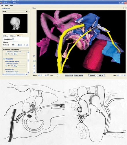
The model used was created from 20 µm thick serial histological sections of a human temporal bone. Sections were digitized, aligned and segmented into anatomical units. The 3D model constitutes a surface rendering of structures of interest, which currently include the temporal bone and air spaces; the perilymphatic and endolymphatic spaces including cochlear aqueduct and endolymphatic duct and sac; the sensory epithelia of the cochlear and vestibular labyrinths; the ossicles and tympanic membrane; the middle-ear muscles; the carotid artery and the auditory, vestibular and facial nerves. For each of these structures, the surface transparency can be individually controlled, thereby revealing the 3D relations between surface landmarks and underlying structures. Moreover, the 3D model can also be re-sectioned in any arbitrary plane resulting in a picture similar to an anatomic section ().
Figure 1. Image enhancement of the anatomical structures allowed with 3D modelling of the visible ear. Top: 3D reconstruction of the middle ear cavities. The ossicular chain appears in blue, the facial nerve in yellow, the cochlea–vestibular apparatus in pink, the carotid artery in red, and the auditory tube in light blue. Bottom: drawings of axial and coronal sections of the middle ear cavities. At least two drawings are required to give information about the 3D arrangement of the structures, whereas the 3D model provides this information directly taking advantage of the 360° rotation to explore several angles of view. Superposition of images or the masking of elements enables the completion or simplification (focussing on, e.g. the auditory ossicles) of the model.

These software packages are currently supported by several systems including Windows, Linux and MacOs.
Evaluation sessions
All evaluations were conducted by the same examiner. Evaluation of the participant-perceived usefulness of the 3D reconstruction tutorial was based on a 6-item questionnaire () given to those first year undergraduate paramedical students who attended the tutorial (i.e. those enrolled in 2008, n = 71) and an 8-item questionnaire given to the residents (n = 19, ). The first year undergraduate students were questioned on their perceived improvement in learning compared to lecture alone, the user-friendliness of the software, and their preferred learning environment for using this software (alone, within a group, or with a teacher). The residents were asked to self-assess their prior anatomical knowledge, measure any anticipated improvement to their academic learning and/or surgical skills from using the software, indicate their level of satisfaction with the tools used previously for temporal bone anatomy teaching, and finally comment on the user-friendliness of the software and their preferred learning environment (alone, within a group, or with a teacher).
Table 1. Results of the questionnaire filled out by first year undergraduates enrolled on speech therapy and hearing aid practitioner courses
Due to the lack of standardized questionnaire specific for temporal bone anatomy learning, we developed our own questionnaires based on these items and adapted to the educational level (undergraduate or graduate students).
Knowledge on temporal bone anatomy was assessed in eight (Montpellier-Nîmes) of the 19 residents prior to the tutorial. Residents had to caption 14 items on a drawing of the middle ear. The results are reported as a percentage of correct answers.
Knowledge gained by the first year undergraduates was evaluated during their final year examination. They had to correctly place the names of several items on a figure with 20 items designated by an arrow. The results are given as a percentage of correct answers. Two different tests were used for the evaluation. The first figure was administrated to 40 speech therapy students in 2007 and 34 hearing aid practitioner students in 2008, whereas the second figure was administrated to 31 hearing aid practitioner students in 2007 and 37 speech therapy students in 2008.
Report and analysis of the data
The results of the questionnaires are reported in and . The results of the final examination of first year students were analysed using stepwise linear logistic regression (Systat 10.0 software, Systat Software Inc., Chicago, IL, USA). Bivariate analysis was performed to identify variables for multivariate analysis. As most of the variables were qualitative, we used the Wilcoxon test to select variables for stepwise logistic regression modelling for multivariate analysis. The percentage of correct answers constituted the dependent variable, and teaching method (with/without 3D reconstruction tutorial), the type of test (1 vs. 2), and undergraduate course (speech therapy vs. hearing aid practitioner) constituted the independent variables. Results were considered statistically significant if the p-value was ≤0.05. Considering their non-Gaussian distribution, scores at the end of first year final examination were compared using a Mann–Whitney test (Systat 10.0 software). Results were considered statistically significant if the p-value was ≤0.05.
Table 2. Results of the questionnaire filled out by the ENT residents
Results
Participant evaluation of the 3D software tutorial
For most of the first year students (), the 3D rendering software was perceived as useful for improving their knowledge of temporal bone anatomy (94.23% of positive opinions), and enhancing their understanding of the traditional teaching via lectures and 2D figures (94.23% of positive opinions). The software was considered as very user-friendly by 15.38% of students and user-friendly by 63.46% of the students. Only 9.62% of students found the software difficult to manipulate. While large percentages of students were willing to use this software either alone or within a group (88.46% and 76.93% strongly or partially agreed, respectively), most would prefer the help of a tutor to complement the instructions given within the software package.
All of the residents () included in this study considered their previously acquired knowledge in temporal bone anatomy insufficient. This was partially confirmed by the pre-test performed on a subset of the residents for whom the average percentage of correct answers was 48.81 ± 11.72 (median 50, minimum 7.14, maximum 85.71); markedly less than the 75% considered to be an acceptable score to be able to perform ear surgery safely in a teaching environment. Only one-fifth of the residents (21.08%) found the classical teaching tools satisfactory for the learning of temporal bone anatomy and all believed that the 3D software could help them improve their academic knowledge and surgical skills. All found the software user-friendly and most (89.47%) would prefer using the software as a self-teaching tool. Use of the software within a group environment or with the help of a tutor was more controversial.
Differences in knowledge acquired among first year students
The results of the final examination correlated well with the type of test (test 1 vs. test 2) and with the form of teaching method (with or without 3D reconstruction tutorial, p < 0.001 for each), but no correlation was found with the choice of course (speech therapy or hearing aid practitioner, p = 0.069). Test 1 was therefore associated with better results than test 2, and the students who received the 3D reconstruction tutorial had better results than those who did not.
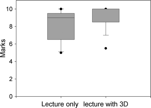
As depicted in , the results obtained in 2008 by the students who received the 3D reconstruction tutorial course were higher than the results of the students of the previous year. Indeed, the average score in 2007 was 80.91 ± 2.18 versus 89.92 ± 1.84 in 2008, with respective median values of 90% and 100%. This difference was statistically significant (p < 0.001, Mann–Whitney test).
Figure 2. Comparison of grades in 2007 (lecture only) and 2008 (lecture and use of 3D reconstruction model). For each box, the line represents the median value, the upper point is the 95th percentile and the lower point is the 5th percentile. Error bars are 25th/75th percentile.

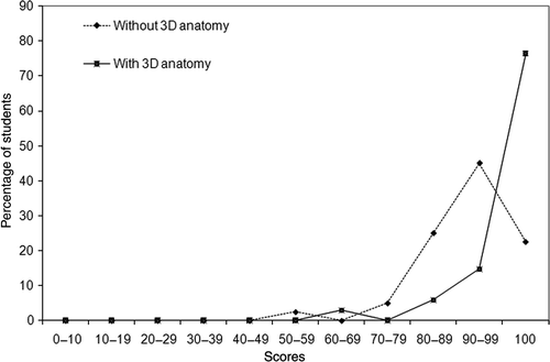
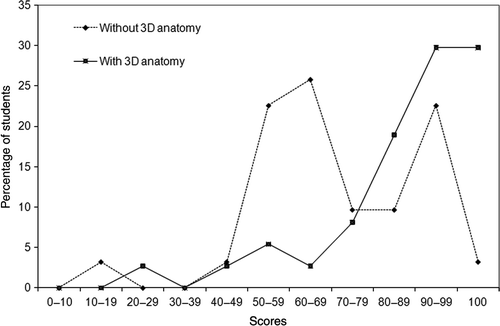
On closer examination of the results obtained for each test ( and ), while an improvement was observed in both tests, it was more obvious for test 2 that was considered to be the most difficult for the students (). Indeed, the bimodal aspect of the curve depicting the results of the students who did not receive the 3D reconstruction tutorial shifted to a more linear aspect with the 3D tutorial. Thus, for test 2 (), the average scores increased from 68.87 ± 3.63 to 83.78 ± 2.99 (median scores of 65% and 90%, respectively), whereas a modest increase from 90.25 ± 1.47 to 93.61 ± 1.39 (median scores of 90% and 100%, respectively), was observed for test 1 (). Nevertheless, the differences observed in both tests following 3D reconstruction tutorial were statistically significant (p < 0.001 for both tests, Mann–Whitney test).
Discussion
The widespread use of information technology has dramatically altered the medical practice over the past three decades. Many applications, sometimes web-based (Petersson et al. Citation2009), have been developed to enhance or replace traditional medical teaching methods, such as lectures, textbooks and laboratory-based work.
The utility of traditional methods of teaching anatomy has also been questioned with the arrival of the information technology era (Spitzer & Scherzinger Citation2006). Indeed, the high level of cost surrounding cadaver dissection has led to a higher number of students per cadaver, making the access difficult for some students. As such, some medical schools have simply stopped offering these forms of teaching (Parker Citation2002). More specifically, dissection would be impossible for most students for the learning of specific anatomical regions, such as the temporal bone.
Virtual 3D anatomical images have vastly enhanced medical imaging and diagnostics (Wiet et al. Citation2005). As such, many teachers have proposed the use of these virtual 3D models as a teaching tool for temporal bone anatomy (Wiet et al. Citation2005; Temkin et al. Citation2006; Smith et al. Citation2007).
While numerous 3D anatomy software packages have now been designed, few studies have reported the impact of their use on learning. Here, we investigated the effect of combining traditional teaching methods with the use of 3D anatomy software in the learning of temporal bone anatomy. We found that students who received the traditional teaching methods (using 2D drawings) enriched with an interactive 3D-reconstruction tutorial, demonstrated a significantly improved retention of information as shown by their overall higher grades during the end of year examination. These results are in agreement with the study of Nicholson et al. (Citation2006), who used a fully interactive model of the middle and inner ear from a magnetic resonance imaging scan of a human cadaver ear. In their study, the students who had been given the tutorial with the 3D model had better scores at the quiz performed shortly after the teaching session (83% in the intervention group vs. 65% in the control group). Similar results have been observed with the use of 3D software designed for the anatomy of complex and small regions, such as the eye or the teeth (Glittenberg & Binder Citation2006; Nance et al. Citation2009).
Our study also suggests a prolonged enhancement of acquired knowledge, lasting up to several months after the teaching session. Indeed, the students included in our study took their examination about 6 months after the teaching session. One explanation for this could be that the students had the possibility to download the software on their own computer, and therefore used it after the teaching session. The individual use of the software was not controlled in our study and therefore this possibility cannot be eliminated. Another explanation for this difference could be the interactive nature of the model. Indeed, this software not only allowed the creation of static 3D images, but also allowed the rotation of images, their slicing in sections and the hiding or enhancement of certain anatomical elements, as was demonstrated during the teaching sessions. It is also reasonable to assume that the novelty of this tool increased the curiosity of the students, thereby increasing their motivation level and their will to become more involved during the teaching session and spend more time on the material. If motivation can explain, at least in part, the differences observed, academic capabilities cannot account for that. Indeed, achievement of students in both groups was similar to comparable rates of failures (7.3% and 6.9% in 2007 and 2008, respectively) at the end of the third year of studies. Whatever the reason behind the difference, the 3D model was shown to improve the middle term results of the students in this study.
The vast majority of students in this study, whether undergraduate or graduate, considered that the traditional teaching tools they had at their disposal were insufficient to learn temporal bone anatomy efficiently. This was most strongly felt among the residents. Despite all of them having received teachings on temporal bone anatomy previously, all deemed their knowledge to be inadequate. This could in part be explained by the gap between their theoretical knowledge, not always proofed by cadaver dissection, and the difficulties experienced in the operating room. This type of 3D model may help bridge this gap and improve the transfer of theoretical knowledge to a more practical use. Indeed, all residents thought that the use of 3D anatomy tools would help enhance their surgical skills. This hypothesis remains to be confirmed by further studies. While 3D anatomy does give precious information about the organization of the structures, it does not however provide the haptic training required for surgery. These students may therefore benefit from the use of 3D models with surgical simulators (Fried et al. Citation2007; O’Leary et al. Citation2008). It is important to remember however that such haptic training can also be acquired by cadaver dissection, which may provide both anatomical learning and surgical training for future surgeons.
The majority of undergraduate and graduate students found the use of the software enjoyable and the interface user-friendly. The main criticism came from the undergraduate students concerning the language of the software. However, this minor issue can easily be addressed by implementing a multilanguage option in future versions of the software. The manner in which to use this type of software was more controversial. This type of software is designed to be a self-teaching tool, and most students agreed with this. However, half to two-thirds of students expressed a will to use the software as part of an interactive tutorial with the help of a teacher. This emphasizes the need for web-based solutions which can be enhanced by or complemented with a comprehensive tutorial with direct access to the teacher, through forum or instant messaging (Petersson et al. Citation2009). Another solution is the use of these tools during group sessions with each student working on his or her own computer under the supervision of a teacher. Despite interesting results, we were unable to test the validity of our questionnaires in an independent study, and additional investigation is required to correlate our results about the friendliness and the type of use of the softwares with other types of efficiency or satisfaction measurements.
Conclusion
These types of softwares, in line with 3D anatomy projects, such as the “Visible Human” (Park et al. Citation2008), are completely interactive allowing students themselves to rotate, slice and visualize the anatomical structures in greater detail, which would normally be performed by someone with experience in dissection. The software also allows the students to enhance their knowledge acquired during traditional lectures with 2D-drawings, thanks to video animations showing the different steps of the virtual dissection. Rendered 3D imaging software may be used by teachers and students of various levels and background as has been demonstrated in this study. Each of their varied needs, from basic anatomy to surgical anatomy, is met using this model. We found that the use of 3D rendering software together with traditional anatomical teaching methods allowed students to achieve better results during their final examination. The range of intrinsic effects of the model, for example an appeal and novelty factor, should be explored in more detail. In addition, the question of whether this type of 3D anatomy software can completely substitute traditional methods of teaching anatomy deserves to be addressed in future evaluations.
Declaration of interest: The authors report no conflicts of interest. The authors alone are responsible for the content and writing of the article.
References
- Crossingham JL, Jenkinson J, Woolridge N, Gallinger S, Tait GA, Moulton CA. Interpreting three-dimensional structures from two-dimensional images: A web-based interactive 3D teaching model of surgical liver anatomy. HPB (Oxford) 2009; 11(6)523–528
- Fried MP, Uribe JI, Sadoughi B. The role of virtual reality in surgical training in otorhinolaryngology. Curr Opin Otolaryngol Head Neck Surg 2007; 15(3)163–169
- Gangata H. An innovative approach to supplement the teaching of the spatial gross anatomy relationships of muscles to undergraduates in health sciences. Clin Anat 2008; 21(4)339–347
- Glittenberg C, Binder S. Using 3D computer simulations to enhance ophthalmic training. Ophthal Physiol Opt 2006; 26(1)40–49
- Nance ET, Lanning SK, Gunsolley JC. Dental anatomy carving computer-assisted instruction program: An assessment of student performance and perceptions. J Dent Educ 2009; 73(8)972–979
- Nicholson DT, Chalk C, Funnell WR, Daniel SJ. Can virtual reality improve anatomy education? A randomised controlled study of a computer-generated three-dimensional anatomical ear model. Med Educ 2006; 40(11)1081–1087
- O’Leary SJ, Hutchins MA, Stevenson DR, Gunn C, Krumpholz A, Kennedy G, Tykocinski M, Dahm M, Pyman B. Validation of a networked virtual reality simulation of temporal bone surgery. Laryngoscope 2008; 118(6)1040–1046
- Park JS, Jung YW, Lee JW, Shin DS, Chung MS, Riemer M, Handels H. Generating useful images for medical applications from the Visible Korean Human. Comput Methods Programs Biomed 2008; 92(3)257–266
- Parker LM. What's wrong with the dead body? Use of the human cadaver in medical education. Med J Aust 2002; 176(2)74–76
- Petersson H, Sinkvist D, Wang C, Smedby O. Web-based interactive 3D visualization as a tool for improved anatomy learning. Anat Sci Educ 2009; 2(2)61–68
- Silen C, Wirell S, Kvist J, Nylander E, Smedby O. Advanced 3D visualization in student-centred medical education. Med Teach 2008; 30(5)e115–124
- Smith DM, Oliker A, Carter CR, Kirov M, McCarthy JG, Cutting CB. A virtual reality atlas of craniofacial anatomy. Plast Reconstr Surg 2007; 120(6)1641–1646
- Spitzer VM, Scherzinger AL. Virtual anatomy: An anatomist's playground. Clin Anat 2006; 19(3)192–203
- Temkin B, Acosta E, Malvankar A, Vaidyanath S. An interactive three-dimensional virtual body structures system for anatomical training over the internet. Clin Anat 2006; 19(3)267–274
- Wang H, Northrop C, Burgess B, Liberman MC, Merchant SN. Three-dimensional virtual model of the human temporal bone: A stand-alone, downloadable teaching tool. Otol Neurotol 2006; 27(4)452–457
- Wiet GJ, Schmalbrock P, Powell K, Stredney D. Use of ultra-high-resolution data for temporal bone dissection simulation. Otolaryngol Head Neck Surg 2005; 133(6)911–915


