Abstract
Abstract: Our objective is to present a comprehensive view of the PDA (prolonged depolarizing afterpotential)-defective Drosophila mutants, nina's and ina's, from the discussion of the PDA and the PDA-based mutant screening strategy to summaries of the knowledge gained through the studies of mutants generated using the strategy. The PDA is a component of the light-evoked photoreceptor potential that is generated when a substantial fraction of rhodopsin is photoconverted to its active form, metarhodopsin. The PDA-based mutant screening strategy was adopted to enhance the efficiency and efficacy of ERG (electroretinogram)-based screening for identifying phototransduction-defective mutants. Using this strategy, two classes of PDA-defective mutants were identified and isolated, nina and ina, each comprising multiple complementation groups. The nina mutants are characterized by allele-dependent reduction in the major rhodopsin, Rh1, whereas the ina mutants display defects in some aspects of functions related to the transduction channel, TRP (transient receptor potential). The signaling proteins that have been identified and elucidated through the studies of nina mutants include the Drosophila opsin protein (NINAE), the chaperone protein for nascent opsin (NINAA), and the multifunctional protein, NINAC, required in multiple steps of the Drosophila phototransduction cascade. Also identified by the nina mutants are some of the key enzymes involved in the biogenesis of the rhodopsin chromophore. As for the ina mutants, they led to the discovery of the scaffold protein, INAD, responsible for the nucleation of the supramolecular signaling complex. Also identified by the ina mutants is one of the key members of the signaling complex, INAC (ePKC), and two other proteins that are likely to be important, though their roles in the signaling cascade have not yet been fully elucidated. In most of these cases, the protein identified is the first member of its class to be so recognized.
The nina and ina mutants have played a major role in the elucidation of the Drosophila photoreceptor signaling cascade (e.g., reviews: Wang & Montell, Citation2007; Katz & Minke, Citation2009; Hardie, Citation2011). nina and ina refer to a group of mutants that were identified from alterations in a component of the light-evoked photoreceptor potential known as the “prolonged depolarizing afterpotential” (PDA). For this reason, these have been referred to as the PDA-defective mutants. There are two classes of PDA-defective mutants, nina and ina, with each class comprising multiple complementation groups. The cellular and molecular functions affected by these mutations are diverse. By and large, the nina class of mutants share varying degrees of deficiency in the amount of the major rhodopsin, Rh1. The defects in the ina class of mutants are more complex, but their ERG (electroretinogram) phenotypes tend to mimic milder versions of that of the phototransduction channel mutant, trp (transient receptor potential). Two of the important components of the Drosophila phototransduction pathway identified through these mutants include the major rhodopsin, NINAE, the first invertebrate rhodopsin to be molecularly characterized (O’Tousa et al., Citation1985; Zuker et al., Citation1985), and INAD, the first scaffold protein identified to orchestrate the formation of a supramolecular signaling complex in a sensory transduction cascade (Huber, Citation2001; Delmas et al., Citation2004).
I. PDA AS A MUTANT SCREENING TOOL
In this review, we will first discuss how we came to use the PDA as a mutant screening strategy and then summarize the information gained from the studies on the nina/ina mutants, isolated using this strategy.
A. The Prolonged Depolarizing Afterpotential (PDA)
We begin with the discussion of the PDA. In many invertebrate eyes, a colored light stimulus, which favors a substantial photoconversion of rhodopsin to its excited form, metarhodopsin (M*), evokes a depolarizing receptor potential that persists in the dark long after the light stimulus is turned off. The PDA refers to this persisting potential in the dark (, arrows). It was first discovered in the lateral ocellus of the barnacle (Hillman et al., Citation1972) and the median ocellus of Limulus (Nolte & Brown, Citation1972). In rhabdomere-based eyes, the absorption maximum of the visual pigment, rhodopsin (R), is well separated from that of metarhodopsin (M*), and, moreover, metarhodopsin is thermally stable at physiological temperatures. In Drosophila, the major rhodopsin present in R1–6 photoreceptors, Rh1, and the corresponding metarhodopsin absorb maximally at ∼485 and ∼575 nm, respectively (Ostroy et al., Citation1974; Pak & Lidington, Citation1974). Thus it is possible to photoconvert rhodopsin to metarhodopsin and back using blue and orange lights. If a sufficiently bright blue light is used to photoconvert >> 20% of rhodopsin to metarhodopsin, the depolarizing potential persists in the dark, i.e., PDA is generated (, arrows); it is then terminated by rephotoconversion of metarhodopsin back to rhodopsin using an orange stimulus (Stephenson et al., Citation1983) The origin of the PDA is now well established (Dolph et al., Citation1993). Arrestin 2 (Arr2), one of the two classes of arrestins (Arrestins 1 and 2) present in the Drosophila eye, binds to M* to terminate its excitation, but Arr2 occurs at 1/5 the concentration of rhodopsin (LeVine et al., Citation1990; Matsumoto & Yamada, Citation1991). If a bright blue stimulus photoconverts Rh1 to M* in molar excess of the available Arr2, M* continues to be active after the cessation of light stimulus and the PDA results. Screening pigments in pigment cells surrounding each ommatidium interfere with the generation of the PDA because they strongly absorb in the blue (Langer, Citation1967; Strother & Casella, Citation1972; Stark, Citation1973). These are genetically removed for ready detection of the PDA.
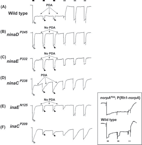
Figure 1. PDA phenotypes revealed in ERG recordings of nina and ina mutants and wild type. The stimulus protocol is shown at the top: three bright blue stimuli (filled rectangles) each of 4 s duration presented at 20-s intervals followed by three bright orange stimuli (unfilled rectangles) also of 4 s duration presented at 20-s intervals. The first blue stimulus generates a large response that lasts the duration of the stimulus (light-coincident component) in flies of all genotypes (unfilled arrowheads). In wild type, the PDA is generated at the termination of the blue stimulus and maintained throughout the two subsequent blue stimuli (Trace A). No PDA is generated in ninaDP245, ninaEP332, or inaEN125 (Traces B, C, and E), and partial PDAs are generated in ninaCP238 and inaCP209 (Traces D and F). During the fully developed PDA in wild type, the R1–6 photoreceptors are inactivated, and only small responses originating from R7/8 photoreceptor are elicited by the second and third blue stimuli (Trace A, filled arrowheads; also see inset). R1–6 photoreceptors of inaEN125 and inaCP209 are also inactivated by the first blue stimulus and generate only small responses to the second and third blue stimuli (Traces E and F, filled arrowheads). Thus, in these ina mutants, the afterpotential (PDA) is not present, but the R1–6 photoreceptors are inactivated, hence the name inactivation but no afterpotential. In strong nina mutants, ninaDP245 and ninaEP332, the PDA is not present and the R1–6 photoreceptors are not inactivated, generating full-amplitude responses to the second and third blue stimuli (Traces B and C, filled arrowheads). Therefore these mutants were named neither inactivation nor afterpotential. The mutant ninaCP238 displays a partial PDA and modest inactivation of R1–6 photoreceptors. The inset illustrates the R7/8 origin of the small responses to the second and third blue stimuli in wild type (Trace A, filled arrowheads). It compares the ERG of wild type (bottom) with that of the transgenic fly (top) carrying wild-type norpA cDNA driven by Rh1 promotor on a norpAP24 mutant background (norpAP24; Rh1-norpA+). The stimulus protocol is shown at the bottom. Since norpAP24 blocks phototransduction and Rh1 drives the expression of wild-type norpA cDNA only in R1–6 cells, phototransduction is blocked in R7/8 cells but the block is rescued in R1–6 cells in this transgenic fly. Note that the small response to the second blue stimulus superposed on the PDA is not present in the transgenic fly. This figure was originally published in the Journal of Biological Chemistry: Pearn, Randall, Shortridge, Burg, & Pak, 1996, J Biol Chem, 271, 4937–4945.
B. Adoption of the PDA-Based Mutant Screening Strategy
Keeping in mind that our goal was to isolate a pool of mutants enriched in phototransduction-defective mutants, it is worthwhile reviewing briefly why we decided to use the PDA in our mutant screening protocol in the first place. As we know, the choice of screening strategy is a compromise between two conflicting demands of mutant screening. On the one hand, the strategy must be sufficiently simple to enable a large number of mutagenized flies to be screened rapidly. On the other hand, it must be sufficiently informative to reveal the phenotypes of desired mutants readily.
We started the screen for phototransduction mutants by phototactic behavioral assay mainly because of its simplicity (Pak et al., Citation1969; review: Pak, Citation2010). After determining that, with optimization of some of the manipulative steps, ERG recordings could be performed on Drosophila relatively simply, we switched to ERG screening to minimize the false positives and negatives inherent in phototactic screening (review: Pak, Citation2010). Clearly, ERG recordings would more directly test for any defects in photoreceptor response than any behavioral assays, since the main component of the ERG corresponds to the extracellularly recorded, mass response of photoreceptors. However, ERG screening will not select only for phototransduction-defective mutants. The ERG represents the voltage developed across the electrodes due to summed extracellular current flow through the tissues in the head generated by light-evoked responses of all responding cells. Defects in the responses of cells other than photoreceptors could affect the ERG. Moreover, any changes that would alter the passive resistive properties of cells in the current path so as to change either the amount or the paths of extracellular current flow would be reflected in the amplitude and waveform of the ERG, even if they arose from reasons totally unrelated to phototransduction. For example, developmental or degenerative changes in extraretinal neurons or glia in the head could alter the properties of resistive current paths.
In the years following the discovery of the PDA (Hillman et al., Citation1972; Nolte & Brown, Citation1972), we became increasingly attracted to it as a potential mutant screening tool that could extend the capabilities of ERG screening. Although the mechanistic understanding of the PDA was rudimentary at the time, it did seem to be closely associated with the photoreceptor potential. If the PDA were an integral component of the receptor potential, any defect in it could be directly attributable to defects in the receptor potential. Moreover, since the PDA is recorded in combination with the “light-coincident” component (, unfilled arrowhead), we thought we could sort out any global effects that affect all components of the ERG, such as those mentioned above, by looking for changes that predominantly or solely affect the PDA with little effect on the light-coincident component.
However, at the time we were considering PDA-based screening, many in the visual physiology community were skeptical whether it represented a physiologically relevant phenomenon at all. If the skeptics were correct, it would be a height of folly to base a major mutagenesis program for the isolation of phototransduction-defective mutants on a phenomenon that has little or no physiological relevance. In 1973, Baruch Minke, who, as a graduate student with Peter Hillman at the Hebrew University, co-discovered the PDA (Hillman et al., Citation1972), came to us as a postdoctoral associate. Baruch clarified some of the issues we had with the PDA. Although the information was still incomplete, and the mechanistic understanding of the PDA did not come until some two decades later (Dolph et al., Citation1993), we decided to go with the PDA-based mutant screening strategy anyway. Although we began using the strategy sporadically as early as 1973/74, it was not until the fall of 1975 that we started using it systematically for the isolation of new mutants. In spite of this initial uncertainly, ultimately, the PDA-based screening turned out to be an efficient means of isolating mutants defective in phototransduction and the rhodopsin chromophore synthesis (see Section II).
C. Wild-Type Phenotype
illustrates a typical PDA protocol we used (filled and unfilled rectangles at the top) and the responses obtained with it from both mutant and wild-type flies. Although we used several variations of this protocol, they all consisted of a series of two or three bright blue stimuli followed by two or three bright orange stimuli. In the protocol illustrated in , three blue stimuli of 4 s duration (, filled rectangles) were followed by three orange stimuli (unfilled rectangles) also of 4 s, all at 20-s interval. Since rhodopsin (Rh1) and metarhodopsin (M*) in R1–6 photoreceptors absorb maximally at ∼485 and ∼575 nm, respectively (see Section IA and , bottom left), the photoequilibrium created by the blue stimulus would strongly favor photoconversion of Rh1 to M*, whereas the photoequilibrium created by the orange stimulus would strongly favor the reconversion of M* to Rh1 in R1–6 photoreceptors. Thus, the blue stimuli would generate the PDA and the orange stimuli would terminate it. Before starting the stimulus series, typically the flies were first exposed to the orange stimulus to photoconvert any M* that might have been present to Rh1, and dark adapted for a few minutes. Because the minority class of photoreceptors, R7/8, have different visual pigments with different absorption spectra (see Section IIA1 for references; reviews: Wang & Montell, Citation2007; Hardie, Citation2011), the same stimulus protocol does not generate the PDA in these photoreceptors. Thus, this protocol would not detect PDA defects specific for R7/8 photoreceptors.
The ERG responses obtained from wild-type flies (marked with w−) using the above protocol are shown in . The first blue stimulus generates a large response that lasts the duration of the stimulus (light- coincident component; , unfilled arrowhead), and after the stimulus is turned off, the response decays to a new level and persists at this new level in the dark. This is the depolarizing afterpotential (PDA) (, arrows). If left alone, it persists for tens of minutes in the dark. A fully developed PDA saturates the depolarizing capacity of the R1–6 photoreceptors, rendering them unresponsive to another stimulus. They are said to be “inactivated.”1 Thus, the second and third blue stimuli generate only small responses superposed on the PDA in extracellular ERG recording (, filled arrowheads). These small responses as well as a comparable portion of the response to the first blue stimulus do not come from R1–6 photoreceptors but from the R7/8 photoreceptors (Minke et al., 1975), in which the PDA is not generated by the stimulus protocol used. In fact, in transgenic flies in which phototransduction is blocked in R7/8 cells but not in R1–6 cells, a similar stimulus protocol would generate ERGs with these superposed responses and the corresponding portion of the first response missing (, inset; Pearn et al., Citation1996). The first orange stimulus, which rephotoconverts M* back to Rh, terminates the PDA and the ERG response returns to the baseline level, i.e., the R1–6 photoreceptors repolarize to the prestimulus level. Because of the broad absorption spectrum of Rh1, the second and third orange stimuli generate responses by activating Rh1 rephotoconverted from M*.
D. Mutant Phenotypes
PDA-defective mutants (or “PDA mutants”) were identified and isolated by looking for those with phenotypes that deviate from the wild-type phenotype. From early on, it was clear that these mutants fell into two broad classes with multiple complementation groups in each (Pak, Citation1979; Stephenson et al., Citation1983). We named these two classes, nina (neither inactivation nor afterpotential) and ina (inactivation but no afterpotential) on the basis of their PDA phenotype, with a capital letter following the class designation (nina or ina) indicating the complementation group.
As the name implies, both classes of mutants are characterized by the absence or underdevelopment of the afterpotential, i.e., the PDA. This is particularly striking in strong nina mutants such as ninaDP245 or ninaEP332 (, arrows). As may be seen, there is no evidence of afterpotentials following the responses to blue stimuli in these mutants. Moreover, unlike in wild type, the second and third blue stimuli generate responses as big or nearly as big as that evoked by the first blue stimulus (, filled arrowheads). Previously, we explained the small responses obtained by the second and third blue stimuli in wild type, consisting exclusively of R7/8 cell responses, to the saturation of depolarizing capacity of R1–6 cells due to the fully developed depolarizing afterpotential induced by the first blue stimulus. Since there is no afterpotential in ninaEP332 or ninaDP245, the R1–6 cells are not saturated and can respond to the second and third blue stimuli with their full capacity. All nina mutants examined to date have been found to be reduced in Rh1 rhodopsin in varying degrees (Stephenson et al., Citation1983). As described earlier, the PDA is generated because of the inability of Arr2 to inactivate M*, which exists in a 5-fold molar excess of Arr2 (Dolph et al., Citation1993). In nina mutants with extremely small amounts of Rh1 (say, ≤ 1%), any M* photoconverted from this small pool of Rh1 can be readily inactivated by the available Arr2 (ninaEP332 or ninaDP245 in ). Hence, there is no afterpotential following the first blue stimulus and the second and third blue stimuli elicit full potentials (no inactivation). In the nina mutants with somewhat larger concentrations of Rh1, such as ninaAP228 with ∼15% Rh1, the afterpotential begins to develop following the cessation of the first blue stimulus but completely decays to the baseline during the interstimulus interval (not shown). In mutants with still larger amounts of Rh1, such as ninaCP238 with ∼35% Rh1, a PDA lasting the entire interstimulus interval develops, but it is much smaller than the fully developed PDA in wild type (, arrows). The second and third blue stimuli generate responses that are distinctly larger than those of wild type, though distinctly smaller than the response to the first blue stimulus (, filled arrowheads). Thus, these mutants show partial PDAs in parallel with partial inactivation. ninaC mutants also exhibit response waveforms different from those of ninaD or ninaE (), indicating that depletion of Rh1 may not be the only phenotype associated with ninaC mutants.
As with nina's, the mutants of the other class, ina, also fail to generate a sustained afterpotential (). Any afterpotential generated by the first blue stimulus decays within a few tens of seconds, the time course depending on the mutant, although the decay (inactivation) kinetics can be complex in some mutants (see ). However, unlike in nina's, the subsequent blue stimuli elicit only small responses, presumably originating from R7/8 cells, even though the afterpotential is small or has even decayed completely to baseline (, filled arrowheads). Thus, although R1–6 cells are inactivated, unlike in nina, the inactivation does not depend on the level of depolarization induced in R1–6 cells by the previous blue stimuli. That is, inactivation of R1–6 photoreceptors is not due to saturation of the depolarizing capacity of these cells caused by the afterpotential generated by the previous blue stimulus, since there is little or no afterpotential.
The origin of inactivation in ina mutants is not well understood but appears to be associated with premature termination of the response that begins almost as soon as the stimulus is turned on. This phenomenon can be seen best in the responses elicited by stimuli of long durations. Shown in are intracellularly recorded responses of several ina mutants (inaCP209, inaEN125, inaFP106x) and wild type to 20-s white stimulus superposed on each other. In wild type, after the initial peak, the response decays to the steady-state level (, filled arrowhead) and remains there for the duration of stimulus, whereas in ina's the response steadily declines during the entire duration of stimulus, the time course and extent of decay depending on the mutant. The premature decay of response during a stimulus is a well-known defining characteristic of the mutants of the phototransduction channel gene trp (Cosens & Manning, Citation1969; Minke et al., 1975), although in general, the response decay in trp is much faster than in most ina mutants (see also Leung et al., Citation2008, ). Another important phenotype of trp mutants is the pronounced refractory period, which prevents generation of full-amplitude response for a minute or more following an initial response to a bright stimulus (Leung et al., Citation2008). The ina mutants that have been examined also display similar refractory periods, although the duration is not as long and the degree of suppression of subsequent responses is not as severe as in trp mutants (Leung et al., Citation2008). In these two important aspects, the ina mutants appear to represent milder forms of trp mutants. Thus, it appears as though in these mutants the response generating mechanism itself steadily inactivates during stimulus and remains inactivated for a time after the stimulus. In the case of trp, Hardie et al. (Citation2001) presented evidence that these defects are attributable to depletion of PIP2 (phosphatidylinositol 4,5-bisphosphate), the substrate for the key effector enzyme for phototransduction, PLCβ (phospholipase Cb). There are two classes of phototransduction channels in Drosophila photoreceptors: highly Ca2+ -permeable TRP and nonselectively cation-specific TRPL (Hardie & Minke, Citation1992). In trp mutants, the only light response remaining is that mediated through the TRPL channels. This response rapidly decays because, in the absence of sufficient Ca2+ influx through the TRP channel to inhibit PLCβ activity, PIP2 is depleted rapidly (Hardie et al., Citation2001). The refractory period presumably represents the time required for regeneration of PIP2. It is not known whether the same or similar mechanisms are responsible for the ina phenotype. Nevertheless, the phenotype suggests that ina mutations affect some fundamental properties of phototransduction, particularly in relation to TRP channel functions.
Figure 2. Intracellularly recorded photoreceptor responses of ina mutants and wild type. Photoreceptor responses to 20-s white stimuli recorded intracellularly from flies of the indicated genotypes are presented superposed on each other. In wild type, the response rapidly declines to a steady-state level (filled arrowhead), which depends on the intensity of the light stimulus, and remains there for the rest of the stimulus. This is a wellknown adaptation response of photoreceptor to the light. In the ina mutants, on the other hand, the decline in amplitude (inactivation)that began at the onset of stimulus continues throughout the stimulus with the time course and the extent of decay depending on the mutant. The kinetics of decay can be complex in some mutants (inaC). Thus, the ina photoreceptor responses tend to terminate prematurely. The premature termination phenotype of a strong ina mutant, such as inaFP106x, closely resembles that of null trp (transient receptor potential) mutants.
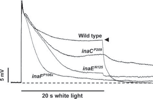
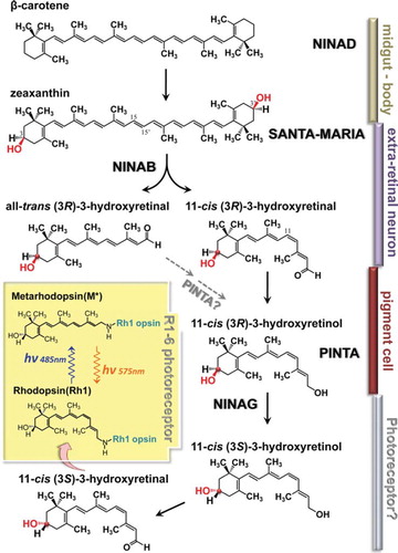
Figure 3. Schematics of putative chromophore biosynthetic pathway. Dietary carotenoids and xanthophils are taken up in the midgut by NINAD and carried by circulation to extra retinal neurons and glia to be taken up there by SANTA MARIA. The dietary β-carotene, once taken up, is reported to be rapidly hydroxylated to form zeaxanthin by unknown mechanisms. In extraretinal neurons and glia, NINAB cleaves and isomerizes zeaxanthin to yield 11-cis (3R)-3-hydroxyretinal and all-trans (3R)-3-hydroxyretinal. Presumably, the chromophore is generated from both products. 11-cis (3R)-3-hydroxyretinal (right branch in ) is converted to its retinol form, 11-cis (3R)-3-hydroxyretinol by a still uncharacterized retinal dehydrogenase. In the mean time, 11-cis (3R)-3-hydroxyretinol may also be generated from all-trans (3R)-3-hydroxyretinal (left branch in ) by conversion to its retinol form and 11-cis isomer. PINTA preferentially binds the retinol form of the retinoids in pigment cells and this binding is required for the chromophore biogenesis, though its precise role is not yet clear. In the final step of chromophore synthesis, 11-cis (3R)-3-hydroxyretinol is converted to its 3S stereoisomer by NINAG to yield 11-cis (3S)-3-hydroxyretinol. This isomerization takes place about the chiral center at the C-3 position of the retinoid ring. C-3 is marked by the red dot and the highlighted OH residue. 11-cis (3R)-3-hydroxyretinol is converted to 11-cis (3R)-3-hydroxyretinal to bind opsin (NINAE) expressed in R1-6 photoreceptors by forming Schiff base linkage with the lysine residue. Inset depicts photo-interconversion of rhodopsin (Rh1) and metarhodopsin (M*).
E. List of PDA Mutants
lists the nina and ina mutants in our collection and the genes identified by them. Most of these mutants were isolated by ethyl methanesulfonate (EMS) mutagenesis in the 6-year period between 1975 and 1981 (see review: Pak, Citation2010), although it took much longer to sort out their complementation groups. Most of them are on the autosomes because they were isolated using autosome-specific mutagenesis schemes (see Pak, Citation1979). In addition to EMS mutagenesis, Joe O’Tousa and Quentin Pye carried out hybrid dysgenic crosses2 in 1982 to isolate alleles of ninaA, ninaB, ninaE, and ninaG. These are indicated by a small “d” following the isolate (allele) number in the superscript in . Gregore Koliantz also used this method in 1987–1988 to isolate nina mutants defining two new complementation groups, ninaJ and ninaI, mapping to the chromosomes X and II, respectively. The first inaF mutant, inaFP105p, was generated by D2–3-mediated P-element mutagenesis in 1997 by Chenjian Li and Lydia Strong (Li et al., Citation1999). Additional alleles were generated in this gene by mobilizing the above allele to induce imprecise excisions.
Table 1. nina and ina mutant alleles in the Pak laboratory.
Some PDA mutants have altered ERG waveforms that can be detected in conventional ERG recordings without using the PDA protocol. Thus, several of our mutants in were actually isolated before the PDA protocol for mutant screening was formally put in place in September 1975. They are inaCP207 (1/74), inaCP209 (3/74), inaDP215 (8/74), inaEP19 (5/69), and ninaCP216 (8/74), with dates of isolation in parentheses. Their PDA phenotype was not known at the time of isolation, and their identity as PDA mutants became known years later through PDA recordings and complementation tests with known PDA mutants.
also lists several mutants isolated in other laboratories, mostly in the John Merriam laboratory at UCLA (University of California–Los Angeles). These are listed in a separate column for each gene. The dates of isolation are not known. The dates we acquired them are also obscure, although we most likely obtained most of them in the early 1980s. As in the case of the previous mutants, they were isolated without using the PDA protocol and were assigned to the respective complementation groups on the basis of genetic complementation tests. In the case of inaEN125, obtained from Martin Heisenberg in Germany, the isolation was based on behavioral optomotor assays (Heisenberg, Citation1971). In all these cases, their identification as PDA mutants came years later. For example, for inaDUS6545, which is homozygous lethal possibly due to a second-site mutation, its assignment to the inaD complementation group did not come until July 1995.
II. CONTRIBUTIONS OF nina/ina MUTANTS TO PHOTORECEPTOR FUNCTION
A. The nina Mutants
We will now discuss how nina/ina mutants have contributed to our knowledge of photoreceptor function. We will start with the nina mutants. The cardinal feature of all nina mutants is an allele-dependent depletion of the visual pigment Rh1 in R1–6 photoreceptors3 (Stephenson et al., Citation1983). In Drosophila, impairments of synthesis of either the protein (opsin) or the chromophore moiety of rhodopsin can lead to rhodopsin depletion. These two mechanisms appear to account for the phenotypes of the majority of nina mutants. Another important mechanism appears to be impairment in maturation and intracellular trafficking of nascent opsin, thereby resulting in depletion of rhodopsin. There are also nina mutants, such as ninaC, in which depletion of Rh1 appears to be only one of the many phenotypes and may not even be their major one.
1. ninaE
The ninaE gene encodes the major class of opsin, Rh1, expressed in R1–6 photoreceptors (O’Tousa et al., Citation1985; Zuker et al., Citation1985). As the gene encoding the first molecule in the phototransduction cascade, this gene was the first to be targeted for molecular cloning. As the number of mutants displaying the nina phenotype accumulated and the complementation groups into which they fell became identified, we came to realize that one common feature all nina mutants seemed to share was Rh1 rhodopsin depletion (Stephenson et al., Citation1983). We hypothesized that one of the five nina complementation groups (ninaA, B, C, D, and E) we had already identified at the time might actually correspond to the Rh1 opsin gene. To test this hypothesis, the available nina mutations were tested for their R1–6 cell-line specificity under the assumption that mutations in any gene encoding R1–6 opsin would only affect R1–6 opsin, and not the minor opsins in R7 or R8. Mutations in two genes, ninaA and ninaE, satisfied this requirement (Larrivee et al., Citation1981; Schinz et al., Citation1982; Stephenson et al., Citation1983). In genetic tests for gene-dosage effect, the ninaE gene (Scavarda et al., Citation1983), but not the ninaA gene (Pak et al., Citation1980), was found to affect the Rh1 rhodopsin content in a dosage-dependent manner, making it very likely that it is the Rh1 opsin gene. This information was crucial for the cloning of this gene.
The ninaE gene was cloned by homology to bovine opsin (O’Tousa et al., Citation1985; Zuker et al., Citation1985). It was the first invertebrate opsin gene to be cloned. At the time, complete sequences were known for only two other opsins, bovine and human (Nathans & Hogness, Citation1983, Citation1984). These were also the first G protein–coupled receptors to have their complete sequences determined. Although the overall amino acid identity between NINAE and bovine opsin was fairly modest (∼36%), NINAE displayed key structural features of opsin. Most notably, it was found to have seven predicted transmembrane segments, with the 7th transmembrane segment containing a conserved lysine residue that would serve as the chromophore linkage site. As with mammalian opsins, there were also several potential phosphorylation sites near the C-terminus.
Cloning of ninaE allowed all five minor classes of opsin to be cloned and their sequences and cellular expressions determined: Rh2 in ocelli (Cowman et al., Citation1986; Mismer et al., Citation1988; Pollock & Benzer, Citation1988), Rh3 and Rh4 in R7 photoreceptors (Zuker et al., Citation1987; Montell et al., Citation1987; Fryxell & Meyerowitz, Citation1987), and Rh5 and Rh6 in R8 photoreceptors (Chou et al., Citation1996; Papatsenko et al., Citation1997; Salcedo et al., Citation1999). A major function of R7 and R8 photoreceptors is color perception. R7 and R8 have shorter rhabdoremeres than R1–6. They are located in the middle of the ommatidium and span only one half of the retina with the R7 rhabdomere located on top of the R8 rhabdomeres. R7 photoreceptors express stochastically either Rh 3 or Rh4 opsins (see above for references), both of which are ultraviolet (UV)-absorbing, in a 30/70 ratio. Expression of Rh3 in R7 is coupled with expression of Rh5 (blue) in R8, whereas expression of Rh4 in R7 is coupled with that of Rh6 (green) in R8 (Chou et al., Citation1996; Papatsenko et al., Citation1997; Chou et al., Citation1999). This pattern of expression defines two spectrally distinct classes of R7/8 photoreceptors, expressing either Rh3/Rh5 or Rh4/Rh6 pair, distributed randomly throughout the eye in a 30/70 ratio. Molecular details of how this coordinate expression of minor opsins in R7 and R8 photoreceptors is achieved have now been largely worked out (reviews: Morante et al., Citation2007; Jukam & Desplan, Citation2010).
It is now widely recognized that mutations in the opsin gene cause degeneration of photoreceptors in both Drosophila and humans. The first indication that mutation in an opsin gene might cause degeneration of photoreceptors came from observations on the Drosophila mutant oraJK84 (Koenig, Citation1975; Koenig & Merriam, Citation1977). It was first reported that oraJK84 mutants are almost completely devoid of rhabdomeres in R1–6 photoreceptors, but not in R7/8 photoreceptors (Koenig, Citation1975; Harris et al., Citation1976). Subsequently, it was found that irregular, loosely packed rhabdomeres do form but quickly degenerate by 1 week post eclosion (Stark & Sapp, Citation1987; O’Tousa et al., Citation1989). O’Tousa et al. (Citation1989) showed (1) that oraJK84 is a double mutant carrying a mutation in the ninaE gene and another mutation in the nearby ort gene and (2) that the mutation in the ninaE gene is solely responsible for the mutant's degeneration phenotype. These findings on Drosophila presaged those then emerging from studies on humans. In humans, a candidate gene approach was used to establish a correlation between a mutation in a gene and retinitis pigmentosa (RP). The rhodopsin gene was the first gene to show such correlation (Dryja et al., Citation1990; Inglehearn et al., Citation1991; Sung et al., Citation1991). It has been estimated approximately 10% of all cases (or approximately 20–25% of autosomal cases) of RP and allied retinal degenerations are due to mutations in the rhodopsin gene (Dryja & Berson, Citation1995).
Some mechanistic understanding has been achieved for some forms of ninaE-mediated degeneration. Retinal degeneration in ninaE null mutants, which include oraJK84, is distinct from those in hypomorphs and appears to be due to rhodopsin being required in rhabdomere morphogenesis (Kumar & Ready, Citation1995). Unlike in hypomorphs, rhabdomere defects in ninaE nulls begin during rhabdomere morphogenesis. As a consequence, structurally normal rhabdomeres never form, and developing rhabdomere membrane, instead of turning into rhabdomeres, catastrophically involutes into the cell as “curtains of apposed membrane.” Even very small amounts of rhodopsin appear to be sufficient to rescue mutants from the above fate (Johnson & Pak, Citation1986; Leonard et al., Citation1992). These hypomorphic ninaE mutants produce smaller than normal rhabdomeres containing structurally normal microvilli, which degenerate over a period of weeks after eclosion (Leonard et al., Citation1992).
Many ninaE mutations are dominant, as are also many mammalian rhodopsin mutations. Two groups developed genetic screens to isolate dominant ninaE mutants (Colley et al., Citation1995; Kurada & O’Tousa, 1995). Some of these mutants carry identical amino acid substitutions as in human autosomal dominant RP (ADRP) patients. In the case of mammals, one of the common mechanisms of dominant rhodopsin-mediated degeneration was found to be faulty maturation and cellular trafficking of nascent rhodopsin (Roof et al., Citation1994; Sung et al., Citation1994). The dominant ninaE mutants were used to address the above question as well as to determine why the dominant ninaE heterozygotes with 50% wild-type rhodopsin molecules also degenerate (Colley et al., Citation1995; Kurada & O’Tousa, 1995; Kurada et al., Citation1998). They found that the mutant proteins interfere with the maturation of the wild-type proteins. These proteins (mutant as well as immature wild-type) do not reach the rhabdomeres and accumulate in the endoplasmic reticulum (ER), causing proliferation of ER cisternae and ultimately degeneration (Colley et al., Citation1995).
Another mechanism of dominant rhodopsin-mediated degeneration in mammals was found to be constitutive activity of rhodopsin (Robinson et al., Citation1992; Dryja et al., Citation1993; Rao et al., Citation1994). The constitutively active dominant ninaE allele, NinaEpp100, was isolated in a genetic screen (Iakhine et al., Citation2004). However, constitutive opening of light-sensitive channels seems to make only a minor contribution to degeneration in this mutant, since near-null mutations in the PLCβ gene (norpA), and the TRP channel gene (trp), suppress the degeneration phenotype only weakly. Instead, the major mechanisms of degeneration appear to be (1) the persistent formation the NINAEpp100-Arr2 complex and (2) the elevated levels of Gqα in the cytosol. Activation of rhodopsin to metarhodopsin causes (1) Arr2 to bind to metarhodopsin to inactivate the latter and (2) Gqα to translocate to the cytosol as a long-term mechanism of adaptation. If rhodopsin is constitutively active, both these events would occur persistently. Stable meta-Arr2 complexes have been shown to be endocytosed and to cause apoptosis of photoreceptors (Alloway et al., Citation2000; Kiselev et al., Citation2000). The persistent localization of Gqα in the cytosol appears to be a novel major cause of degeneration, independent of the Arr2-dependent mechanism.
Recently, a completely new function has been discovered for Rh1 rhodopsin, temperature sensing in Drosophila larvae (Shen et al., Citation2011). Generally, temperature sensing in animals is mediated through direct activation of transient receptor potential (TRP) cation channels (Caterina, Citation2007; Bandell et al., Citation2007). In Drosophila larvae, however, discrimination between 18°C temperature, which is optimal for their survival, and slightly higher temperatures (19–24°C) is accomplished indirectly through a signal transduction cascade similar to that used in Drosophila phototransduction (Kwon et al., Citation2008). The receptor molecule initiating this cascade appears to be Rh1 rhodopsin, since mutations in the ninaE gene eliminate this thermosensory response (Shen et al., Citation2011).
2. ninaA
Early on, the ninaA gene sparked interest because mutations in this gene, along with those in the ninaE gene, were found to reduce the rhodopsin content specifically in R1–6 photoreceptors (Larrivee et al., Citation1981; Stephenson et al., Citation1983). The molecular basis of the R1–6 cell-line specificity of ninaE was elucidated with the demonstration that ninaE encodes Rh1 opsin (apoprotein of rhodopsin) specifically expressed in R1–6 cells (previous section). However, the specificity of ninaA for R1–6 cells was not clear.
The ninaA gene was found to encode a 237-aa (amino acid), cyclophilin homolog (Schneuwly et al., Citation1989; Shieh et al., Citation1989). Because cyclophilins were shown to be peptidyl prolyl cis-trans isomerases (Takahashi et al., Citation1989; Fischer et al., Citation1989) implicated in catalyzing protein folding, it was first thought that the NINAA protein might be required for the proper folding and stability of Rh1. However, the following lines of evidence demonstrated that NINAA acts as a chaperone in intracellular trafficking of nascent rhodopsin from the endoplasmic reticulum (ER), where opsin is synthesized, to the rhabdomeres. NINAA is expressed in the ER and secretory vesicles, and, in ninaA mutants, transport of Rh1 out of the ER is strongly inhibited, leading to dramatic Rh1 retention in ER and accumulations of ER cisternae (Colley et al., Citation1991) and eventual degeneration of photoreceptors (Colley et al., Citation1995). The NINAA protein forms a stable and specific complex with Rh1, and maturation of Rh1 rhodopsin depends quantitatively on the amount of NINAA available (Baker et al., Citation1994). The specificity of ninaA mutations for Rh1 was shown to arise from the substrate specificity of NINAA for Rh1 opsin (Stamnes et al., Citation1991).
The NINAA protein was the first membrane- associated accessory protein identified to play an important role in the maturation of a G protein–coupled receptor (GPCR: rhodopsin, in this case) (Colley et al., Citation1991; review: Bermak & Zhou, Citation2001). In searching the bovine genome for NINAA-homologous genes highly expressed in the retina, Ferreira et al. (Citation1995) identified the RanBP2 (Ran-binding protein 2) gene, encoding a large protein that binds the GTPase Ran (Yokoyama et al., Citation1995; Wu et al., Citation1995). Two contiguous domains of this multidomain protein, Ran- binding domain 4 (RBD4) and the adjacent cyclophilin domain, were shown to act in concert as a chaperone to facilitate the biogenesis of the red/green visual pigment in the dichromatic bovine retina (Ferreira et al., Citation1995). In Caenorhabditis elegans, ODR-4 (abnormal odorant response 4) was shown to be required for the proper targeting of the odorant receptor ODR-10 to olfactory cilia (Dwyer et al., Citation1998). ODR-4 is a 445-aa protein expressed exclusively in the olfactory organ with a similar structural topology but no obvious sequence similarity to NINAA. It may be that many GPCRs require specific chaperone/accessory proteins of their own for proper folding/maturation.
In vertebrates, export of GPCRs from the ER and transport to the Golgi and the cell surface has been extensively studied and found to be a highly regulated dynamic process requiring many players at several different stages of regulation (reviews: Bermak and Zhou, Citation2001; Dong et al., Citation2007). A crucial step in this regulation is interactions of GPCRs with chaperone proteins in the ER. Among the best characterized ER chaperone proteins for GPCRs are calnexin, calreticulin, and BiP (Kleizen & Braakman, Citation2004; Williams, Citation2006). In the Drosophila eye, the protein products of one or the three known calnexin genes, cnx99A, were found to be required as chaperones specifically for Rh1 maturation, much like NINAA (Rosenbaum et al., Citation2006). CNX99A also acts as a regulator of Ca2+ entering the photoreceptors during phototransduction. Mutations in this gene cause light-enhanced retinal degeneration.
3. ninaB, D, G, santa maria, and pinta: Genes Required for Chromophore Synthesis
Animals depend on dietary intake of carotenoids and xanthophills (mainly β-carotene) for the production of the chromophore. Although retinal (the aldehyde of vitamin A) is commonly used as the chromophore in most vertebrates, chemically related compounds are also utilized for this purpose in various species throughout the animal kingdom. For example, (3,4)-didehydroxyretinal is found in fish and amphibians (Suzuki et al., Citation1984) and 3-hydroxyretinal, derived from vitamin A3 (3-hydroxyretinol), is found in insects (Vogt, Citation1989). The two enantiomers of 3-hydroxyretinal appear to be differentially utilized in insects. Whereas the lower order dipterans use (3R)-3-hydroxyretinal, members of the higher Diptera, Cyclorrapha, which include Drosophila, use (3S)-3-hydroxyretinal (Seki et al., Citation1994), at least for the majority class of photoreceptors, R1–6 (Ahmad et al., Citation2006).
It was realized since the early 1980s that, in a substantial fraction of nina mutants, defects in chromophore synthesis rather than opsin synthesis was the basis of rhodopsin depletion (Stephenson et al., Citation1983). Thus, mutants in two of the five nina genes tested by these authors, ninaB and D, could be rescued by dietary supplementation with carotenoids or retinoids. Moreover, ninaB and D mutants exhibited different substrate preferences for rescue. Whereas ninaD mutants could be rescued by a broad range of carotenoids and retinoids, ninaB seemed to show a preference for retinal, suggesting that the ninaB gene product may act downstream of the ninaD product. Since that time, three other genes that function in chromophore synthesis have been identified, santa maria (scavenger receptor acting in neural tissue and majority of rhodopsin is absent) (Wang et al., Citation2007), pinta (PDA is not apparent) (Wang & Montell, Citation2005), and ninaG (Sarfare et al., Citation2005; Ahmad et al., Citation2006). The santa maria and pinta mutants were isolated by the Craig Montell group at Johns Hopkins using a mutant selection scheme similar to the “PDA protocol” we have described. The first ninaG mutant, ninaGP330, was isolated in 1979, as part of the original PDA-based screen. The remaining alleles were isolated in hybrid dysgenesis screen in 1982 by Joe O’Tousa and Quentin Pye, both of whom were in the Pak laboratory at the time. Studies of mutants in these five genes form the basis of our current understanding of the chromophore synthetic pathway in Drosophila.
The first of the chromophore-related genes to be characterized was ninaB. It encodes a β,β-carotene-15,15′-monooxygenase (BCO), which was shown in an in vitro assay to symmetrically cleave β-carotene at the 15-15’ double bond to yield two molecules of retinaldehyde (von Lintig & Vogt, Citation2000). The NINAB protein was the first invertebrate member of the β-carotene cleavage oxygenase I (BCO1) family to be characterized. In living flies, however, dietary β-carotene is reported to be preferentially hydroxylated to form zeaxanthin (3,3′-dihydroxy β,β′-carotene) (Giovanuucci & Stephenson, 1999; Voolstra et al., Citation2006). Detailed enzymatic analyses of both moth and Drosophila NINAB suggest that NINAB can use zeaxanthin as a substrate to yield two molecules of 3-dihydroxyretinaldhyde (Oberhauser et al., Citation2008; Voolstra et al., Citation2010). This would occur outside the retina, since NINAB is expressed in extraretinal neurons and glia in the head (Wang et al., Citation2007).
NINAB has been reported to function also as an isomerase. NINAB, like other the members of the carotenoid cleavage oxygenase family, shows significant sequence homology to mammalian RPE65 (retinal pigment epithelium 65-kDa protein), which is important in the reisomerization of all-trans retinol in mammals (Jin et al., Citation2005). Indeed, a NINAB homolog in the moth Galleria mellonella was found to exhibit both oxygenase and isomerase activities in that it catalyzes the conversion of zeaxanthin (3,3′-dihydroxy β,′-carotene) into equal parts of all-trans and 11-cis dihydroxy retinaldehydes (Oberhauser et al., Citation2008). The combined oxygenase and isomerase activities of NINAB may also be the basis of the earlier observation that Drosophila can biosynthesize 11-cis 3-hydroxyretinal from carotenoid in the dark (Seki et al., Citation1986; Isono et al., Citation1988).
Two genes that encode proteins likely to be involved in the uptake of dietary β-carotene have been identified, ninaD and santa maria (Kiefer et al., Citation2002; Wang et al., Citation2007). Kiefer et al. (Citation2002) showed that ninaD encodes a Drosophila homolog of class B scavenger receptor type 1 (SRB1). Genes of the SRB1 family are conserved throughout metazoans and encode proteins implicated in lipid binding and transport, particularly cholesterol transport in mammals (Acton et al., Citation1996). It was therefore suggested NINAD likely plays a role in the uptake of β-carotene. However, since NINAD is expressed in the midgut, whereas NINAB is expressed in extraretinal neurons and glia in the head (Wang et al., Citation2007), it seemed unlikely that carotenoids taken up in the midgut via NINAD is the substrate of NINAB for centric cleavage. Wang et al. (Citation2007) isolated a mutant in a new locus, santa maria, encoding another SRB1. In contrast to NINAD, SANTA MARIA is co-expressed with NINAB in glia and extraretinal neurons. Thus, the picture that emerges is that dietary β-carotene is taken up in the midgut via NINAD and carried by circulation to glia and extraretinal neurons to be taken up there via SANTA MARIA, probably as zeaxanthin, the hydroxylated form, and NINAB uses it as substrate for cleavage ().
The protein products of two other genes have been proposed to participate in the subsequent steps of chromophore synthesis, pinta (Wang & Montell, Citation2005) and ninaG (Sarfare et al., Citation2005; Ahmad et al., Citation2006) (see ). The pinta gene encodes a retinoid-binding protein, with preference for all-trans retinol, expressed and required in the retinal pigment cells for the production of the chromophore. This finding was the first to implicate the retinal pigment cells in the production of the chromophore in Drosophila. However, the precise cellular function of PINTA has not yet been elucidated. Two possibilities, which are not mutually exclusive, have been proposed (Wang & Montell, Citation2005): (1) it sequesters all-trans retinol in pigment cells thereby generating a concentration gradient that favors uptake of retinol, and/or (2) it facilitates the presentation of all-trans retinols to the enzymes involved in the next step of chromophore synthesis. In light of the previous discussion, however, the retinoids PINTA binds may actually be 3-hydroxyretinoids rather than retinoids (without the hydroxyl residue). PINTA binding of 3-hydroxyretinoids was not investigated by these authors.
The ninaG gene encodes a glucose-methanol- choline (GMC) oxidoreductase proposed to act in the conversion of (3R)-3-hydroxyretinol to the 3S enantiomer in the retina (; Ahmad et al., Citation2006). The conversion of 3R to 3S form occurs through isomerization about the chiral center at the C3 position of the retinoid ring (The C3 position is marked with a red dot in ). In ninaG mutants, synthesis of the chromophore (3S)-3-hydroxyretinal is blocked (Sarfare et al., Citation2005). Moreover, in transgenic flies ectopically expressing Rh4 visual pigment in the majority class photoreceptors, R1–6, when placed on a ninaG mutant background, large amounts of 11-cis3-hydroxyretinol accumulate, suggesting that 11-cis3-hydroxyretinol is an intermediate just upstream of the ninaG block. They thus proposed that the accumulating intermediate is in the 3R form and NINAG acts in the final step of chromophore production, the conversion of (3R)-3- hydroxyretinol to the 3S enantiomer (Sarfare et al., Citation2005; Ahmad et al., Citation2006) (). In the Drosophila eye, only Rh1 rhodopsin expressed in R1–6 photoreceptors use (3S)-3-hydroxyretinal as its chromophore, and the rhodopsins expressed in the minor class of photoreceptors, such as Rh4, have (3R)-3-hydroxyretinal–based chromophore. Thus, only Rh1 rhodopsin is depleted in ninaG mutants. In the experiment mentioned above, the authors used transgenic flies ectopically expressing Rh4 in R1–6 photoreceptors to avoid the possibility that any alterations in the amount of retinoids that they observe might be secondary effects of rhodopsin depletion.
4. ninaC
The ninaC gene encodes a class III myosin expressed predominantly in the eye (Montell & Rubin, Citation1988). Myosin III differs from all other classes of myosins in having an N-terminal kinase domain. The two Drosophila NINAC isoforms were the first class III myosins to be identified. Since then, a NINAC homolog has been identified in the Limulus eye (Battele et al., 2001), in striped sea bass (Dosé et al., Citation2003), and humans (Dosé and Burnside, Citation2000; Berg et al., Citation2001). Human myosin III and Drosophila NINAC have been shown to bind actin in an ATP-dependent manner (Hicks et al., Citation1996; Dosé et al., Citation2003), and human myosin III has been shown to perform plus-end–directed motor activity (Komaba et al., Citation2003; Kambara et al., Citation2006). One of the two human MyoIII isoforms, MyoIIIA, is highly expressed in the retina and cochlea and mutation in MyoIIIA gene has been reported to cause non-syndromic hearing loss (Walsh et al., Citation2002). However, little is known about cellular functions of vertebrate MyoIII, and to date most of the details of in vivo functions of MyoIII have come from the work on the Drosophila eye. Unlike human myosin III, motor function has not yet been directly demonstrated for NINAC.
The ninaC gene encodes two splice variants: a 174-kDa protein (p174) localized to the rhabdomeres and a 132-kDa protein (p132) localized to the cell body (Montell & Rubin, Citation1988; Porter et al., Citation1992). The NINAC protein is a multifunctional protein, and strong ninaC mutants exhibit a wide range of phenotypes. These include reduced visual pigment contents (Stephenson et al., Citation1983), ultrastructural microvillar abnormality (Matsumoto et al., Citation1987; Hicks & Williams, Citation1992), defects in response termination and adaptation (Porter et al., Citation1992, Citation1993), loss of rhabdomeral calmodulin (Porter et al., Citation1993), and light-dependent retinal degeneration (Porter et al., Citation1992), etc. For many of these phenotypes, it is not yet clear what their molecular bases are or how they are related to each other. We will discuss some of these below.
Reduction in visual pigment content was one of the first phenotypes to be noted in this mutant (Stephenson et al., Citation1983). Also noted early was the loss of the central axial cores of the rhabdomeral microvilli in strong ninaC mutants in electron microscopy (Matsumoto et al., Citation1987). The axial cytoskeleton, consisting of actin filaments, appears as a single structure with side arm linkages to the microvillar plasma membrane in electron microscopy (EM) (Arikawa et al., Citation1990). Hicks et al. (Citation1996) showed that ninaC mutants lacking the p174 isoform display a fragmented axial structure without side arm linkage even before eclosion, eventually resulting in degeneration of rhabdomeres. They suggested that p174 may have a role in forming the structural linkage between the axial cytoskeleton and the microvillar membrane, in a manner similar to that of brush border myosin I in the intestinal microvilli (Matsudaira & Burgess, Citation1979). This hypothesis, if correct, could provide common explanations for some of the ninaC phenotypes.
One of the salient characteristics of the ERG of strong ninaC mutants is the slow response termination following light off (slow deactivation—see footnote 1) (Porter et al., Citation1992, Citation1993). Deactivation of the light response is at least in part Ca2 + -regulated and appears to be mediated through the Ca2 + -binding regulatory protein, calmodulin (CaM). In Drosophila photoreceptors, CaM is highly enriched in the rhabdomeres (Porter et al., Citation1993). The rhabdomeral localization of CaM is determined by the NINAC isoform p174, which localizes specifically to rhabdomeres itself. Thus, the CaM localization to the rhabdomeres is severely reduced in mutants that do not express the p174 isoform or in which CaM binding sites in p174 are eliminated (Porter et al., Citation1993, Citation1995). Moreover, the elimination of CaM sites results in slow termination of light response (ibid.). Similarly, hypomorphic mutants of the CaM gene, cam, display slow prolonged response termination (Scott et al., Citation1997).
The NINAC protein has also been implicated in the light-dependent shuttling of signaling proteins into and out of the rhabdomeres during long-term adaptation. Arrestin 2 (Arr2) (Kiselev et al., Citation2000; Satoh & Ready, Citation2005), the α subunit of G protein (Gqα) (Kosloff et al., Citation2003; Cronin et al., Citation2004), and the TRPL channel (Bähner et al., Citation2002) have all been shown to translocate in a light-dependent manner to regulate the sensitivity of the phototransduction machinery during light and dark illumination conditions. Thus, Gqα and TRPL, both of which contribute to activation of phototransduction, shuttle out of the rhabdomeres during illumination and translocate back into the rhabdomeres in the dark, whereas Arr2, which inactivates phototransduction, does the opposite. Lee et al. (Citation2003) reported that translocation of Arr2 into the rhabdomeres requires phosphoinositide (PI)-mediated interactions of Arr2 with NINAC. However, this finding was disputed by another group, which found no evidence of NINAC-dependent shuttling of either Arr1 or Arr2 (Satoh & Ready, Citation2005). Translocation of Gqα into the rhabdomeres has also been reported to require NINAC (Cronin et al., Citation2004). In both these cases, NINAC is required for shuttling into the rhabdomeres. Assuming that NINAC is a plus-end–directed motor, as the human MyoIII was shown to be (Komaba et al., Citation2003; Kambara et al., Citation2006), this direction of movement is consistent with the polarity of actin filaments, which are arranged with the plus ends toward the distal end of the microvilli (Arikawa et al., Citation1990). However, in the case of TRPL shuttling, ninaC null mutation is reported to disrupt movements out of the rhabdomeres (Meyer et al., Citation2006). Unlike the other two signaling proteins, TRPL is a membrane protein. It is possible that TRPL does not utilize the NINAC-based movement. The above authors suggested that the observed disruption of translocation in ninaC may be a secondary effect of degeneration in ninaC.
NINAC is also involved in Ca2+-dependent inactivation of metarhodopsin (M*) (Liu et al., Citation2008). Following photoconversion of rhodopsin to M*, M* is inactivated by Arr2 binding. Liu et al. (Citation2008) measured the lifetime of M* using a two-flash technique in which the first flash photoactivates rhodopsin and the second flash activates M*. If the second flash hits M* before it is inactivated by Arr2, photoconversion of M* to rhodopsin occurs. On the other hand, if it hits M* after it has inactivated, no photoconversion can occur. They found that M* inactivation is very rapid (τ ∼ 20 ms) under physiological conditions and highly Ca2+-dependent, being ∼10-fold slower in Ca2+-free solutions. Moreover, they found that this process requires calmodulin (CaM) and NINAC, because mutations in cam and ninaC genes essentially eliminate the Ca2+ dependence. They proposed that under low-Ca2+ conditions, Arr2's within microvilli are bound to NINAC hindering their access to M*. Following Ca2+ influx, Ca-CaM binds to NINAC, causing NINAC to release Arr2, which can now diffuse and inactivate M*. They argued that this strategy of Ca2+ acting via CaM and NINAC to accelerate Arr2-M* binding promotes “quantum efficiency, temporal resolution, and fidelity of visual signaling.”
Considering the multifunctional nature of the NINAC protein, it would not be surprising if NINAC performed many of its functions through interacting protein partners. Recently, such a NINAC-interacting protein has been identified, Retinophilin (Retin/RTP), a poorly characterized protein conserved from flies to humans. Retin/RTP is a retinal phosphoprotein, which is phosphorylated in the dark and dephosphorylated by light, and is identical to the 23-kDa phosphoprotein first identified in two-dimensional (2-D) gel analysis (Matsumoto & Pak, Citation1984). Two groups have shown recently that this protein is highly expressed in the rhabdomeres and associates with NINACp174, and this interaction is mutually required for the stability of both proteins (Mecklenburg et al., Citation2010; Venkatachalam et al., Citation2010). Mecklenburg et al. (Citation2010) showed that mutants in this gene display high rates of spontaneous dark noise, as do ninaC mutants (Hofstee et al., Citation1996), leading to the suggestion that the role of RTP is to suppress random dark noise to promote signaling fidelity (Mecklenburg et al., Citation2010). Venkatachalam et al. (Citation2010), on the other hand, focused on the age-dependent slow termination of light response in retin/rtp mutants, which seems to parallel the loss of eye-enriched protein kinase C (PKC) (INAC) (see Section IIB1). They proposed that loss of Retin/RTP leads in turn to instability and reductions in levels of p174, the scaffold protein INAD (see Section IIB2), and ePKC, and that this ePKC decline underlies the slow termination defect.
B. The ina Mutants
Unlike the nina mutants, the rhodopsin levels are normal in most ina mutants. In the discussion of mutant phenotypes (Section ID), we noted certain similarities in key features of the ERG between the ina mutants and the trp mutants of the phototransduction channel, TRP. The ina mutant ERGs tend to behave as though they were milder versions of trp ERG, although some ina mutants, such as inaFP106x, exhibit phenotypes as strong as that of a null trp. The cellular and molecular functions that have been uncovered for the INA proteins to date are diverse. Nevertheless, one common feature that these proteins share is that they are all somehow related to TRP channel functions. Thus, for example, INAC is an eye-enriched PKC whose major substrate is TRP, INAE is a potential key enzyme in the production of messenger(s) to TRP, and INAF is required for the normal expression (and perhaps function) of TRP. As for INAD, it orchestrates the formation of a supramolecular signaling complex whose key member is TRP. We now discuss each of the ina genes in more detail.
1. inaC
The first inaC mutants were isolated before the PDA mutant screening protocol was put in place in 1975 because their slow response deactivation phenotype is detectable even without this protocol. However, their identification as ina mutants came much later using the protocol.
The inaC gene encodes an eye-enriched protein kinase C (ePKC) (Smith et al., Citation1991), which is expressed primarily in the rhabdomeres (Schaeffer et al., Citation1989; Smith et al., Citation1991). It exhibits 53% identity to human PKCβ1 and contains C1 and C2 domains for binding diacylglycerol (DAG) and Ca2+, respectively (Shieh et al., Citation2002).
As to be discussed in the next section (Section IIB2), some of the key components of the Drosophila phototransduction cascade are arranged in supramolecular signaling complexes formed by the scaffold protein, INAD, presumably to facilitate speed and efficiency of signaling. Along with PLCβ (NORPA), a key activator of the transduction cascade, and TRP, the transduction channel, ePKC is a core member of this signaling complex (Huber et al., Citation1996; Shieh & Zhu, Citation1996; Chevesich et al., Citation1997; Tsunoda et al., Citation1997). This fact alone suggests its importance in phototransduction, although details of its function still remain to be elucidated.
Null or strong mutants in this gene exhibit a severe defect in response termination (deactivation) (Ranganathan et al., Citation1991; Smith et al., Citation1991) and adaptation (Hardie et al., Citation1993). The deactivation defect is traceable to slow termination of quantum bumps, responses to single photons. In whole-cell recordings, these decay as current noise of unknown origin undergoing damped oscillations (Hardie et al., Citation1993; Henderson et al., Citation2000). The TRP channel protein is a major target of INAC-mediated phosphorylation (Huber et al., Citation1998; Liu et al., Citation2000), and proper termination of light response depends at least in part on TRP phosphorylation, since transgenic flies lacking the INAC-dependent phosphorylation site, Ser982 of TRP, display slow response termination (Popescu et al., Citation2006; see Section IIB2). Other reported substrates of INAC include the scaffold protein INAD (see Section IIB2) (Huber et al., Citation1996, Citation1998; Liu et al., Citation2000) and NINAC myosin III (Li et al., Citation1998). Phosphorylation of these proteins by INAC has also been implicated in proper termination of light response (Huber et al., Citation1996; Li et al., Citation1998).
2. inaD
The first inaD mutant, inaDP215, isolated in August 1974, was one of the first PDA-defective mutants to be isolated. It was the only mutant available in this gene at the time and formed the basis of much of the early work on inaD.
The inaD gene was cloned and sequenced by Shieh and Niemeyer (Citation1995). The encoded protein was found to be a scaffold protein consisting of five protein-binding PDZ motifs (Postsynaptic Density 95, Discs Large, Zona Occludens 1) (Tsunoda et al., Citation1997) and to nucleate the formation of a signaling complex by binding several key components of the phototransduction cascade, TRP, NORPA (PLCβ), and INAC (ePKC) (Huber et al., Citation1996; Shieh & Zhu, Citation1996; Chevesich et al., Citation1997; Tsunoda et al., Citation1997) in stoichiometric ratios (Huber et al., Citation1996). INAD is required for the stability and normal localization to the rhabdomeres of the other three proteins (Chevesich et al., Citation1997; Tsunoda et al., Citation1997). The complex between INAD and the two soluble proteins, NORPA and INAC, is preformed before the complex reaches the rhabdomeres (Tsunoda et al., Citation2001). The relationship between INAD and TRP, however, is mutual. Not only does TRP require INAD for its stability and rhabdomeral localization but also INAD needs TRP for the same. Thus, INAD rhabdomeral localization is severely disrupted in trp null mutants or in transgenic flies expressing TRP with deleted INAD binding site, resulting in the destruction of the entire signaling complex (Li & Montell, Citation2000; Tsunoda et al., Citation2001). The mutual requirement between TRP and INAD is not for targeting of either protein to the rhabdomeres but for long-term retention in the rhabdomeres. That is, either protein can reach the rhabdomeres independently of the other but does not remain there without the other.
Other reported binding partners of INAD include TRPL, rhodopsin (Rh1), NINAC, and calmodulin (CaM) (Xu et al., Citation1998; Wes et al., Citation1999). These do not depend on INAD for rhabdomeral localization, and the binding is thought to be dynamic. Nevertheless, the INAD binding site in NINAC was reported to target a heterologous protein to the rhabdomeres (Wes et al., Citation1999). Since then, the immunophilin, FKBP59 (Goel et al., Citation2001), and INAF (Cheng & Nash, Citation2007; see Section IIB4) have also been shown to bind INAD by Western blotting of head extracts. A NINAC-binding protein, retinophilin (Retin/RTP) interacts with INAD indirectly through NINAC (see Section IIA4), and this interaction is reported to be required for the long-term stability of INAD (Venkatachalam et al., Citation2010). However, Mecklenburg et al. (Citation2010) did not find any noticeable difference in the amount of INAD in their retin/rtp mutant.
A number of functions have been ascribed to the INAD scaffolding of signaling proteins. One of the more obvious is the rhabdomeral localization of the key signaling proteins in high concentrations and stoichiometric ratios and the preservation of their stability. The preassembly of the INAD-NORPA-INAC complex would help ensure that the assembled proteins are in the proper composition and correct stoichiometry (Tsunoda et al., Citation2001). In addition, it has been argued that a major function of INAD complexes is to promote speed and efficiency of transduction by bringing key proteins in close proximity to each other (Huber et al., Citation1998). INAD and TRP have been reported to be targets of INAC (ePKC)-dependent phosphorylation (Huber et al., Citation1998; Liu et al., Citation2000). Thus, the INAD scaffolding would bring the transduction channel, TRP, into the immediate vicinity of its activator, PLCβ, and its potential regulator, ePKC. Popescu et al. (Citation2006) showed that TRP is phosphorylated by ePKC at Ser982 both in vitro and in vivo and that this phosphorylation depends on INAD in vitro. Moreover, mutation of this site leads to slow response termination kinetics, in support of INAD's role in promoting speed. Other evidence in support of the above argument includes the following: (1) genetic elimination of INAC (ePKC) results in slow response termination that is observed at the bump level (Hardie et al., Citation1993; Henderson et al., Citation2000) (see Section IIB1 on inaC) and (2) a similar phenotype is also observed in inaDP215 mutants (Henderson et al., Citation2000), in which INAD-TRP binding is defective (Shieh & Niemeyer, Citation1995; Shieh & Zhu, Citation1996). However, it has also been reported that transgenic flies expressing TRP with deleted INAD-binding site show normal response kinetics (Li & Montell, Citation2000).
Recent evidence suggests that the INAD scaffold may have a dynamic role in signaling in addition to its role as a scaffold for the formation of signaling complexes. Mishra et al. (Citation2007) carried out crystal structure studies of the recombinant peptide of INAD PDZ-5, which is a PLC-binding domain (Shieh et al., Citation1997; van Huizen et al., Citation1998). Their studies suggested that PDZ-5 exists in two redox-dependent conformations—a reduced state maintained in the dark and an oxidized state switched on by light-dependent formation of an intramolecular Cys-Cys disulfide bond (Mishra et al., Citation2007). The formation of the disulfide bond would distort the PLC-binding groove to potentially regulate signaling. Mutants in which the formation of the disulfide bond is disrupted exhibit defects in response termination, the bump refractory period, and escape behavior. Insights into the potential mechanism of light-dependent cycling of the redox state of PDZ5 were provided by Liu et al. (Citation2011). They showed that whereas isolated PDZ5 is stable in the oxidized state, its interaction with the neighboring PDZ4 to form a PDZ4-5 supramolecule allosterically raises its redox potential to lock it in the reduced state. A light stimulus uncouples the PDZ4-5 interactions to switch PDZ5 to the oxidized state. The uncoupling is suggested to be mediated by acidification of the microvillar microenvironment by protons released during light-mediated hydrolysis of PIP2 by PLCβ (Huang et al., Citation2010).
When we started screening for mutants to study phototransduction (review: Pak, Citation2010), one of the major criticisms of the approach at the time was that phototransduction was much too fast to involve a large number of proteins, as would be the assumption in large-scale forward genetic mutant screens. If signal transfer from an activated molecule to its downstream target occurs simply by diffusion and random collisions, it would indeed take a long time to get through multistep processes. However, little was known about the microenvironment in which phototransduction takes place. As we have related above, the study of mutants generated in the screen have now provided a reasonable and likely answer to what was then a vexing conundrum.
3. inaE
The inaE gene encodes a sn-1–specific diacylglycerol lipase (DAGL) expressed abundantly in photoreceptors in addition to other tissues (Leung et al., Citation2008). It was the first Drosophila DAGL to be characterized. The importance of inaE-encoded DAGL (INAE) lies in its potential involvement in the activation of TRP/TRPL channels. The mechanism(s) of activation of Drosophila phototransduction channels is not known and is currently under active investigation (see reviews: Katz & Minke, Citation2009; Hardie, Citation2011). At least five models of TRP/TRPL channel activation have been proposed. We will discuss two of these here that are directly relevant to INAE.
The Drosophila phototransduction cascade generates two potential second messengers to the channels, inositol 1,4,5-trisphosphate (IP3) and diacylglycerol (DAG), from the hydrolysis of phosphatidylinositol 4,5-bisphosphate (PIP2) by PLCβ (NORPA) (). IP3 has been excluded as a candidate messenger by genetic evidence (Acharya et al., Citation1997; Raghu et al., Citation2000a). DAG and its hydrolysis products, polyunsaturated fatty acids (PUFAs), have both been proposed as agents of channel activation. Exogenous application of PUFAs has been shown to activate TRP/TRPL channels in intact photoreceptors, heterologous expression systems (Chyb et al., Citation1999), and more recently inside-out patches of rhabdomeral microvillar membrane (Delgado & Bacigalupo, Citation2009). However, no genetic or biochemical evidence exists to support the observations. DAG's proposed role as an excitatory agent is largely based on the observation that in rdgA mutants TRP/TRPL channels are constitutively active (Raghu et al., Citation2000b). The rdgA mutation blocks the conversion of DAG to phosphatidic acid (PA) (the first step in resynthesis of PIP2) and thus is expected to raise the basal level of DAG. If DAG is excitatory to the channel, a rise in its basal levels would activate TRP/TRPL channels and thus could explain the constitutive activity of TRP/TRPL channels. However, since DAG would be hydrolyzed by DAG lipase, rdgA mutations are expected to raise the basal levels not only of DAG but also of its hydrolysis products (). Thus, inaE, encoding DAGL, could be important in providing insights into the potential roles of DAG vs. its metabolites ().
Figure 4. PIP2 hydrolysis in the phototransduction cascade. Upon light stimulation, PIP2 is hydrolyzed by PLCβ (NORPA) to yield diacylglyceorol (DAG) and inositol trisphosphate (IP3). DAG is hydrolyzed by the sn-1 DAG lipase, INAE, to yield 2-monoacylglycerol (2-MAG) and free fatty acid liberated from the sn-1 position of the DAG substrate. 2-MAG is hypothesized to be further hydrolyzed by MAG lipase to yield polyunsaturated fatty acid (PUFA) and glycerol. Note that Drosophila photoreceptor PIP2 is likely to be composed of acyl chains of varying lengths and that the sn-1 and -2 positions are preferentially occupied by the saturated and unsaturated acyl chains, respectively
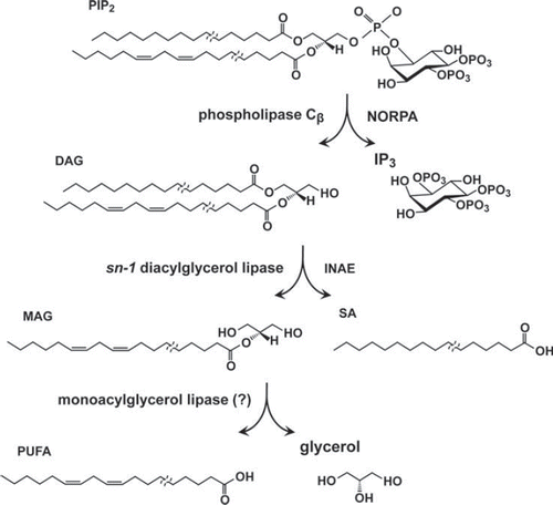
Strong mutations generated in inaE were lethal and had to be examined as eye mosaics, in which only the eye tissues were homozygous for the mutation (Leung et al., Citation2008). These mutations severely reduced the responses of photoreceptor to light in an allele-dependent manner, leading to the suggestions that hydrolysis products of DAG generated by DAGL activity, not DAG itself, are responsible for TRP channel excitation. This work represented a first step in an attempt to understand lipid regulation of TRP channels and raised a number of questions. The first of these is that the INAE protein localizes primarily to submicrovillar cisternae and photoreceptor cell bodies with just traces of staining in the rhabdomeres (Leung et al., Citation2008). The second problem is that INAE is a type sn-1 DAGL. It releases 2-monoacylglycerol (2-MAG) and mostly saturated fatty acid from DAG and does not release PUFA directly (). PUFA can be released from 2-MAG by the action of MAG lipase (MAGL) in a subsequent reaction. However, invertebrate MAGLs are a poorly understood class of lipases, and no direct orthologs of mammalian or bacterial MAGL appear to be present. Using a combined bioinformatic, biochemical, and molecular approach, we are now focusing on a candidate MAGL gene with a desired expression profile.
A new model of phototransduction has been put forward by Huang et al. (Citation2010). They have proposed that the two consequences of light-induced hydrolysis of PIP2 by PLCβ, namely, the reduction in the levels of PIP2 (and other phosphoinositides) and the release of protons, may act combinatorially to activate the TRP/TRPL channels. In support of this hypothesis, they showed, using pH indicator dyes, a light-dependent acidification of ∼0.1 pH unit in photoreceptors in the time scale comparable to that of phototransduction. Moreover, following thorough depletion of PIP2, TRP/TRPL channels could be activated by the metabolic poison, 2,4-dinitrophenol, which is a protonophore. Their hypothesis is novel and striking and merits serious consideration. Its long-term significance is not yet clear.
4. inaF
Unlike the other mutants discussed herein, inaF mutants were not the products of the original EMS (ethyl methanesulfonate) mutagenesis of the Pak laboratory (review: Pak, Citation2010). Instead, they were isolated in P element mutagenesis of an unrelated gene long after the early mutagenesis efforts had ceased (Li et al., Citation1999). The inaF gene encodes a small, eye-enriched protein, initially thought to be encoded by a 241-aa ORF (open reading frame) in the second exon of the transcript. Subsequently, Cheng and Nash (Citation2007) showed that the protein critical to inaF function (designated INAF-B) is encoded by an 81-aa ORF in the first exon of the transcript. The null inaF phenotype includes greatly reduced levels of the TRP channel protein (∼5–10%), without any effect on other phototransduction proteins tested, and ERG responses closely resembling those of trp null mutants (Li et al., Citation1999). The mRNA levels, however, are unaffected by inaF mutations (Cheng & Nash, Citation2007). The INAF-B protein is a single-pass, integral membrane protein that colocalizes and interacts with TRP in the rhabdomere (ibid.). The interaction between INAF-B and TRP is required for their mutual survival and persists even in a inaD null mutant background. INAF-B also interacts with the scaffold protein INAD, suggesting that it may be a member of the signaling complex formed by INAD (see Section IIB3 above). The INAF-B coding sequence contains a 32-aa motif (inaF motif) conserved from flies and worms to fish and humans (Cheng & Nash, Citation2007). The TRPL channels appear to function normally in inaF, and the residual TRP is still localized to the rhabdomeres (Li et al., Citation1999). Li et al. (Citation1999) assessed the effects of inaF null on the ERG waveform, the refractory period, and the ability to rescue the degeneration phenotype of the constitutively active trp mutant (TrpP365/ +) and compared them to the effects of comparable depletions in the amount of TRP protein. They argued that the inaF phenotype could not be explained by the reduction in the amount of the TRP protein alone and suggested that the INAF protein may have a role in TRP channel function. The subsequent biochemical findings on INAF-B by Cheng and Nash tend to reinforce this suggestion. However, INAF cellular function remains unknown.
ANECDOTAL STORY AND CONCLUDING REMARKS
At times a seemingly ordinary event can profoundly alter the course of development of a field. This is one such story. In the spring of 1984, one of us was on sabbatical at the Department of Biological Chemistry, Harvard Medical School. Gerald Rubin had once been on the faculty at Harvard Medical School. In the spring of 1984, he came back to Harvard Medical School as a guest speaker. Rubin sought out Pak to express his interest in the phototransduction mutants Pak had been isolating. Rubin explained that he had just moved to University of California at Berkeley and would be organizing a seminar course on Drosophila neurogenetics the following semester and would like to invite Pak as one of the speakers. Pak was surprised that Rubin knew about his mutants at all. As best as Pak can recall, Rubin called him in the fall of 1984 at Biocentrum, University of Basel, where he was doing the second half of his sabbatical, to arrange for his visit to Berkeley the following March. In addition, Rubin asked for the following nina and ina mutants: ninaA, B, C, D, and E and inaA, B, C, and D. Fairly detailed characterizations of ninaA, B, C, D, and E mutants were published in a symposium volume (Stephenson et al., Citation1983). As for the ina mutants, except for brief mentions of the fact that some of them were being isolated in a review and a symposium volume (Pak, Citation1979; Pak et al., Citation1980), nothing had yet been published on any of them; nevertheless, Pak sent all the requested mutants. Pak went to Berkeley in March 1985 and gave two talks introducing the audience to the field of Drosophila phototransduction and the mutants he had been isolating. Subsequently, two of Rubin's talented postdoctoral associates, Craig Montell and Charles Zuker, decided to go into the field of Drosophila phototransduction. Glancing through the references makes it clear, these two investigators and their associates and students went on to make major contributions to this field.
Perusal through the References also makes it clear that many other talented investigators from all over the world also contributed to the progress in this field. Although the nina/ina mutants may be the common thread running through this story, the story is really about the contributions of all these investigators.
In this review, we have attempted to provide a reasonably comprehensive view of the PDA-defective mutants starting with the rationale for the PDA-based mutant screening strategy to summaries of knowledge gained from the mutants isolated through this strategy. The PDA-based strategy turned out to be a surprisingly effective and efficient means of identifying genes important in photoreceptor function. Almost every gene that has been identified turned out to be interesting, and the importance of the proteins encoded by many of these genes in cellular signaling is already being recognized, with ramifications of their significance extending far beyond the field of photoreceptor function. Many of the proteins encoded by the nina/ina genes so far studied turned out to be the first member of their class to be identified. Thus, NINAE was the first invertebrate opsin to be identified and characterized; NINAA was the first chaperone protein for a GPCR (G protein–coupled receptor) to be identified; NINAC was the first class III myosin to be identified; NINAB was the first invertebrate β-carotene oxygenase to be identified; and INAD led to the discovery of the first supramolecular signaling complex in sensory transduction cascades. The discovery of the INAD-based signaling complexes promises to be particularly important in our understanding of sensory signaling cascades in general.
Although much has been achieved, we have not yet fully tapped the potential of the nina/ina mutants. As may be seen in , a sizable number of nina/ina mutants remain uncharacterized. Moreover, the PDA-based mutagenesis was never carried out to saturation. There are many more nina and ina mutants to be identified and isolated. Our expectation is that these mutants will continue to contribute to our understanding of cellular signaling for some time to come.
ACKNOWLEDGMENTS
We thank Dr. Chun-Fang Wu for asking us to write this review. We are deeply indebted to the following individuals who, in various capacities, contributed to the isolation and initial characterizations of the PDA-defective mutants over many years: Postdocs: Michael Deland, Rudolf Schinz, Robert Stephenson, Hiroyuki Matsumoto, Joe O’Tousa, Kunio Isono, Gregore Koliantz, Stephan Schneuwly; Graduate students: Mei-Ven Lo, Denis Larrivee, Michael Wilcox, Ruthann Nichols, Nancy Scavarda, Edwin Johnson, Debra S. Leonard, Paulo Ferreira; Undergrad: Marco Floreani; Technicians: Sherry Conrad (1973–78), Mary Look (1975–77), Marjorie Busching (1976–77), Jessica R. Miller (1977–79), Mary Ann Scott (1977–78), Carol M. Jones (1978–80), Lydia R. Strong (1979–99), Frances Piaszynski (1980–83), Quentin Pye (1980–86), John Bowman (1987–2001). We thank Vicki Croy for help with the manuscript and Don Ready and Marion Pak for their helpful comments on the manuscript.
The mutagenesis project was supported by grants from the National Science Foundation and the National Eye Institute (T32EY07008; R01EY000033).
Declaration of interest: The authors report no conflicts of interest. The authors alone are responsible for the content and writing of the paper.
REFERENCES
- Acharya, J. K., Jalink, K., Hardy, R. W., Hartenstein, V., &Zuker, C. S. (1997). InsP3 receptor is essential for growth and differentiation but not for vision in Drosophila. Neuron, 18, 881–887.
- Acton, S., Rigotti, A., Landschulz, K. T., Xu, S., Hobbs, H. H., & Krieger, M. (1996). Identification of scavenger receptor SR-BI as a high density lipoprotein receptor. Science, 271, 518–520.
- Ahmad, S. T., Joyce, M. V., Boggess, B., & O’Tousa, J. E. (2006). The role of DrosophilaninaG oxidoreductase in visual pigment chromophore biogenesis. J Biol Chem, 281, 9205–9209.
- Alloway, P. G., Howard, L., & Dolph, P. J. (2000). The formation of stable rhodopsin-arrestin complexes induces apoptosis and photoreceptor cell degeneration. Neuron, 28, 129–138.
- Arikawa, K., Hicks, J. L., & Williams, D. S. (1990). Identification of actin filaments in the rhabdomeral microvilli of Drosophila photoreceptors. J Cell Biol, 110, 1993–1998.
- Bähner, M., Frechter, S., Da Silva, N., Minke, B., Paulsen, R., & Huber, A. (2002). Light-regulated subcellular translocation of Drosophila TRPL channels induces long-term adaptation and modifies the light-induced current. Neuron, 34, 83–93.
- Baker, E. K., Colley, N. J., & Zuker, C. S. (1994). The cyclophilin homolog NinaA functions as a chaperone, forming a stable complex in vivo with its protein target rhodopsin. EMBO J, 13, 4886–4895.
- Bandell, M., Macpherson, L. J., & Patapoutian, A. (2007). From chills to chilis: Mechanisms for thermosensation and chemesthesis via thermoTRPs. Curr Opin Neurobiol, 17, 490–497.
- Battelle, B. A., Dabdoub, A., Malone, M. A., Andrews, A. W., Cacciatore, C., Calman, B. G., . (2001). Immunocytochemical localization of opsin, visual arrestin, myosin III, and calmodulin in Limulus lateral eye retinular cells and ventral photoreceptors. J Comp Neurol, 435, 211–225.
- Berg, J. S., Powell, B. C., & Cheney, R. E. (2001). A millennial myosin census. Mol Biol Cell, 12, 780–794.
- Bermak, J. C., & Zhou, Q. Y. (2001). Accessory proteins in the biogenesis of G protein-coupled receptors. Mol Interv, 1, 282–287.
- Caterina, M. J. (2007). Transient receptor potential ion channels as participants in thermosensation and thermoregulation. Am J Physiol Regul Integr Comp Physiol, 292, R64–R76.
- Cheng, Y., & Nash, H. A. (2007). Drosophila TRP channels require a protein with a distinctive motif encoded by the inaF locus. Proc Natl Acad Sci U S A, 104, 17730–17734.
- Chevesich, J., Kreuz, A. J., & Montell, C. (1997). Requirement for the PDZ domain protein, INAD, for localization of the TRP store-operated channel to a signaling complex. Neuron, 18, 95–105.
- Chou, W. H., Hall, K. J., Wilson, D. B., Wideman, C. L., Townson, S. M., Chadwell, L. V., . (1996). Identification of a novel Drosophila opsin reveals specific patterning of the R7 and R8 photoreceptor cells. Neuron, 17, 1101–1115.
- Chou, W. H., Huber, A., Bentrop, J., Schulz, S., Schawb, K., Chadwell, L. V., (1999). Patterning of the R7 and R8 photoreceptor cells of Drosophila: Evidence for induced and default cell-fate specification. Development, 126, 607–616.
- Chyb, S., Raghu, P., & Hardie, R. C. (1999). Polyunsaturated fatty acids activate the Drosophila light-sensitive channels TRP and TRPL. Nature, 397, 255–259.
- Colley, N. J., Baker, E. K., Stamnes, M. A., & Zuker, C. S. (1991). The cyclophilin homolog ninaA is required in the secretory pathway. Cell, 67, 255–263.
- Colley, N. J., Cassill, J. A., Baker, E. K., & Zuker, C. S. (1995). Defective intracellular transport is the molecular basis of rhodopsin-dependent dominant retinal degeneration. Proc Natl Acad Sci U S A, 92, 3070–3074.
- Cosens, D. J., & Manning, A. (1969). Abnormal electroretinogram from a Drosophila mutant. Nature, 224, 285–287.
- Cowman, A. F., Zuker, C. S., & Rubin, G. M. (1986). An opsin gene expressed in only one photoreceptor cell type of the Drosophila eye. Cell, 44, 705–710.
- Cronin, M. A., Diao, F., & Tsunoda, S. (2004). Light-dependent subcellular translocation of Gqalpha in Drosophila photoreceptors is facilitated by the photoreceptor-specific myosin III NINAC. J Cell Sci, 117, 4797–4806.
- Delgado, R., & Bacigalupo, J. (2009). Unitary recordings of TRP and TRPL channels from isolated Drosophila retinal photoreceptor rhabdomeres: Activation by light and lipids. J Neurophysiol, 101, 2372–2379.
- Delmas P., Crest M., Brown D. A. (2004). Functional organization of PLC signaling microdomains in neurons. Trends Neurosci, 27, 41–47.
- Dolph, P. J., Ranganathan, R., Colley, N. J., Hardy, R. W., Socolich, M., & Zuker, C. S. (1993). Arrestin function in inactivation of G protein-coupled receptor rhodopsin in vivo. Science, 260, 1910–1916.
- Dong, C., Filipeanu, C. M., Duvernay, M. T., & Wu, G. (2007). Regulation of G protein-coupled receptor export trafficking. Biochim Biophys Acta, 1768, 853–870.
- Dosé, A. C., & Burnside, B. (2000). Cloning and chromosomal localization of a human class III myosin. Genomics, 67, 333–342.
- Dosé, A. C., Hillman, D. W., Wong, C., Sohlberg, L., Lin-Jones, J., & Burnside, B. (2003). Myo3A, one of two class III myosin genes expressed in vertebrate retina, is localized to the calycal processes of rod and cone photoreceptors and is expressed in the sacculus. Mol Biol Cell, 14, 1058–1073.
- Dryja, T. P., & Berson, E. L. (1995). Retinitis pigmentosa and allied diseases. Implications of genetic heterogeneity. Invest Ophthalmol Vis Sci, 36, 1197–1200.
- Dryja, T. P., Berson, E. L., Rao, V. R., & Oprian, D. D. (1993). Heterozygous missense mutation in the rhodopsin gene as a cause of congenital stationary night blindness. Nat Genet, 4, 280–283.
- Dryja, T. P., McGee, T. L., Reichel, E., Hahn, L. B., Cowley, G. S., Yandell, D. W., . (1990). A point mutation of the rhodopsin gene in one form of retinitis pigmentosa. Nature, 343, 364–366.
- Dwyer, N. D., Troemel, E. R., Sengupta, P., & Bargmann, C. I. (1998). Odorant receptor localization to olfactory cilia is mediated by ODR-4, a novel membrane-associated protein. Cell, 93, 455–466.
- Engels, W. R. (1996). P elements in Drosophila. Curr Top Microbiol Immunol, 204, 103–123.
- Ferreira, P. A., Hom, J. T., & Pak, W. L. (1995). Retina-specifically expressed novel subtypes of bovine cyclophilin. J Biol Chem, 270, 23179–23188.
- Fischer, G., Wittmann-Liebold, B., Lang, K., Kiefhaber, T., & Schmid, F. X. (1989). Cyclophilin and peptidyl-prolyl cis-trans isomerase are probably identical proteins. Nature, 337, 476–478.
- Fryxell, K. J., & Meyerowitz, E. M. (1987). An opsin gene that is expressed only in the R7 photoreceptor cell of Drosophila. EMBO J, 6, 443–451.
- Giovannucci, D. R., & Stephenson, R. S. (1999). Identification and distribution of dietary precursors of the Drosophila visual pigment chromophore: Analysis of carotenoids in wild type and ninaD mutants by HPLC. Vision Res, 39, 219–229.
- Goel, M., Garcia, R., Estacion, M., & Schilling, W. P. (2001). Regulation of Drosophila TRPL channels by immunophilin FKBP59. J Biol Chem, 276, 38762–38773.
- Hardie, R. C. (2011). A brief history of trp: Commentary and personal perspective. Pflugers Arch, 461, 493–498.
- Hardie, R. C., & Minke, B. (1992). The trp gene is essential for a light-activated Ca2+ channel in Drosophila photoreceptors. Neuron, 8, 643–651.
- Hardie, R. C., Peretz, A., Suss-Toby, E., Rom-Glas, A., Bishop, S. A., Selinger, Z., . (1993). Protein kinase C is required for light adaptation in Drosophila photoreceptors. Nature, 363, 634–637.
- Hardie, R. C., Raghu, P., Moore, S., Juusola, M., Baines, R. A., & Sweeney, S. T. (2001). Calcium influx via TRP channels is required to maintain PIP2 levels in Drosophila photoreceptors. Neuron, 30, 149–159.
- Harris, W. A., Stark, W. S., & Walker, J. A. (1976). Genetic dissection of the photoreceptor system in the compound eye of Drosophilamelanogaster. J Physiol, 256, 415–439.
- Heisenberg, M. (1971). Isolation of mutants lacking the optomotor response. Dros Inf Serv, 46, 68.
- Henderson, S. R., Reuss, H., & Hardie, R. C. (2000). Single photon responses in Drosophila photoreceptors and their regulation by Ca2+ . J Physiol, 524, 179–194.
- Hicks, J. L., Liu, X., & Williams, D. S. (1996). Role of the ninaC proteins in photoreceptor cell structure: Ultrastructure of ninaC deletion mutants and binding to actin filaments. Cell Motil Cytoskeleton, 35, 367–379.
- Hicks, J. L., & Williams, D. S. (1992). Distribution of the myosin I–like ninaC proteins in the Drosophila retina and ultrastructural analysis of mutant phenotypes. J Cell Sci, 101, 247–254.
- Hillman, P., Hochstein, S., & Minke, B. (1972). A visual pigment with two physiologically active stable states. Science, 175, 1486–1488.
- Hofstee, C. A., Henderson, S., Hardie, R. C., Stavenga, D. G. (1996). Differential effects of ninaC proteins (p132 and p174) on light-activated currents and pupil mechanism in Drosophila photoreceptors. Visual Neurosci, 13, 897–906.
- Huang, J., Liu, C. H., Hughes, S. A., Postma, M., Schwiening, C. J., & Hardie, R. C. (2010). Activation of TRP channels by protons and phosphoinositide depletion in Drosophila photoreceptors. Curr Biol, 20, 189–197.
- Huber, A., Sander, P., Bahner, M., & Paulsen, R. (1998). The TRP Ca2 + channel assembled in a signaling complex by the PDZ domain protein INAD is phosphorylated through the interaction with protein kinase C (ePKC). FEBS Lett, 425, 317–322.
- Huber, A., Sander, P., Gobert, A., Bahner, M., Hermann, R., & Paulsen, R. (1996). The transient receptor potential protein (Trp), a putative store-operated Ca2+ channel essential for phosphoinositide-mediated photoreception, forms a signaling complex with NorpA, InaC and InaD. EMBO J, 15, 7036–7045.
- Huber A. (2001). Scaffolding proteins organize multimolecular protein complexes for sensory signal transduction. Eur J Neurosci, 14, 769–776.
- Iakhine, R., Chorna-Ornan, I., Zars, T., Elia, N., Cheng, Y., Selinger, Z., . (2004). Novel dominant rhodopsin mutation triggers two mechanisms of retinal degeneration and photoreceptor desensitization. J Neurosci, 24, 2516–2526.
- Inglehearn, C. F., Bashir, R., Lester, D. H., Jay, M., Bird, A. C., & Bhattacharya, S. S. (1991). A 3-bp deletion in the rhodopsin gene in a family with autosomal dominant retinitis pigmentosa. Am J Hum Genet, 48, 26–30.
- Isono, K., Tanimura, T., Oda, Y., & Tsukahara, Y. (1988). Dependency on light and vitamin A derivatives of the biogenesis of 3-hydroxyretinal and visual pigment in the compound eyes of Drosophila melanogaster. J Gen Physiol, 92, 587–600.
- Jin, M., Li, S., Moghrabi, W. N., Sun, H., & Travis, G. H. (2005). Rpe65 is the retinoid isomerase in bovine retinal pigment epithelium. Cell, 122, 449–459.
- Johnson, E. C., & Pak, W. L. (1986). Electrophysiological study of Drosophila rhodopsin mutants. J Gen Physiol, 88, 651–673.
- Jukam, D., & Desplan, C. (2010). Binary fate decisions in differentiating neurons. Curr Opin Neurobiol, 20, 6–13.
- Kambara, T., Komaba, S., & Ikebe, M. (2006). Human myosin III is a motor having an extremely high affinity for actin. J Biol Chem, 281, 37291–37301.
- Katz, B., & Minke, B. (2009). Drosophila photoreceptors and signaling mechanisms. Front Cell Neurosci, 3, 2.
- Kiefer, C., Sumser, E., Wernet, M. F., & Von Lintig, J. (2002). A class B scavenger receptor mediates the cellular uptake of carotenoids in Drosophila. Proc Natl Acad Sci U S A, 99, 10581–10586.
- Kiselev, A., Socolich, M., Vinos, J., Hardy, R. W., Zuker, C. S., & Ranganathan, R. (2000). A molecular pathway for light-dependent photoreceptor apoptosis in Drosophila. Neuron, 28, 139–152.
- Kleizen, B., & Braakman, I. (2004). Protein folding and quality control in the endoplasmic reticulum. Curr Opin Cell Biol, 16, 343–349.
- Koenig, J., & Merriam, J. R. (1977). Autosomal ERG mutants. Dros Inf Serv, 52, 50–51.
- Koenig, J. H. (1975). The isolation and preliminary characterization of autosomal electroretinogram defective mutants in Drosophilamelanogaster. Dissertation.
- Komaba, S., Inoue, A., Maruta, S., Hosoya, H., & Ikebe, M. (2003). Determination of human myosin III as a motor protein having a protein kinase activity. J Biol Chem, 278, 21352–21360.
- Kosloff, M., Elia, N., Joel-Almagor, T., Timberg, R., Zars, T. D., Hyde, D. R., . (2003). Regulation of light-dependent Gqalpha translocation and morphological changes in fly photoreceptors. EMBO J, 22, 459–468.
- Kumar, J. P., & Ready, D. F. (1995). Rhodopsin plays an essential structural role in Drosophila photoreceptor development. Development, 121, 4359–4370.
- Kurada, P., & O’Tousa, J. E. (1995). Retinal degeneration caused by dominant rhodopsin mutations in Drosophila. Neuron, 14, 571–579.
- Kurada, P., Tonini, T. D., Serikaku, M. A., Piccini, J. P., & O’Tousa, J. E. (1998). Rhodopsin maturation antagonized by dominant rhodopsin mutants. Vis Neurosci, 15, 693–700.
- Kwon, Y., Shim, H. S., Wang, X., & Montell, C. (2008). Control of thermotactic behavior via coupling of a TRP channel to a phospholipase C signaling cascade. Nat Neurosci, 11, 871–873.
- Langer H. (1967). Über die Pigmentgranula im Facettenauge von Calliphora erythrocephala. Z Vergl Physiol, 55, 354–377.
- Larrivee, D. C., Conrad, S. K., Stephenson, R. S., & Pak, W. L. (1981). Mutation that selectively affects rhodopsin concentration in the peripheral photoreceptors of Drosophila melanogaster. J Gen Physiol, 78, 521–545.
- Lee, S. J., Xu, H., Kang, L. W., Amzel, L. M., Montell, C. (2003). Light adaptation through phosphoinositide-regulated translocation of Drosophila visual arrestin. Neuron, 39, 121–132.
- Leonard, D. S., Bowman, V. D., Ready, D. F., & Pak, W. L. (1992). Degeneration of photoreceptors in rhodopsin mutants of Drosophila. J Neurobiol, 23, 605–626.
- Leung, H. T., Tseng-Crank, J., Kim, E., Mahapatra, C., Shino, S., Zhou, Y., . (2008). DAG lipase activity is necessary for TRP channel regulation in Drosophila photoreceptors. Neuron, 58, 884–896.
- LeVine, H., ., 3rd, Smith, D. PWhitney, M., Malicki, D. M., Dolph, P. J., Smith, G. F., . (1990). Isolation of a novel visual-system-specific arrestin: An in vivo substrate for light-dependent phosphorylation. Mech Dev, 33, 19–25.
- Li, C., Geng, C., Leung, H. T., Hong, Y. S., Strong, L. L., Schneuwly, S., . (1999). INAF, a protein required for transient receptor potential Ca2+ channel function. Proc Natl Acad Sci U S A, 96, 13474–13479.
- Li, H. S., & Montell, C. (2000). TRP and the PDZ protein, INAD, form the core complex required for retention of the signalplex in Drosophila photoreceptor cells. J Cell Biol, 150, 1411–1422.
- Li, H. S., Porter, J. A., & Montell, C. (1998). Requirement for the NINAC kinase/myosin for stable termination of the visual cascade. J Neurosci, 18, 9601–9606.
- Liu, C. H., Satoh, A. K., Postma, M., Huang, J., Ready, D. F., & Hardie, R. C. (2008). Ca2+ -dependent metarhodopsin inactivation mediated by calmodulin and NINAC myosin III. Neuron, 59, 778–789.
- Liu, M., Parker, L. L., Wadzinski, B. E., & Shieh, B. H. (2000). Reversible phosphorylation of the signal transduction complex in Drosophila photoreceptors. J Biol Chem, 275, 12194–12199.
- Liu, W., Wen, W., Wei, Z., Yu, J., Ye, F., Liu, C. H., . (2011). The INAD scaffold is a dynamic, redox-regulated modulator of signaling in the Drosophila eye. Cell, 145, 1088–1101.
- Matsudaira, P. T., & Burgess, D. R. (1979). Identification and organization of the components in the isolated microvillus cytoskeleton. J Cell Biol, 83, 667–673.
- Matsumoto, H., Isono, K., Pye, Q., & Pak, W. L. (1987). Gene encoding cytoskeletal proteins in Drosophila rhabdomeres. Proc Natl Acad Sci U S A, 84, 985–989.
- Matsumoto, H., & Pak, W. L. (1984). Light-induced phosphorylation of retina-specific polypeptides of Drosophila in vivo. Science, 223, 184–186.
- Matsumoto, H., & Yamada, T. (1991). Phosrestins I and II: Arrestin homologs which undergo differential light-induced phosphorylation in the Drosophila photoreceptor in vivo. Biochem Biophys Res Commun, 177, 1306–1312.
- Mecklenburg, K. L., Takemori, N., Komori, N., Chu, B., Hardie, R. C., Matsumoto, H., . (2010). Retinophilin is a light-regulated phosphoprotein required to suppress photoreceptor dark noise in Drosophila. J Neurosci, 30, 1238–1249.
- Meyer, N. E., Joel-Almagor, T., Frechter, S., Minke, B., & Huber, A. (2006). Subcellular translocation of the eGFP-tagged TRPL channel in Drosophila photoreceptors requires activation of the phototransduction cascade. J Cell Sci, 119, 2592–2603.
- Minke B, Wu. C.-F., Pak W. L. (1975). Isolation of light-induced response of the central retinula cells from the electroretinogram of Drosophila. J Comp Physiol, 98, 345–355.
- Mishra, P., Socolich, M., Wall, M. A., Graves, J., Wang, Z., & Ranganathan, R. (2007). Dynamic scaffolding in a G protein-coupled signaling system. Cell, 131, 80–92.
- Mismer, D., Michael, W. M., Laverty, T. R., & Rubin, G. M. (1988). Analysis of the promoter of the Rh2 opsin gene in Drosophilamelanogaster. Genetics, 120, 173–180.
- Montell, C., Jones, K., Zuker, C., & Rubin, G. (1987). A second opsin gene expressed in the ultraviolet-sensitive R7 photoreceptor cells of Drosophilamelanogaster. J Neurosci, 7, 1558–1566.
- Montell, C., & Rubin, G. M. (1988). The DrosophilaninaC locus encodes two photoreceptor cell specific proteins with domains homologous to protein kinases and the myosin heavy chain head. Cell, 52, 757–772.
- Morante, J., Desplan, C., & Celik, A. (2007). Generating patterned arrays of photoreceptors. Curr Opin Genet Dev, 17, 314-319.
- Nathans, J., & Hogness, D. S. (1983). Isolation, sequence analysis, and intron-exon arrangement of the gene encoding bovine rhodopsin. Cell, 34, 807–814.
- Nathans, J., & Hogness, D. S. (1984). Isolation and nucleotide sequence of the gene encoding human rhodopsin. Proc Natl Acad Sci U S A, 81, 4851–4855.
- Nolte, J., & Brown, J. E. (1972). Ultraviolet-induced sensitivity to visible light in ultraviolet receptors of Limulus. J Gen Physiol, 59, 186–200.
- O’Tousa, J. E., Baehr, W., Martin, R. L., Hirsh, J., Pak, W. L., & Applebury, M. L. (1985). The DrosophilaninaE gene encodes an opsin. Cell, 40, 839–850.
- O’Tousa, J. E., Leonard, D. S., & Pak, W. L. (1989). Morphological defects in oraJK84 photoreceptors caused by mutation in R1–6 opsin gene of Drosophila. J Neurogenet, 6, 41–52.
- Oberhauser, V., Voolstra, O., Bangert, A., von Lintig, J., & Vogt, K. (2008). NinaB combines carotenoid oxygenase and retinoid isomerase activity in a single polypeptide. Proc Natl Acad Sci U S A, 105, 19000–19005.
- Ostroy, S. E., Wilson, M., & Pak, W. L. (1974). Drosophila rhodopsin: Photochemistry, extraction and differences in the norpAP12 phototransduction mutant. Biochem Biophys Res Commun, 59, 960–966.
- Pak, W. L. (1979). Study of photoreceptor function using Drosophila mutants. In X. O. Breakfield. Neurogenetics: Genetic approaches to the nervous system (pp 67–99). New York: Elsevier.
- Pak, W. L. (2010). Why Drosophila to study phototransduction? J Neurogenet, 24, 55–66.
- Pak, W. L., Conrad, S. K., Kremer, N. E., Larrivee, D. C., Schinz, R. H., & Wong, F. (1980). Photoreceptor function. Basic Life Sci, 16, 331–346.
- Pak, W. L., Grossfield, J., & White, N. V. (1969). Nonphototactic mutants in a study of vision of Drosophila. Nature, 222, 351–354.
- Pak, W. L., & Lidington, K. J. (1974). Fast electrical potential from a long-lived, long-wavelength photoproduct of fly visual pigment. J Gen Physiol, 63, 740–756.
- Papatsenko, D., Sheng, G., & Desplan, C. (1997). A new rhodopsin in R8 photoreceptors of Drosophila: Evidence for coordinate expression with Rh3 in R7 cells. Development, 124, 1665–1673.
- Pearn, M. T., Randall, L. L., Shortridge, R. D., Burg, M. G., & Pak, W. L. (1996). Molecular, biochemical, and electrophysiological characterization of DrosophilanorpA mutants. J Biol Chem, 271, 4937–4945.
- Pollock, J. A., & Benzer, S. (1988). Transcript localization of four opsin genes in the three visual organs of Drosophila; RH2 is ocellus specific. Nature, 333, 779–782.
- Popescu, D. C., Ham, A. J., & Shieh, B. H. (2006). Scaffolding protein INAD regulates deactivation of vision by promoting phosphorylation of transient receptor potential by eye protein kinase C in Drosophila. J Neurosci, 26, 8570–8577.
- Porter, J. A., Hicks, J. L., Williams, D. S., & Montell, C. (1992). Differential localizations of and requirements for the two DrosophilaninaC kinase/myosins in photoreceptor cells. J Cell Biol, 116, 683–693.
- Porter, J. A., Minke, B., & Montell, C. (1995). Calmodulin binding to Drosophila NinaC required for termination of phototransduction. EMBO J, 14, 4450–4459.
- Porter, J. A., Yu, M., Doberstein, S. K., Pollard, T. D., & Montell, C. (1993). Dependence of calmodulin localization in the retina on the NINAC unconventional myosin. Science, 262, 1038–1042.
- Raghu, P., Colley, N. J., Webel, R., James, T., Hasan, G., Danin, M., . (2000a). Normal phototransduction in Drosophila photoreceptors lacking an InsP3 receptor gene. Mol Cell Neurosci, 15, 429–445.
- Raghu, P., Usher, K., Jonas, S., Chyb, S., Polyanovsky, A., & Hardie, R. C. (2000b). Constitutive activity of the light-sensitive channels TRP and TRPL in the Drosophila diacylglycerol kinase mutant, rdgA. Neuron, 26, 169–179.
- Ranganathan, R., Harris, G. L., Stevens, C. F., & Zuker, C. S. (1991). A Drosophila mutant defective in extracellular calcium-dependent photoreceptor deactivation and rapid desensitization. Nature, 354, 230–232.
- Rao, V. R., Cohen, G. B., & Oprian, D. D. (1994). Rhodopsin mutation G90D and a molecular mechanism for congenital night blindness. Nature, 367, 639–642.
- Robinson, P. R., Cohen, G. B., Zhukovsky, E. A., & Oprian, D. D. (1992). Constitutively active mutants of rhodopsin. Neuron, 9, 719–725.
- Roof, D. J., Adamian, M., & Hayes, A. (1994). Rhodopsin accumulation at abnormal sites in retinas of mice with a human P23H rhodopsin transgene. Invest Ophthalmol Vis Sci, 35, 4049–4062.
- Rosenbaum, E. E., Hardie, R. C., & Colley, N. J. (2006). Calnexin is essential for rhodopsin maturation, Ca2+ regulation, and photoreceptor cell survival. Neuron, 49, 229–241.
- Salcedo, E., Huber, A., Henrich, S., Chadwell, L. V., Chou, W. H., Paulsen, R., . (1999). Blue- and green-absorbing visual pigments of Drosophila: Ectopic expression and physiological characterization of the R8 photoreceptor cell-specific Rh5 and Rh6 rhodopsins. J Neurosci, 19, 10716–10726.
- Sarfare, S., Ahmad, S. T., Joyce, M. V., Boggess, B., & O’Tousa, J. E. (2005). The DrosophilaninaG oxidoreductase acts in visual pigment chromophore production. J Biol Chem, 280, 11895–11901.
- Satoh, A. K., & Ready, D. F. (2005). Arrestin1 mediates light-dependent rhodopsin endocytosis and cell survival. Curr Biol, 15, 1722–1733.
- Scavarda, N. J., O’Tousa, J., & Pak, W. L. (1983). Drosophila locus with gene-dosage effects on rhodopsin. Proc Natl Acad Sci U S A, 80, 4441–4445.
- Schaeffer, E., Smith, D., Mardon, G., Quinn, W., & Zuker, C. (1989). Isolation and characterization of two new Drosophila protein kinase C genes, including one specifically expressed in photoreceptor cells. Cell, 57, 403–412.
- Schinz, R. H., Lo, M. V., Larrivee, D. C., & Pak, W. L. (1982). Freeze-fracture study of the Drosophila photoreceptor membrane: Mutations affecting membrane particle density. J Cell Biol, 93, 961–967.
- Schneuwly, S., Shortridge, R. D., Larrivee, D. C., Ono, T., Ozaki, M., & Pak, W. L. (1989). DrosophilaninaA gene encodes an eye-specific cyclophilin (cyclosporine A binding protein). Proc Natl Acad Sci U S A, 86, 5390–5394.
- Scott, K., Sun, Y., Beckingham, K., & Zuker, C. S. (1997). Calmodulin regulation of Drosophila light-activated channels and receptor function mediates termination of the light response in vivo. Cell, 91, 375–383.
- Seki, T., Fujishita, S., Ito, M., Matsuoka, N., Kobayashi, C., & Tsukida, K. (1986). A fly, Drosophilamelanogaster, forms 11-cis 3-hydroxyretinal in the dark. Vision Res, 26, 255–258.
- Seki, T., Isono, K., Ito, M., & Katsuta, Y. (1994). Flies in the group Cyclorrhapha use (3S)-3-hydroxyretinal as a unique visual pigment chromophore. Eur J Biochem, 226, 691–696.
- Shen, W. L., Kwon, Y., Adegbola, A. A., Luo, J., Chess, A., & Montell, C. (2011). Function of rhodopsin in temperature discrimination in Drosophila. Science, 331, 1333–1336.
- Shieh, B. H., & Niemeyer, B. (1995). A novel protein encoded by the InaD gene regulates recovery of visual transduction in Drosophila. Neuron, 14, 201–210.
- Shieh, B. H., Parker, L., & Popescu, D. (2002). Protein kinase C (PKC) isoforms in Drosophila. J Biochem, 132, 523–527.
- Shieh, B. H., Stamnes, M. A., Seavello, S., Harris, G. L., & Zuker, C. S. (1989). The ninaA gene required for visual transduction in Drosophila encodes a homologue of cyclosporin A-binding protein. Nature, 338, 67–70.
- Shieh, B. H., & Zhu, M. Y. (1996). Regulation of the TRP Ca2+ channel by INAD in Drosophila photoreceptors. Neuron, 16, 991–998.
- Shieh, B. H., Zhu, M. Y., Lee, J. K., Kelly, I. M., Bahiraei, F. (1997). Association of INAD with NORPA is essential for controlled activation and deactivation of Drosophila phototransduction in vivo. Proc Natl Acad Sci U S A, 94, 12682–12687.
- Smith, D. P., Ranganathan, R., Hardy, R. W., Marx, J., Tsuchida, T., & Zuker, C. S. (1991). Photoreceptor deactivation and retinal degeneration mediated by a photoreceptor-specific protein kinase C. Science, 254, 1478–1484.
- Stamnes, M. A., Shieh, B. H., Chuman, L., Harris, G. L., & Zuker, C. S. (1991). The cyclophilin homolog ninaA is a tissue-specific integral membrane protein required for the proper synthesis of a subset of Drosophila rhodopsins. Cell, 65, 219–227.
- Stark, W. S. (1973). The effect of eye colour pigments on the action spectrum of Drosophila. J Insect Physiol, 19, 999–1006.
- Stark, W. S., & Sapp, R. (1987). Ultrastructure of the retina of Drosophilamelanogaster: The mutant ora (outer rhabdomeres absent) and its inhibition of degeneration in rdgB (retinal degeneration-B). J Neurogenet, 4, 227–240.
- Stephenson, R. S., O’Tousa, J., Scavarda, N. J., Randall, L. L., & Pak, W. L. (1983). Drosophila mutants with reduced rhodopsin content. Symp Soc Exp Biol, 36, 477–501.
- Strother, G. K., & Casella, A. J. (1972). Microspectrophotometry of arthropod visual screening pigments. J Gen Physiol, 59, 616–636.
- Sung, C. H., Davenport, C. M., Hennessey, J. C., Maumenee, I. H., Jacobson, S. G., Heckenlively, J. R., . (1991). Rhodopsin mutations in autosomal dominant retinitis pigmentosa. Proc Natl Acad Sci U S A, 88, 6481–6485.
- Sung, C. H., Makino, C., Baylor, D., & Nathans, J. (1994). A rhodopsin gene mutation responsible for autosomal dominant retinitis pigmentosa results in a protein that is defective in localization to the photoreceptor outer segment. J Neurosci, 14, 5818–5833.
- Suzuki, T., Makino-Tasaka, M., & Eguchi, E. (1984). 3-Dehydroretinal (vitamin A2 aldehyde) in crayfish eye. Vision Res, 24, 783–787.
- Takahashi, N., Hayano, T., & Suzuki, M. (1989). Peptidyl-prolyl cis-trans isomerase is the cyclosporin A-binding protein cyclophilin. Nature, 337, 473–475.
- Tsunoda, S., Sierralta, J., Sun, Y., Bodner, R., Suzuki, E., Becker, A., . (1997). A multivalent PDZ-domain protein assembles signalling complexes in a G-protein-coupled cascade. Nature, 388, 243–249.
- Tsunoda, S., Sun, Y., Suzuki, E., & Zuker, C. (2001). Independent anchoring and assembly mechanisms of INAD signaling complexes in Drosophila photoreceptors. J Neurosci, 21, 150–158.
- van Huizen, R., Miller, K., Chen, D. M., Li, Y., Lai, Z. C., Raab, R. W., . (1998). Two distantly positioned PDZ domains mediate multivalent INAD-phospholipase C interactions essential for G protein-coupled signaling. EMBO J, 17, 2285–2297.
- Venkatachalam, K., Wasserman, D., Wang, X., Li, R., Mills, E., Elsaesser, R., . (2010). Dependence on a retinophilin/myosin complex for stability of PKC and INAD and termination of phototransduction. J Neurosci, 30, 11337–11345.
- Vogt, K. (1989). Distribution of insect visual chromophores: Functional and phylogenetic aspects (pp. 134–151). xx: Springer.
- von Lintig, J., & Vogt, K. (2000). Filling the gap in vitamin A research. Molecular identification of an enzyme cleaving beta-carotene to retinal. J Biol Chem, 275, 11915–11920.
- Voolstra, O., Kiefer, C., Hoehne, M., Welsch, R., Vogt, K., & von Lintig, J. (2006). The Drosophila class B scavenger receptor NinaD-I is a cell surface receptor mediating carotenoid transport for visual chromophore synthesis. Biochemistry, 45, 13429–13437.
- Voolstra, O., Oberhauser, V., Sumser, E., Meyer, N. E., Maguire, M. E., Huber, A., . (2010). NinaB is essential for Drosophila vision but induces retinal degeneration in opsin-deficient photoreceptors. J Biol Chem, 285, 2130–2139.
- Walsh T., Walsh V., Vreugde S., Hertzano R., Shahin H., Haika S., (2002). From flies’ eyes to our ears: Mutations in a human class III myosin cause progressive nonsyndromic hearing loss DFNB30. Proc Natl Acad Sci U S A, 99, 7518–7523.
- Wang, T., Jiao, Y., & Montell, C. (2007). Dissection of the pathway required for generation of vitamin A and for Drosophila phototransduction. J Cell Biol, 177, 305–316.
- Wang, T., & Montell, C. (2005). Rhodopsin formation in Drosophila is dependent on the PINTA retinoid-binding protein. J Neurosci, 25, 5187–5194.
- Wang, T., & Montell, C. (2007). Phototransduction and retinal degeneration in Drosophila. Pflugers Arch, 454, 821–847.
- Wes, P. D., Xu, X. Z., Li, H. S., Chien, F., Doberstein, S. K., & Montell, C. (1999). Termination of phototransduction requires binding of the NINAC myosin III and the PDZ protein INAD. Nat Neurosci, 2, 447–453.
- Williams, D. B. (2006). Beyond lectins: The calnexin/calreticulin chaperone system of the endoplasmic reticulum. J Cell Sci, 119, 615–623.
- Wu, J., Matunis, M. J., Kraemer, D., Blobel, G., & Coutavas, E. (1995). Nup358, a cytoplasmically exposed nucleoporin with peptide repeats, Ran-GTP binding sites, zinc fingers, a cyclophilin A homologous domain, and a leucine-rich region. J Biol Chem, 270, 14209–14213.
- Xu, X.-Z. S., Choudhury, A., Li, X., & Montell, C. (1998). Coordination of an array of signaling proteins through homo- and heteromeric interactions between PDZ domains and target proteins. J Cell Biol, 142, 545–555.
- Yokoyama, N., Hayashi, N., Seki, T., Pante, N., Ohba, T., Nishii, K., . (1995). A giant nucleopore protein that binds Ran/TC4. Nature, 376, 184–188.
- Zuker, C. S., Cowman, A. F., & Rubin, G. M. (1985). Isolation and structure of a rhodopsin gene from D. melanogaster. Cell, 40, 851–858.
- Zuker, C. S., Montell, C., Jones, K., Laverty, T., & Rubin, G. M. (1987). A rhodopsin gene expressed in photoreceptor cell R7 of the Drosophila eye: Homologies with other signal-transducing molecules. J Neurosci, 7, 1550–1557.