Abstract
A commercial diagnostic ultrasound scanner (Octoson) was modified for performing hyperthermia treatments. The temperature elevations were induced in tissues by four large, focused ultrasonic transducers whose common focal zone was scanned along a computer controlled path as determined from B-scan images. The system is described and the results of preliminary tests demonstrating some of its capabilities are given. Extensive tests with canine thighs and kidneys were performed. The blood flow to the kidneys was controllable, and thus tumours having different blood perfusion rates could be simulated. The results showed that the system is capable of inducing a local temperature maximum deep in tissues (up to 10 cm was tested) and that tissues with high perfusion rates could be heated.
Introduction
Due to the good penetration of ultrasound at frequencies associated with short wavelengths (when compared with the dimensions of the treated volumes), ultrasound has the potential to produce therapeutic temperature distributions in deep-seated tumours. In previous ultrasound efforts, single, unfocused transducers have been found to be relatively useful in the treatment of superficial tumours Citation1, Citation2, with the main disadvantage of these systems being the pain caused if the tumour was lying above a bone. Similarly, patient pain limited the usefulness of a system utilizing six, overlapping, unfocused, low frequency (356 kHz) beams which were able to elevate the temperature in deep-lying tumours Citation3. However, pain was not a problem when a sharply focused, higher frequency ultrasound beam was mechanically scanned to heat tumours Citation4. The in vivo animal and patient data also showed that that device was capable of heating a desired tissue volume to the target temperature Citation4. Another related method to induce a controllable temperature distribution is to use multiple, stationary, focused fields Citation5, Citation6. An ultrasound beam can also be focused and scanned electrically. The disadvantages of electrical focusing are limited focusing capabilities, and the large amount of electronics required with two-dimensional phased arrays. However, recent new approaches to reduce the cost of the electrical scanning systems have been reported and the results are promising Citation7–9.
In order to develop an ultrasound hyperthermia system capable of elevating the temperature of deep target volumes to 43°C or above, while leaving the surrounding tissues at lower temperatures we have designed our system by combining the most useful features of previous approaches. Thus, we have utilized multiple (four), large, focused transducers, operating at 1 MHz and capable of being mechanically scanned under computer control. Also, in order to locate and image a target volume and any critical internal structures, such as bones or gas, a commercial ultrasound diagnostic scanner was included. The system has been constructed and tested in a number (30) of animal experiments. Both it and the main results of those experiments are described in this paper.
System design
General
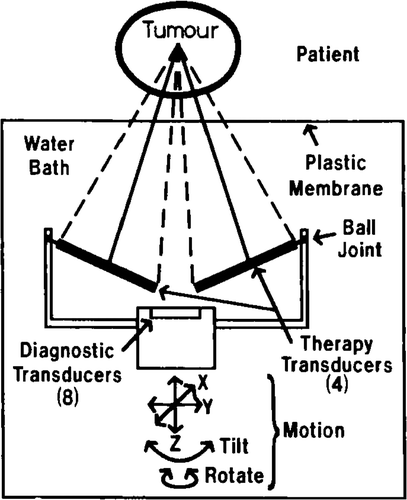
A commercial, diagnostic ultrasound scanner (Octoson; Ausonics Inc., Australia) was modified to become a hyperthermia unit. The Octoson has eight, focused, diagnostic, ultrasonic transducers mounted in a circular arc on a gantry that can be moved in the x, y and z directions and can also be rotated and tilted by stepper motors. The transducer gantry is immersed in a waterbath on top of which the patient lies (). Two, high-power, focused transducers were added on each side of the gantry (four transducers total) as shown in . The direction of the central line of each transducer can be varied independently and thus the relative location of the transducers’ foci can be selected as desired. Once the directions of these four axes are chosen, the transducers are fixed in position, and once set the transducer orientation with respect to the gantry remains constant during the treatment. In our initial animal experiments the beams have always been overlapping at a single acoustical focus as shown in .
Figure 1. A diagram of the transducer arrangement in the Octoson.
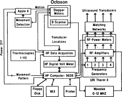
The basic diagnostic system has been supplemented by a computerized data acquisition and control system, and a computerized motor control system (). The data acquisition control unit is a Hewlett-Packard 9836 desk top computer utilizing 1.7 MB of memory and a floating point processor. Generally, all calibration and experimental programs were written in Basic and the patient treatment programs written in Pascal (due to the higher execution speed).
Figure 2. A block diagram of the equipment used in the scanned focused ultrasound hyperthermia unit.
Generation of ultrasound
Radio frequency square wave signals for the generation of ultrasound are provided by four oscillators in a URI-THERM-X, L-500 power amplifier system or by a separate frequency generator (Wavetek 271). The URI oscillators can provide only three frequencies (0.50, 1.0 and 1.5 MHz), whereas the Wavetek is programmable from 0 to 12 MHz. The signal is amplified by a four channel amplifier (URI THERM-X L-500). The forward and reflected power in the four channels are measured by the same URI unit. The output voltage of each amplifier can be varied from 0 to 100 V in 16 steps, while the duty cycle of each channel can be independently varied from 0.0 to 1.0 in 16 steps. The cycle frequency is 60 Hz. To supplement the limited duty cycle choice available on the URI system, the external frequency generator can be used. At present the external frequency generator does not allow the duty cycle of all four RF signals to be varied independently, whereas the URI system does. The voltage and duty cycle settings can be changed by the HP9836 computer via a RS-232 interface in the URI amplifier, and in the case of the external frequency generator, duty cycle changes can be made via an IEEE-488 interface bus.
Three different sets of focused transducers have been tested (). The transducers were air-backed and matched to the 50 Ω output load by an external matching network. The total acoustical power was calculated from the radiation force on an absorbing target measured with a balance (Mettler AE 160). The weight changes on the target due to the ultrasound radiation force were recorded by the HP9836 computer via the IEEE-488 interface. The warming of the target and the convergence of the beam were estimated and taken into account in the program that calculated the total acoustical power from the measurements Citation10. At high output powers, low duty cycles were used in order to reduce water streaming. The repeatability of the measurements was better than 1 per cent and the absolute accuracy, when compared with a calibrated standard source, was about 5 per cent.
Table I. The physical characteristics of the focused ultrasonic transducers used in these studies.
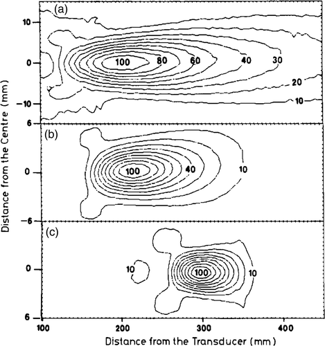
The ultrasound field distributions were measured in a water tank using a thermocouple coated with polyurethane Citation11 which was stepped through the field via stepper motors under computer control. The relative intensity value was calculated at each location from the rate of temperature rise during a 20 ms sound pulse. The typical step length was 0.5 mm across the beam and 5 mm along the axis of a single focused beam. For measuring the interference patterns a step of 0.5 mm was used in all directions. The ultrasonic field distribution from the multiple transducers in their actual configuration was measured using the stepper motors of the Octoson with the water bath and thermocouple mounted in the normal patient location. Examples of intensity distributions measured in water are presented in .
Figure 3. The intensity contours in the axial plane measured in a water bath: (a) FOC 1: f = 0.5 MHz, D = 70 mm, R = 350 mm; (b) FOC 3: f = 1.0 MHz, D = 70 mm, R = 250 mm; (c) FOC 5: f = 1.0 MHz, D = 130 mm, R = 340 mm.
The spatial peak intensity at the acoustical focus was measured using thermocouples in castor oil Citation12. The intensities were measured at low powers and low duty cycles in order to avoid streaming in the oil and then extrapolated to higher power values based on the total acoustical power. The absorption changes in the oil due to the temperature elevation during the pulse were taken into account in this computerized calibration routine.
Temperature measurements
The temperatures were measured using multi-sensor, copper–constantan or manganin–constantan thermocouple probes which were made by either soldering or welding the junctions. The wires were electrically insulated and the two thermocouple wires were twisted around each other. In each probe there were seven separate thermocouples, located such that the junctions were at different depths (1 cm spacing) from the tip of the probe. The probes were put into fused silica tubing (outside diameter 0.7 mm) to electrically insulate and mechanically strengthen the sensors. The fused silica was selected in order to avoid absorption effects in the ultrasound field Citation13. During the measurements the thermocouples were switched in sequence to the DVM (HP3456A) by a data acquisition system (HP3497A) and the measured voltages transferred to the computer. The temperatures were calculated from the voltages using a standard copper–constantan polynomial formula. The cold junction temperatures were measured by four calibrated thermistors.
The accuracy and repeatability of the temperature reading system was compared against a calibrated high precision thermistory. During the calibration both the thermocouples and the thermistors were in a metal block immersed in a thermostated water-bath Citation14. When uninsulated junctions were used, large variations in the thermocouples’ temperatures (<3°C) were measured. Though the noise and the variation between the probes was reduced by insulating the probes, the resultant accuracy of 0.5°C was still not good enough for the hyperthermia treatments. Thus, a computerized waterbath calibration routine against the high precision thermistor was developed which gave a resultant accuracy of 0.5°C. A reading interval of 40 ms was used in order to reach a stable reading, since at higher reading speeds the temperature values drifted during sequential reading.
In order to avoid the artifact induced by ultrasound-sensor interaction during the hyperthermia treatments, often called viscous heating Citation12, Citation15, the sound was switched off for 0.7 s before the first thermocouple feading. Thus, the sound was off for a total of 3.0 s when 56 thermocouple sensors were read (our usual number of sensors in the experiments). At present up to 112 thermocouples can be measured by the system.
Computerized location of treatment volume/thermocouples
The treatment volume can be located using the diagnostic imaging capabilities of the Octoson. From each two-dimensional B-scan display the boundaries of tumour (or the target volume for the canine kidney or thigh in our experiments), bones, gas and skin can be traced using a trackball. This boundary information is transferred to the HP computer and stored on a floppy disk together with the current gantry location. By repeating the scan in suitable sequential vertical planes, a three-dimensional map of the treatment volume can be obtained. This took approximately 15 min in our kidney experiments. After receiving the above outline information, the computer can display the vertical plane outlines and calculate and display the outline of any horizontal cross-section of the treatment volume. The geometrical window through which the ultrasound beams will pass can also be displayed and thus absorption in bones or any other critical organs can be minimized by using these displays during treatment planning. In order to allow automated treatment planning, the computer fits a symmetric octagon around the tumour boundary at any selected depth. The location and dimensions of this octagon can be interactively changed, and once the desired pattern is chosen, the centre and corner locations of the octagon are transferred to the Apple IIe computer which controls the gantry motors.
The insertion of the thermocouple probes can be done under ultrasound guidance if the thermocouples are inserted perpendicular to the imaging beams, i.e. horizontally. By tracing them from the scans, their locations with respect to the treatment volume can be obtained. If the thermocouples are parallel with the beams, they cannot be seen. In addition, by scanning a sharply focused, low power, ultrasound beam throughout the treatment volume and recording the temperature in the thermocouples, the locations of the sensors can be accurately obtained from the temperature maximum which occurs when the beam is focused on each junction. This is done automatically via a computerized scanning algorithm.
The scanning mechanism/program
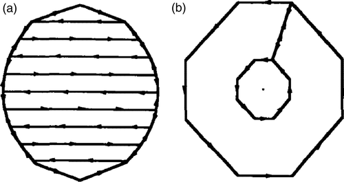
The actual scanning path coordinates, i.e. the sequential commands to the Octoson Stepper Motors, are calculated by the Apple II computer based on the information (oxtagon centre and corner locations and dimensions) obtained from the HP computer. At present the programs are able to drive the transducers along an octagonal path, the width and length of which can be specified independently. One can specify up to five concentric octagons at each scanning depth. Each of these octagons can be repeatedly scanned any number of times before scanning the next size octagon. These specifications are repeated at any number of depths. Thus, a three-dimensional volume can be covered as desired. In addition, a separate program is available to perform similar scanning path calculations for a raster scan pattern (). The scanning speed can be varied from zero to the maximum motor speed values—100, 50 and 40 mm/s in x, y and z (vertical) directions, respectively. The variable speed has been obtained by using programmable stepper motor indexers (Anaheim Automation Model CL 1397-2) to feed the stepper motor drivers of the Octoson instead of using its own fixed frequency oscillators. The communication between the Apple and the indexers is via a RS-232 interface. The minimum step lengths are 1/16, 1/34, and 1/70 mm in x, y and z directions, respectively.
Figure 4. Examples of the different scanning patterns tested, (a) A raster scan: the number of lines and the width of each path segment can be specified independently, (b) An octagonal scan: the width and length for each octagon, the number of concentric octagons, and the number of repetitions of each octagon can be specified.
During sonication the Apple computer also measures the resistance of potentiometers mounted on the gantry in order to detect its motion in all directions. The actual motion is compared with the programmed value and if a significant difference appears the computer will stop the sonication immediately.
Feedback control algorithm
During the treatments the power levels can be set manually by the operator or by the computer. In the feedback mode the temperatures from a prespecified set of thermocouples are read at desired intervals and the thermocouple with the maximum temperature detected. The difference between this value and the target temperature determines the duty cycle of the RF signal via a standard proportional controller.
Animal experiments
The following description of experimental results are provided to illustrate the capabilities of our system.
Thighs
The system was tested initially by heating dogs’ thighs using extensive thermometry (56 thermocouples). The dogs (15) were anaesthetized with halothane and eight, seven-sensor thermocouple probes were inserted vertically into the thigh in a row. The spacing between the probes was 10 mm and the distance between the sensors in the probes was 10 mm as well. The first sensor was on the skin and the last at a depth of 60 mm in the muscle. Thus temperature information was obtained from a vertical plane 70 mm wide and 60 mm deep. The focal region of the beam was located on a thermocouple sensor at a desired depth at one of the central probes by driving the transducers at low power and moving the gantry until the maximum temperature elevation was achieved. The scans used this location as their centre point. The scans were done in a horizontal plane, perpendicular to the plane of the thermocouple array.
In the investigations of deep heating, both thighs were coupled together using acoustic gel. The seven-sensor thermocouple probes were inserted horizontally in a vertical plane between the depths of 20 and 120 mm with suitable spacing. Thus, the temperatures were recorded in a vertical plane 60 mm wide and 100 mm deep starting at the depth of 20 mm. Again, the horizontal scanning plane was perpendicular to the plane of the thermocouple array.
The effect of scanning pattern on temperature was investigated by using transducers FOC 1–4 with the acoustical foci of the beams overlapping at the depth of 30 mm in the thigh of the dog. With the two, large focal zone, 1 MHz beams (FOC 3, 4) stationary, a sharp temperature maximum was produced at the depth of 30 mm (). When regular octagonal scans 20 mm in diameter were performed, the whole scan area was heated to a relatively uniform temperature (results not shown). However, if the scanning diameter was increased to 30 mm or more, the temperature at the centre region remained lower than the temperatures under the scanning path (results not shown). In order to elevate the temperature in a volume 50 mm in diameter, two concentric scans were performed. A reasonable result was obtained when two octagons, one 50 mm in diameter and one internal 20 mm in diameter, were scanned in the repeated sequence of two external and one internal octagon ( show these results for the two paths of different frequency, less focused transducers). In these cases, the temperatures were relatively uniform at depths between 10 and 60 mm, i.e. extending beyond our measurement area.
Figure 5. The temperature elevations measured in steady state in dog thigh using the small diameter transducers, (a) Two stationary transducers (FOC 3, 4), the beams overlapping at the depth of 30 mm. Total acoustical power was 15 W. (b) Two focused transducers (FOC 1, 2) scanned along two external (50 mm in diameter) and one internal (20 mm in diameter) octagons centred around the 35 mm distance position. The overlapping region was at the depth of 30 mm. Total acoustical power was 67 W. (c) Two transducers (FOC 3, 4) scanned along the same path as in . The total acoustical power was 34 W.
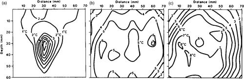
To illustrate the increased heating at depth that can be obtained, the four large transducers (FOC 5–8) were kept stationary, and were used to produce a maximum temperature at depths up to 100 mm (which was the maximum depth tested in our experiments) (). Also, by scanning the beams along an octagonal path 20 mm in diameter the maximum temperature was still produced at the focal depth, but the temperature elevation started about 20 mm in front of the overlapping region. Beyond the focal region, the temperatures decayed rapidly as illustrted in , which was obtained for a single 20 mm scan at a focal depth of 50 mm.
Figure 6. The isotherms measured in dog thigh using the four large focused transducers (FOC 5–8). (a) Stationary transducers focused at 100 mm depth. Total acoustic power was 7 W. The baseline temperature was 38–39°C. (b) A single 20 mm octagonal scan at a focal depth of 100 mm and centred at the 30 mm distance position. Total acoustic power was 27 W. The baseline temperature was 38–39°C. (c) A single 20 mm octagonal scan at a focal depth of 50 mm and centred at the 30 mm distance position. Total acoustic power was 22 W. The baseline temperature was 38–39°C.
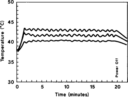
Finally in , the temperatures of three thermocouples as a function of time are presented during a feedback control experiment. The was kept within ±0–3°C even during vasodilation in the thigh.
Figure 7. The temperature as a function of time in three locations in dog thigh during feedback controlled sonication. The target temperature was 43°C.
Kidneys
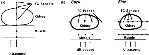
To study the effect of blood perfusion in a controlled manner, a doppler flow probe and an occluder were put around the renal artery of six dogs as described by Citation16. This allows control over and measurement of the total blood flow rate to the kidney. The kidney was fixed to the body wall during the surgery. Ten days after this operation the dogs were anaesthetized again and up to three, seven-sensor thermocouple probes were inserted into the kidney—in most of the experiments under ultrasound guidance. shows the two different thermocouple configurations used. The effect of transducer choice was again studied by comparing results obtained with the four smaller diameter transducers (FOC 1–4), and with four larger diameter, more highly focused transducers (FOC 5–8). The effect of scanning patterns was also studied with these transducer sets.
Figure 8. Diagrams illustrating the thermocouple placement in the kidney experiments. (a) For the experimental results given in and . (b) For the experimental results given in .
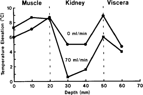
With the four small-diameter focused transducers (FOC 1–4) and a single 20 mm scan it was not possible to heat the kidney with full blood flow, due to the induction of high temperatures in the proximal muscle and visceral tissue distal to the kidney (). Once the blood supply to the kidney is cut off a significant temperature elevation was detected in the kidney but the maximum temperatures were still at the edges of the kidney. The situation was worse with larger scans, for example, with the two 50 mm external and one 20 mm internal scans, the ratio between the kidney temperature elevation and the muscle temperature elevation was 1/5 and 2/5 with kidney blood flows of 70 ml/min and 0 ml/min, respectively.
Figure 9. The steady-state temperature distributions produced with the four less highly focused transducers (FOC 1–4) along the centre of an octagonal scan 20 mm in diameter. The blood flow values to the kidney are shown in the graph. The total acoustic power was 30 W.
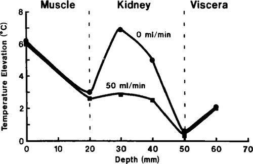
The results of using only two of the large transducers (FOC 5, 6) are presented in , where the temperature elevation along the central axis of a scanning path (2, 40 mm external and 1, 20 mm internal octagonal scans) are shown. The temperature elevation in the muscle was about twice that of the kidney when the kidney was receiving its full blood flow. However, when the blood supply was reduced to zero the maximum temperature occurred in the kidney (where the beams are overlapping). With a single 20 mm diameter scan with full kidney blood perfusion, the muscle temperatures were still higher than those in the kidney (results not shown).
Figure 10. The same as except the sonication was done with two larger focused transducers (FOC 5, 6).
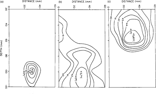
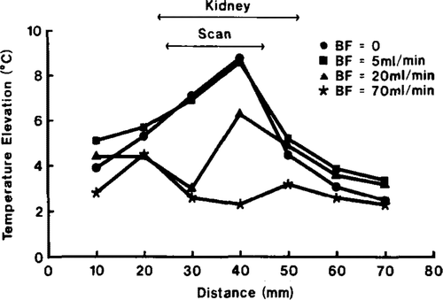
When all four large transducers (FOC 5–8) were used with the two 40 mm and one 20 mm octagon scan, almost equal temperature elevations were induced in both the kidney and the muscle at all kidney flow rates above approximately 5 ml/min (). At a flow rate of 5 ml/min the kidney temperature was, on the average, slightly higher than in the muscle, and with zero flow the kidney temperature was about 2°C above the muscle temperature. With single 20 mm scans, improved kidney heating was obtained and equal temperature elevations were detected in the whole heating field with full blood to the kidney (). The kidney temperature increased with decreasing blood flow and thus preferential heating of the kidney was obtained at lower flow rates (). In the temperature distribution across the kidney during a 20 mm scan is presented at various flow rates. At full flow the maximum temperature occurred at the edges of the kidney, but was elevated more at the centre with restricted flow.
Figure 11. The average (symbol), maximum (top of bar) and the minimum (bottom of bar) measured temperatures at various positions in the muscle and kidney as a function of the blood flow to the kidney (34 g) during two external (40 mm) and one internal 20 mm scans with the four large transducers (FOC 5–8). The total acoustic power was 37 W. The number of thermocouples is given in parentheses.
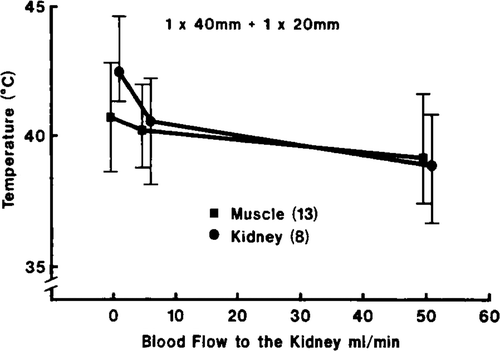
Figure 12. The same as in , but the scanning path was a single octagon 20 mm in diameter. The total acoustical power was 37 W.
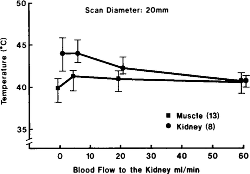
Figure 13. The steady-state temperature profile across the centre of a single 20 mm scanning path (using transducers FOC 5–8) at several flow rates into the kidney (34 g). The total acoustic power was 37 W.
Discussion
Temperature distributions
The system we have developed has been used to study the detailed temperature distributions in animals. Our previous report Citation17 described the effect of scanning speed, power level and blood perfusion on the temperature oscillations that occurred during repeated scans. The experimental results described in the present paper are illustrative of the effects of transducer choice, scanning pattern and blood perfusion rate on the temporal average temperature distributions produced.
In uniformly perfused tissues, like the thigh, the temperature distributions could be made relatively uniform in the heated region when a proper scanning pattern was used. The temperatures decreased rapidly outside the scan and also beyond the focal region if sharply focused transducers (FOC 5–8) were used. The maximum temperature could be reached at depths up to 10 cm in our experiments (the size of the dogs used limited attempts to heat deeper) providing that bones or air did not prevent the beams from penetrating. In clinical situations the size of the acoustical window through which the sound beams can propagate, will determine if particular deep-seated tumours can be heated. This may even lead to a situation, where only a small part of the tumour can be treated during one treatment and the other parts must be heated in the subsequent treatments. Technically this is possible with our system since the energy deposition can be accurately controlled.
The smaller diameter, less sharply focused transducers (FOC 1–4) were able to induce uniform temperature elevations that are contained within the outer scan diameter, but extended a few centimetres beyond the focal region. This was due to the long focal zones of the transducers () and is an obvious drawback if the tumour is small, or above a bone. However, if this is not the case, these types of longer focal zone transducers can be used to heat larger, superficial tumours with simpler two-dimensional scans. Thus, more weakly focused transducers can be used in some situations allowing simpler scanning mechanics and control algorithms, but generally more sharply focused transducers give better control over the temperature and also deeper penetration. Similarly, if large blood perfusion rate variations occur in the treatment volume or if a well-perfused target volume lies under a poorly perfused tissue layer, e.g. in our experiments the kidney was under a muscle layer, sharply focused transducers are required to overcome the blood perfusion difference and to induce the temperature maximum in the well-perfused volume. This was demonstrated in our attempts to heat the kidney with the small, weakly focused transducers, where the long focal zones caused the maximum temperatures in the poorly perfused surrounding tissues. However, with the larger, more sharply focused transducers and small scans, the muscle and kidney region could be heated to the same temperature with full blood flow to the kidney. If surface cooling had been applied, the temperature maximum would have been reached in the kidney. When the kidney blood flow was restricted to 20 ml/min, which is a perfusion rate of about 66 ml/100 g/min (if uniform perfusion is assumed throughout the kidney)—a value close to perfusion of the liver and higher than most other organs—the maximum temperature could be induced in the kidney (). These results show that the present system is capable of overcoming most of the blood perfusion disadvantages to be expected to occur during hyperthermia treatments, and can produce adequate heating in the target volume despite high perfusion rates.
Another important observation concerns the effect of perfusion on the temperature fields, which are not uniform in the volume scanned with constant power when large perfusion differences are present, as in the kidney experiments. Thus, in those cases a feedback system capable of controlling the power deposition distributions (i.e. varying the power along the scan) based on the temperature measurements is required. Our simple single point feedback control routine worked satisfactorily under large blood flow variations. However, it was not designed to control the temperature distribution and a more complex three-dimensional program will be needed in the future. This kind of feedback system (currently under development) is possible with our system due to the knowledge of the thermocouple locations with respect to the scanning pattern and the ability to vary power as a function of position along the scanning path.
The location of the treatment volume and thermocouples
The diagnostic ultrasound images of the Octoson provided a tool to identify internal anatomical structures. From the sequential vertical images, a three-dimensional map of the treatment volume, skin-water boundary and the location of bones and gas could be built into the computer memory. The heating of bones and attempted insonication through gas could be avoided and a suitable treatment window selected based on this imaging information. In addition, the treatment geometry can be compared with CT-scans by using markers locatable in both ultrasound and X-ray images. This planning capability, combined with the ability to scan the high focal zone around the desired treatment volume allows controlled, preferential power deposition in the desired treatment volume and thus provides control over the temperature distribution. The ultrasound images can also be used to guide the thermocouple probes into desired locations.
Summary
In summary, a scanned, focused ultrasound hyperthermia system has been constructed and tested extensively in animal trials. The system has multiple, focused transducers and the ability to image the treatment area and automatically calculate and control the scanning motor positions based on the anatomy of the desired treatment area. This system can induce maximum temperature elevations up to 10 cm deep (in our experiments the maximum depth was limited to 10 cm by anatomical size) in uniformly perfused tissues (thigh). In such uniformly perfused tissues, uniform temperature elevations can be produced in relatively large regions (up to 5–6 cm diameter regions were tested in this study) with a proper choice of scanning pattern. Large differences in the blood perfusion rate of adjacent tissues can be compensated for by using sharply focused transducers and thus therapeutic temperatures can be induced in organs with high perfusion rates even when the ultrasound must pass through adjacent layers of poorly perfused tissues, like muscle. Finally, the treatment geometry including the thermocouple locations could be determined in the actual treatment position by using the diagnostic ultrasound images. Thus, the ultrasound beams could be guided accurately to the treatment volume, and overheating of bones or other critical organs could be avoided. The question of feeling pain—so common with plane, stationary transducer systems—cannot be tested in animals, but only human trials can answer if it really limits the usefulness of scanned, focused ultrasound in the induction of hyperthermia. Since this has not been a problem with a previous scanned, focused single transducer system Citation4, we do not anticipate problems with our system.
Acknowledgments
We would like to thank Peggy Kundrat, Beverly Curtis, Michael Carlton, Terry Hoover, Eduardo Moros and David Edwards for their assistance in the construction and testing of this device. Ausonics, Inc. has been very generous in their loan of an Octoson system, the construction of the power transducers, and in general maintainance of the Octoson. This research has been supported by NIH Grant CA 33922.
Declaration of interest: The authors report no conflicts of interest. The authors alone are responsible for the content and writing of the paper.
References
- Marmor JB, Hahn GM. Ultrasound heating in previously irradiated sites. Int J Radiat Oncol Biol Phys 1978; 4: 1029–1032
- Corry PM, Barlogie B, Tilchen EJ, Armour EPR. Ultrasound-induced hyperthermia for the treatment of human superficial tumors. Int J Radiat Oncol Biol Phys 1982; 8: 1225–1229
- Fessenden P, Lee ER, Anderson TL, Strohbehn JW, Meyer JL, Samulski TV, Marmor JB. Experience with a multitransducer ultrasound system for localized hyperthermia of deep tissues. IEEE Trans Biomed Eng 1984; 31: 126–135
- Lele PP. Physical aspects and clinical studies with ultrasound hyperthermia. Hyperthermia in Cancer Therapy, FC Storm. Hall Medical Publishers, Boston 1983; 333–367
- Hynynen K, Watmough DJ, Shammari M, Wilmot G, Murthy MSN, Mallard JR, Fuller M, Sarkar T. A clinical hyperthermia unit utilizing an array of seven focused ultrasonic transducers. Proceedings of the IEEE Ultrasonics Symposium 1983a; 816–821
- Shapiro E, Seppi E, Zitelli L, Henderson S, Wehlau A, Wu G, Dittmer C. A large aperture ultrasonic array system for hyperthermia treatment of deep-seated tumors. Proceedings of the IEEE Ultrasonics Symposium 1985; 942–948
- Ocheltree KB, Benkeser PJ, Frizzell LA, Cain CA. An ultrasonic phased array applicator for hypothermia. IEEE Trans. Sonics and Ultras. 1984; 31: 526–531
- Benkeser PJ, Ocheltree KB, Frizzell LA, Cain CA. Ultrasound phased array hyperthermia applicator. Proceedings of the 7th Annual Conference of IEEE Engineering in Medicine and Biology Society. 1985; 337–340
- Cain CA, Umemura S. Annular and sector phased array applicators for ultrasound hyperthermia. Proceedings of the IEEE Ultrasonics Symposium 1985; 936–941
- Stewart HF. Ultrasonic measurement techniques and equipment output levels. Essentials of Medical Ultrasound, MH Repacholi, DA Benwell. Humana Press, Clifton, NJ 1982; 77–116
- Martin CJ, Law ANR. Design of thermistor probes for measurement of ultrasound intensity distributions. Ultrasonics 1983; 21: 85–90
- Fry WJ, Fry RB. Determination of absolute sound levels and acoustic absorption coefficients by thermocouple probes-experiment. J Acoustical Soc Am 1954; 26: 311–317
- Cristensen DA, Kuhn PK. Temperature errors caused by probe heating in ultrasound hyperthermia. Proceedings of the 33rd Annual Meeting of the Radiation Research Society. 1985; 61
- Cetas TC, Connor WG. Thermometry considerations in localized hyperthermia. Med Phys 1978; 5: 79–91
- Hynynen K, Martin CJ, Watmough DJ, Mallard JR. Errors in temperature measurement by thermocouple probes during ultrasound induced hyperthermia. Br J Radiol 1983b; 56: 969–970
- Cetas TC. Use of dog kidneys as in vivo thermal models. 7th Annual Conference of IEEE Engineering in Medicine and Biology Society. September, 1985
- Hynynen K, Roemer RB, Moros E, Johnson C, Anhalt D. The effect of scanning speed on temperature and equivalent thermal exposure distributions during ultrasound hyperthermia in vivo. IEEE Trans Microwave Theory Tech 1986; 34: 552–559