Abstract
For superficial hyperthermia a custom-built multi-applicator multi-amplifier superficial hyperthermia system operating at 433 MHz is utilised. Up to 6 Lucite Cone applicators can be used simultaneously to treat an area of 600 cm2. Temperatures are measured continuously with fibre optic multi-sensor probes. For patients with non-standard clinical problems, hyperthermia treatment planning is used to support decision making with regard to treatment strategy. In 74% of our patients with recurrent breast cancer treated with a reirradiation scheme of 8 fractions of 4 Gy in 4 weeks, combined with 4 or 8 hyperthermia treatments, a complete response is achieved, approximately twice as high as the CR rate following the same reirradation alone. The CR rate in tumours smaller than 30 mm is 80–90%, for larger tumours it is 65%. Hyperthermia appears beneficial for patients with microscopic residual tumour as well. To achieve high CR rates it is important to heat the whole radiotherapy field, and to use an adequate heating technique.
Introduction
Reirradiation combined with hyperthermia is an effective treatment for recurrent breast cancer. Results from five randomised trials have shown that the complete response (CR) rate in breast cancer recurrences increases from 41% to 59% when hyperthermia is added to radiotherapy Citation[1]. For the subgroup of patients treated within the ESHO 5-88 trial with a reirradiation schedule of eight fractions of 4 Gy, applied twice weekly, the CR rate even increased from 38% after radiation alone to 78% after combined treatment.
In Rotterdam, the first patient with recurrent breast cancer was treated with hyperthermia in 1978. Over the years, many alterations in hyperthermia and thermometry equipment, in treatment procedure, registration and treatment scheme were made. In this review we take you through some of the history of hyperthermia in our department, and present the resulting treatment procedure and a summary of our clinical results.
History of equipment used
Heating equipment
In 1978 we started our clinical research on hyperthermia with the Pomp-Siemens whole body hyperthermia cabin which included several applicators for local hyperthermia Citation[2]. These were condenser plates operating at 27 MHz and dipole antennas operating at 433 or 2450 MHz. With these applicators, originally designed for physiotherapy, we treated patients who were reirradiated for palliative reasons, among whom were patients with recurrent breast cancer. The first applicators developed in our department were air-filled waveguides operating at 2450 MHz with aperture sizes of 8 × 4 and 8 × 6 cm2. The rectangular shapes allowed us to use combinations of up to eight applicators at the same time. The number of amplifiers was still limited so that up to four applicators were coupled to one power supply, without the possibility to control power supply to the individual applicators. Surface cooling, when necessary, was performed by directing air currents under the applicators.
In 1985 we started using custom built water-filled waveguides operating at 433 MHz with a radiating opening of 10 × 10 cm2. Until 1987 we could use maximally two applicators simultaneously, thereafter five and some time later six. Each applicator was supplied with independent power control Citation[3]. These standard waveguides were replaced in 1996 by Lucite cone applicators (LCA), with a larger effective field size (EFS). The EFS of the LCA is approximately 100 cm2, which is considerably larger than the 33 cm2 of the standard waveguide Citation[4]. The performance of both waveguide types was tested in the clinical setting by treating patients alternately by standard waveguides and LCA arrays. The average invasive temperature was 0.28°C higher with the LCAs than with the standard waveguides, which was primarily the result of higher temperatures in the periphery of the treatment field. With the 433 MHz waveguide applicators, a perfused water bolus was used to control surface temperatures.
Water bolus dimensions and selection of water bolus temperature
The water bolus placed between water-filled waveguides and skin has two functions: improvement of coupling between the applicators and tissue and control of superficial temperature.
We investigated the effects of water bolus configuration on the EFS of the LCA Citation[5]. Placement of the LCA near the water bolus edge reduced the EFS considerably. With water bolus heights of more than 20 mm the EFS became more sensitive to distance to the water bolus edge. Based on the results, the guideline now is that the height of the water bolus should not exceed 20 mm and the water bolus should extend the LCA aperture at least 25 mm, especially at the Lucite windows.
The two main parameters used for optimising the temperature distribution are the electromagnetic power and the water bolus temperature. A 3-D model was set up to simulate an abstraction of the treatment. In the model a convection coefficient for the water bolus to skin surface was employed, which was measured for water boluses of different sizes. The effect of perfusion and fat layer thickness were investigated in a layered model. The performance of the general model was verified against clinical data. The model was found to predict the temperature distribution well on a global view, and was used to set up guidelines, specific for our equipment, for the water bolus temperature selection for various target depths and applicator arrays Citation[6].
Thermometry
We started with thermocouples, either single sensor probes within a needle, or multi-sensor probes within a catheter. These probes had to be placed perpendicular to the direction of the electric field (E-field) and temperatures were measured every 5 min with the power shut off. Since 1987 temperatures have been measured continuously during treatments by a 24-channel fibre-optic system, with which five multi-sensor probes (up to four sensors) and four single sensor probes are available (Takaoka FT1210). Closed-tip catheters are placed interstitially immediately before the first treatment and left in place till after the last treatment. The aim is to have both interstitial and superficial thermometry under each applicator. Usually these catheters cause no clinical problems Citation[7].
Use of hyperthermia treatment planning
Hyperthermia treatment planning tools have a significant potential to further improve the quality of hyperthermia treatments by providing insight into the 3-D absorbed power distributions. In some patients with non-standard clinical problems, SEMCAD-X Citation[8], Citation[9] hyperthermia treatment planning was successfully used to support decision making with regard to the treatment strategy. Two cases are shortly described here.
A patient with recurrent breast cancer had undergone open heart surgery in the past, and sternal cerclage wires were within the target volume for hyperthermia. Treatment planning showed that the distortion of the electromagnetic field by the cerclage wires was negligible with the E-field direction perpendicular to the cerclage wires. This patient was treated without problems with the applicator configuration advised by the planning.
The tumour of a patient with a recurrent breast cancer in the infraclavicular region was located at a depth of 37 to 54 mm below the skin, which is beyond the superficial system's standard maximum target depth of 40 mm. However, the subcutaneous fat layer in this patient was above average: 29 mm. Because the effective conductivity of fatty tissue is relatively low, it could be anticipated that power absorption in the fat layer would be limited, and that the remaining power at depth would be sufficient within the tumour. This was confirmed by hyperthermia treatment planning, and during hyperthermia treatment the intratumour temperature reached therapeutic levels.
Treatment scheme
When we started combining reirradiation with hyperthermia, the tolerance limits for reirradiation were not known. We started cautiously, with total doses of 12–25 Gy, in fraction sizes of 2 to 4 Gy. To avoid thermotolerance we chose a treatment scheme of hyperthermia twice weekly with at least 3-day intervals. In order to sensitise every radiation fraction, radiation was also given twice weekly, in fractions of 4 Gy. Hyperthermia was given after radiotherapy on the basis of experimental studies showing that maximum therapeutic benefit can be obtained with that sequence Citation[10–14].
When we did the first evaluation of the results of reirradiation and hyperthermia in 97 patients with recurrent breast cancer, we found a large influence of the applied reirradiation dose on CR rate. With a total dose of less than 29 Gy the CR rate was 24%, while it was 58% after a dose of 30 to 32 Gy. Time till progression was median 4 months after a partial response (PR) and 26 months for CR Citation[15]. The reirradation schedule of eight fractions of 4 Gy appeared safe, effective and well tolerated and was therefore selected as the standard scheme.
In 1996 we had a capacity problem for superficial hyperthermia. Taking in mind the results of several published randomised studies comparing a low with a high number of hyperthermia treatments, usually one versus two treatments per week, which showed no difference in results Citation[16–21], we decreased the number of hyperthermia sessions to four. The number of patients was insufficient to do a randomised trial ourselves and we planned to evaluate the results after treating a sufficient number of patients with the new schedule.
We did a first analysis of results in patients treated with four hyperthermia sessions in 2004 Citation[22] and compared these to those in patients who received eight hyperthermia sessions: 40 patients received four and 132 patients eight hyperthermia treatments. For patients with a maximum tumour diameter ≤30 mm, CR rate was 86% after eight treatments and 82% after four treatments (not significant). For patients with larger tumours, CR rate was 59% after eight treatments and 65% after four treatments (not significant). The preliminary conclusion is that a decrease in number of hyperthermia treatments does not lead to inferior results. On the other hand, the hoped-for decrease in hyperthermia toxicity was not observed as well. A problem with this comparison is that, at around the same time that we changed the number of hyperthermia treatments, we also replaced the standard waveguide with the Lucite Cone Applicator, with which we achieved average 0.28°C higher temperatures. Although it is unlikely that a 0.28°C higher temperature compensates for 240 min treatment duration, we will perform a detailed analysis of prognostic factors including thermal dose parameters in these patients.
Lessons learned
No electromagnetically induced hyperthermia in anaesthetised patient
We started our clinical hyperthermia research with the idea to apply local hyperthermia during whole body hyperthermia, in order to achieve a more homogeneous temperature distribution. In the first patient in whom we tried whole body hyperthermia, it appeared that a core temperature >40°C was not tolerated by the conscious patient. We therefore gave subsequent treatments under general anaesthesia. During the third treatment, core temperature was increased to a temperature of 41.6°C and the recurrent tumour at the chest wall was simultaneously heated with 433 MHz. One of the thermometry probes suddenly showed a steep temperature increase to maximum 47°C. After the treatment, the patient developed a severe third degree burn of the thoracic wall with a diameter of 100 mm and including ribs Citation[23].
We have seen similar toxicity in two other patients who were treated under general anaesthesia. During normothermic regional isolation perfusion of the leg for multiple skin metastases of malignant melanoma, local hyperthermia was given to one of the metastases. Hyperthermia was given with a 2450 MHz dipole antenna with a diameter of 80 mm and interstitial temperatures were average between 39.2° and 40.9°C. In two of three patients treated this way, in whom the measured maximum temperature had been 41.4° and 40.3°C, a third-degree burn developed.
Unnoticed hot spots resulting in toxicity can occur in conscious patients as well, at sites of decreased sensitivity due to previous surgery, but usually some sensation of pain limits the extent of the damage.
No stray irradiation near linear accelerator
For a short period of time we treated our patients in an orthovoltage room which was located next to a linear accelerator. The microwave equipment at that time consisted of a circular field dipole antenna connected to a generator operating at 433 MHz. In the Netherlands, 433 MHz can be used without shielding. The linear accelerator was a CGR-MeV Sagittaire. We found that the stray microwave radiation, at intensities of about 0.4 mW cm−2 in the control room of the accelerator interfered with the beam energy settings. The microwave interference caused an increase in beam energy. At maximum power output this was a change from 25.5 MeV to 29.1 MeV Citation[24]. The most practical solution to this problem was to transfer the hyperthermia treatment to another room.
Heating technique is important for treatment outcome
In the first evaluation of treatment results in the group of patients treated with eight fractions of 4 Gy and hyperthermia, we found a CR rate of 58% Citation[15]. When we evaluated later a larger group of patients, the CR rate was 71%. A multivariate analysis showed that two factors were independent and significantly associated with local control probability: tumour size (maximum diameter ≤ 30 mm or >30 mm) and equipment used (2450 MHz or 433 MHz equipment) Citation[25]. The better overall results were the effect of a large improvement in CR rate in the larger tumours: 31% with 2450 MHz heating and 65% with 433 MHz heating. In tumours ≤30 mm the results of 2450 and 433 MHz heating were not different; approximately 90% CR. The CR rate achieved with 2450 MHz in the larger tumours was similar to the results of reirradiation with 8 × 4 Gy without hyperthermia: 28% in the RTOG study Citation[26] and 38% in the ICHG study Citation[1]. Apparently, 2450 MHz heating was inadequate for the larger tumours. A disadvantage of using 2450 MHz compared to 433 MHz is the smaller penetration depth and thereby a smaller heated volume. From this experience we learned that patients should not be accepted for hyperthermia treatment if we expect that we cannot adequately heat the whole target volume.
Whole reirradiation volume is target for hyperthermia
Until July 1987, the aim of treatment was to heat the macroscopic tumour. With that approach, we observed in a few patients tumour regrowth within the radiation field, outside the margin of the hyperthermia field. At the same time, there was no regrowth within the combined treated field. Since then we choose an applicator set-up such that the radiation field is widely covered.
Further experience suggests that hyperthermia is an effective additional treatment for microscopic tumour. The patient population in which we found better outcomes after 433 MHz heating compared to 2450 MHz heating included a total of 15 patients with microscopic disease. Three patients treated with 2450 MHz equipment all developed in-field tumour regrowth 10–12 months after the start of treatment. In 12 patients treated with 433 MHz equipment only, two in-field re-recurrences occurred, 10 and 13 months after start treatment. Three patients died with a locally controlled tumour after median 10 months, and seven patients were still alive with a locally controlled tumour 16–70 months after treatment. This is a significant difference, suggesting that good-quality hyperthermia is effective here as well Citation[25].
A tumour near the eye can be treated successfully
A patient was referred with a metastatic lesion of breast cancer in the lower eyelid, recurring after two radiation treatments with partially overlapping fields. The tumour was progressive under second-line hormonal therapy and she was unfit for chemotherapy. The first local treatment of this tumour had been irradical resection (positive surgical margins) and radiotherapy, 10 × 3 Gy plus boost of 10 × 2 Gy. The tumour recurred 9 months later at the margin of the radiotherapy field, was treated again with irradical surgery and 15 × 2 Gy. Four months later the tumour recurred again. Two treatment options were discussed with the patient: surgery, including enucleation of the eye, or reirradiation with hyperthermia, with unknown risk of toxicity such as eyelid fibrosis, retina damage, cataract and glaucoma. The patient preferred radiotherapy and hyperthermia. During radiotherapy the eyeball was shielded with a lead contact lens. We applied eight fractions of 4 Gy and four hyperthermia treatments of 60 min. The tumour regressed fast with a complete regression established one week after the last treatment. During follow-up, local tumour control was maintained. The only side effect was a dry eye for which the patient used eye drops. Vision was unchanged. The patient died 22 months after the last treatment of a cerebrovascular accident unrelated to breast cancer Citation[27].
No excessive toxicity in patients with tissue transfers
Between 1992 and 2009, 36 patients were treated for a total of 37 tissue transfers, including split skin grafts (15), transverse rectus abdominis myocutaneous (1), latissimus dorsi (14) and rhomboid flaps (1) or a combination of grafts and flaps. The guidelines for treating these patients were no different from those for other patients. Hyperthermia toxicity (according to CTC-AE version 3) grade 2 (minimal medical intervention required, no interference with activities of daily life (ADL) occurred in four patients and grade 3 (surgical intervention required and/or interference with ADL) in three. The incidence of toxicity appears not much different from that observed in patients without tissue transfer and is acceptable Citation[28].
Current treatment procedure
Patient selection
In the Netherlands, national guidelines prescribe reirradiation and hyperthermia for recurrent breast cancer after previous irradiation in the same area, when the expected survival is 6 months or more. This concerns inoperable tumours, irradically resected tumours (tumour positive surgical margins), or radically resected tumours with a high risk of re-recurrence (multifocal recurrences or second recurrences).
The aim of the treatment is a complete response, which means that it must be possible to heat the whole target volume. The target volume should be within 40 mm of the skin surface, but on occasion subcutaneous fat can be subtracted from this distance. It must be feasible to place the applicators parallel to the surface of the treatment area. When the area is larger than 20 × 30 cm2, two (or more) hyperthermia applications are scheduled for one treatment. We consider a pacemaker a contraindication for hyperthermic treatment. Metallic implants larger than surgical clips may give problems, e.g. a portacath has to be removed. In case of doubt we will model a treatment with SEMCAD-X.
The patients receive a detailed explanation of the treatment procedure and information on their own role in monitoring the temperature distribution, specifically instructions concerning the mentioning of hot spot-induced complaints.
Preparation before first treatment
The treatment team of physician or nurse practitioner, physicist and technician examine the treatment area and decide which applicator set-up will be used. The aim is to cover the whole radiotherapy field with the combined applicator footprint with an overlap of 10 mm all around. Thereafter the closed tip catheters are placed under local anaesthesia, with the aim to have interstitial thermometry under each of the applicators. The catheters are fixed to the skin with Histoacryl® tissue adhesive (Braun, Melsungen, Germany) and Tegaderm® transparent dressing (3M, St. Paul, Minnesota). The interstitial length and depth of each catheter are measured. A life-size drawing of the treatment area is made on a transparent sheet including some anatomical landmarks (prominent bones, scars, and birth marks), the radiotherapy field margins, the location of macroscopic tumour, the location of the applicators, and the location of interstitial and superficial thermometry probes Citation[29]. All necessary information is loaded into the computer program for treatment monitoring, steering and registration.
Treatment
The patient is positioned on the treatment bed in a position as comfortable as possible, and up to 23 thermometry probes are placed within the catheters and on the skin. Multi-sensor probes with four measuring points at 20 mm spacing are placed in the catheters, some of the superficial probes are placed on scars. The applicator position is indicated on a gauze which is placed on the surface of the treatment area and then wetted. The water bolus is placed such that it extends the planned position of the applicators with at least 25 mm. The water bolus temperature is selected depending on the size of the water bolus and the depth of the target volume. Usually the applicators are placed ‘clockwise’: adjacent applicators have their E-field direction perpendicular to each other Citation[4].
The treatment is administered by the technician. All necessary information is visible on PC screens during treatment: the position of all applicators and thermometry probes in relation to the patient's anatomy, the power output and reflected power per applicator, the course of temperature under each applicator and the current temperature at each measuring site (, , and ). The first treatment starts with a power of 30 W per applicator. The increase of power per applicator depends on the steepness of temperature increase under the specific applicator. The aim is to have all interstitial temperatures above 40°C. An interstitial temperature of maximum 43°C is allowed during the first 30 minutes, thereafter maximum 44°C. In tumour tissue at a distance of more than 10 mm from normal tissue higher temperatures are allowed. The treatment duration is 60 min with power on.
Figure 1. The superficial hyperthermia treatment set-up. The technician observes both the patient and the temperature and power information. The PC screens show, from left to right (A) the course of temperatures over time, per applicator; (B) the drawing of the treated area with location of applicators and actual temperature per measuring site (details are shown in ), and (C) the power output per applicator.
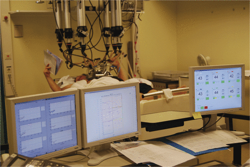
Figure 2. Close-up of the applicators placed above the thoracic wall, on top of the perfused water bolus.
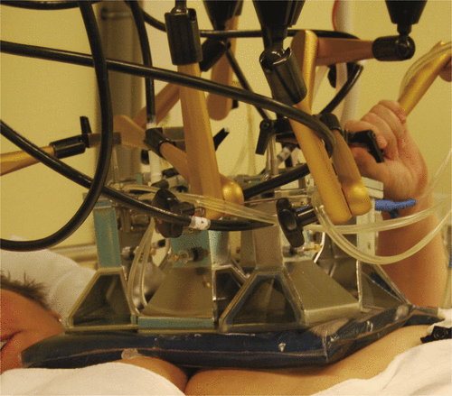
Figure 3. (A) A patient under treatment, with recurrent breast cancer on the ventral and lateral thoracic wall. This patient was treated with two applications.
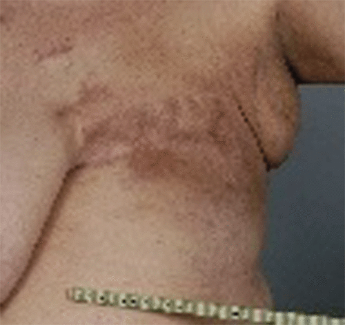
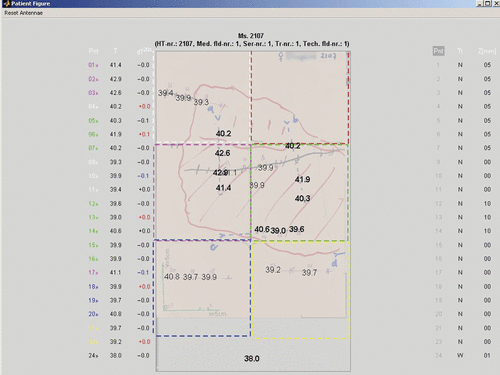
Figure 3. (B) An example of the middle PC screen during treatment with the drawing of the ventral treatment area. The drawing shows the tumour tissue, the mastectomy scar, four interstitially placed catheters, the footprints of the applicators, and actual interstitial (in bold) and superficial temperatures.
Evaluation
Between treatments, the course of the previous treatment is discussed with the whole team and adjustments for the application of the next treatment determined. Special attention is paid to cold spots (average temperature below 40°C), treatment-limiting hot spots, and the temperature distribution in depth. When a cold or a hot spot can be explained by the expected SAR distribution, the position of the applicators or their E-field direction is changed. When superficial temperatures were power-limiting during treatment, the water bolus temperature is decreased. When the treatment quality is limited by tumour-related pain, or general stress or anxiety, appropriate medication will be given during subsequent treatments. A detailed description of this evaluation can be found in De Bruijne et al. Citation[30].
Results
Effect on tumour
Since we use 433 MHz for the application of hyperthermia, the results are rather stable. In 1999 we published a CR rate of 74% for the total group of patients treated with eight fractions of 4 Gy and eight hyperthermia treatments, 87% for patients with tumours smaller than 30 mm and 65% for patients with larger tumours Citation[25]. With the same reirradiation schedule combined with four hyperthermia treatments, the overall CR rate is 73%; 82% for small tumours and 65% for larger tumours Citation[22]. The median duration of local tumour control is 32 months. In patients treated for a microscopic tumour residual, the local tumour control rate till death or date of last follow up was 83% for the patients who received eight hyperthermia treatments and 84% for those receiving four hyperthermia treatments.
Toxicity
Acute radiation toxicity usually is no problem with this schedule with an incidence of epidermolysis in 11% of the patients Citation[25]. In the randomised trial no increase in radiation toxicity was found Citation[1]. One case report even suggested a decrease in late toxicity (telangiectasis) with the addition of hyperthermia Citation[31].
Hyperthermia toxicity is rather frequent in these patients. In 1999 we reported second-degree burns in 19% of the patients and third-degree burns in 7% and subcutaneous burns in 3% after eight 433 MHz treatments. In 71 patients who received four treatments we observed second-degree burns in 31%, third-degree burns in 10% and subcutaneous burns in 7%. These side-effects usually are grade 2 or less according to the Common Terminology Criteria for Adverse Events version 3.0 scoring system. The hyperthermia-induced burns generally cause no pain because of their occurrence at sites of decreased sensitivity.
Late radiation toxicity was evaluated in 121 patients treated with reirradiation (8 × 4 Gy) and hyperthermia (eight treatments) between 1992 and 2000. The overall incidence of late radiation toxicity was 12%: a skin ulcer in six patients, bone necrosis or fracture in seven patients, and both an ulcer and bone fracture in one patient. The incidence of late radiation toxicity, however, increased with longer follow up durations. In 38 patients with a follow up duration >3 years it was 18%, and in eight patients followed longer than 5 years it was 38%. The median follow up of all patients was between 1 and 2 years (Van der Zee unpublished results).
Discussion
In approximately three quarters of our patients, reirradiation with eight fractions of 4 Gy combined with hyperthermia results in a CR, which lasts for a median duration of 32 months. In over 80% of the patients treated for microscopic disease, local tumour control lasts till death or date of last follow up. We do not expect that a locally controlled chest wall recurrence will influence overall survival. Nevertheless, the absence of symptomatic local tumour can result in an improvement in quality of life Citation[32]. In our view, the achievement of a partial response is less worthwhile, since regrowth is observed after median 4 months and we find it unlikely that hyperthermia influences time to progression. That is the reason that we do not accept patients for hyperthermia treatment when we can heat only part of the target volume.
We have not included a test heating session in the patient selection procedure. Patients are selected on the basis of tumour location and extension in depth. We assume that a target volume can be adequately heated when the depth is limited to 40 mm from the skin surface and the applicators can be placed parallel to the surface over the whole target area. The results of the randomised trial of Jones et al., for which patients were eligible after a test treatment had shown ‘heatability’ (the achievement of a hyperthermia dose of 0.5 CEM43°CT90) Citation[33], has triggered us to evaluate retrospectively this thermal dose parameter in our patients. CR rates were the same for patients who were heatable and unheatable, and for patients who received more or less than 10 CEM43°CT90 during the whole treatment series Citation[34].
Many clinical studies on hyperthermia in addition to radiotherapy included patients with recurrent breast cancer after previous irradiation. The results in this specific subgroup, however, are not always reported separately. summarises CR rates in this subgroup which are available from published experience Citation[1], Citation[21], Citation[26], Citation[35–52]. This table includes three studies in which not all, but the majority of patients were reirradiated, and one study reporting CR combined with partial response with >80% regression. Four studies included a control group treated with the same radiation alone: three randomised studies and one study in which patients with multiple lesions received radiation alone to one lesion and combined treatment to another. CR rates following reirradiation and hyperthermia vary widely, from 20% to 95%. This is not surprising, since the used radiotherapy schedule varies between studies and also within studies, and the prognostic variables of included patients will differ between studies as well. A summation of the data results in 61% CR after combined treatment and 32% after radiotherapy alone. In the majority of studies, hyperthermia is combined with radiotherapy only and applied after radiation. Unusual approaches are simultaneous combination of radiation and hyperthermia and the addition of chemotherapy. Myerson et al. Citation[46] tested the simultaneous combination of radiotherapy and hyperthermia in 15 patients and achieved a CR in 79%. Feyerabend et al. Citation[47] applied once weekly epirubicin and ifosfamide, simultaneously with hyperthermia, in the period of radiotherapy. Kouloulias et al. Citation[48] applied once-monthly liposomal doxorubicin 3 to 40 h before hyperthermia, once during the period of radiotherapy and five times thereafter. The complete response rates in the last two studies were lower than in all other studies: 22% and 20% respectively.
Table I. Results of reirradiation and hyperthermia in recurrent breast cancer
We find the schedule of two fractions of 4 Gy per week attractive in view of the palliative aim of the treatment. The overall duration of a treatment series is 3.5 weeks, during which period patients have to come to the hospital only twice weekly for around two h; the inconvenience is limited. On the other hand, the incidence of late toxicity can be expected to be lower with a radiation schedule with smaller fraction sizes Citation[53], Citation[54]. Oldenburg et al. recently reported a 40% incidence of ≥ grade 3 toxicity at three years in 78 patients treated with 8 × 4 Gy and hyperthermia for microscopic disease Citation[55]. In our patients it was 38% at five years follow up. For the majority of patients late toxicity will not be a problem in view of the limited overall survival, but for the patients with a longer expected overall survival smaller fraction sizes may be considered.
We are now investigating the potential use of predicted 3D-SAR (specific absorption rate) coverage as a prognostic indicator for treatment outcome. Patient-specific treatment planning is done with SEMCAD X Citation[8]. Predicted SAR-volume histograms, total absorbed energy per tissue type and calculated temperatures will be compared with measured temperatures, and we will analyse whether the predicted treatment quality is correlated with treatment outcome. A correlation of predicted treatment quality with measured temperatures and/or with clinical outcome would allow the abandonment of interstitial thermometry, to prescribe treatments of a certain quality, and to apply reproducible treatments.
In conclusion, it is feasible to achieve CR rates of 65% to 90% in breast cancer recurrences when reirradiation is combined with hyperthermia. The burden to the patient can be limited to four 2-h visits to the clinic. To achieve high CR rates it is important to heat the whole radiotherapy field, and to choose an adequate heating technique. In special cases hyperthermia treatment planning can be applied to support clinical decisions.
Declaration of interest: The authors report no conflicts of interest. The authors alone are responsible for the content and writing of the paper.
References
- International Collaborative Hyperthermia Group (CC Vernon, JW Hand, SB Field, D Machin, JB Whaley, J van der Zee, WLJ van Putten, GC van Rhoon, JDP van Dijk, D Gonzalez Gonzalez, F-F Liu, P Goodman and M Sherar). Radiotherapy with or without hyperthermia in the treatment of superficial localized breast cancer–results from five randomised controlled trials. Int J Radiation Oncology Biol Phys 1996;35:731–744
- Reinhold HS, Van der, Zee J, Faithfull NS, Van Rhoon GC, Wike-Hooley J. Utilization of the Pomp-Siemens hyperthermia cabin. Natl Cancer Inst Monogr 1982; 61: 371–375
- Van Rhoon GC, Rietveld PJM, Van der Zee J. A 433 MHz Lucite cone waveguide applicator for superficial hyperthermia. Int J Hyperthermia 1998; 14: 13–27
- Rietveld PJM, Van Putten WLJ, Van der, Zee J, Van Rhoon GC. Comparison of the clinical effectiveness of the 433 MHz Lucite cone applicator with that of a conventional wave guide applicator in applications of superficial hyperthermia. Int J Radiation Oncology Biol Phys 1999; 43: 681–687
- De, Bruijne M, Samaras T, Bakker JF, Van Rhoon GC. Effects of water bolus size, shape and configuration on the SAR distribution pattern of the Lucite cone applicator. Int J Hyperthermia 2006; 22: 15–28
- Van der Gaag ML, De Bruijne M, Samaras T, Van der Zee J, Van Rhoon GC. Development of a guideline for the water bolus temperature in superficial hyperthermia. Int J Hyperthermia 2006; 22: 637–656
- van der Zee J, van Rhoon GC, Broekmeyer-Reurink MP, Reinhold. HS. The use of implanted closed-tip catheters for the introduction of thermometry probes during local hyperthermia treatment series. Int J Hyperthermia 1987; 3: 337–345
- SEMCAD Reference Manual. Zürich, Switzerland: Schmid & Partner Engineering AG; 2004. Available at: http//www.semcad.com (accessed 24 November 2005)
- Bruijne De, Wielheesen M, Van der Zee DHM, Chavannes J, Van Rhoon N, GC. Benefits of superficial hyperthermia treatment planning: five case studies. Int J Hyperthermia 2007; 23: 417–429
- Gillette EL, Ensley BA. Effect of heating order on radiation response of mouse tumor and skin. Int J Radiat Oncol Biol Phys 1979; 5: 209–213
- Hill SA, Denekamp J. The response of six mouse tumours to combined heat and X rays: Implications for therapy. Br J Radiol 1979; 52: 209–218
- Hiraoka M, Miyakoshi J, Jo S, Takahashi M, Abe M. Effects of step-up and step-down heating combined with radiation on murine tumor and normal tissues. Jpn J Cancer Res 1987; 78: 63–67
- Overgaard J, Overgaard M. Hyperthermia as an adjuvant to radiotherapy in the treatment of malignant melanoma. Int J Hyperthermia 1987; 3: 483–501
- Van der Zee J, De Bruijne M, Van Rhoon GC. Hyperthermia dose and schedule: no evidence yet for changing treatment design. Int J Hyperthermia 2006; 22: 433–447 , (Letter)
- Van der Zee J, Treurniet-Donker AD, The SK, Helle PA, Seldenrath JJ, Meerwaldt JH, Wijnmaalen AJ, Van den Berg AP, Van Rhoon GC, Broekmeyer-Reurink MP, Reinhold HS. Low dose reirradiation in combination with hyperthermia: A palliative treatment for patients with breast cancer recurring in previously irradiated areas. Int J Radiat Biol Phys Oncol 1988; 15: 1407–1413
- Arcangeli G, Nervi C, Cividalli A, Lovisolo GA. Problem of sequence and fractionation in the clinical application of combined heat and radiation. Cancer Res 1984; 44: S4857–4863
- Valdagni R. Two versus six hyperthermia treatments in combination with radical irradiation for fixed metastatic neck nodes: progress report. Recent Results Cancer Res 1988; 107: 123–128
- Kapp DS, Petersen IA, Cox RS, Hahn GM, Fessenden P, Prionas SD, Lee ER, Meyer JL, Samulski TD, Bagshaw MA. Two or six hyperthermia treatments as an adjunct to radiation therapy yield similar tumor responses: Results of a randomized trial. Int J Radiat Oncol Biol Phys 1990; 19: 1481–1495
- Emami B, Myerson RJ, Cardenes H, Paris KG, Perez CA, Straube W, Leybovich L, Mildenberger M, Kuske RR, Devineni R, Kucik N. Combined hyperthermia and irradiation in the treatment of superficial tumors: Results of a prospective randomized trial of hyperthermia fractionation (1/wk versus 2/wk). Int J Radiat Oncol Biol Phys 1992; 24: 145–152
- Engin K, Tupchong L, Moylan DJ, Alexander GA, Waterman FM, Komarnicky L, Nerlinger RE, Leeper DB. Randomized trial of one versus two adjuvant hyperthermia treatments per week in patients with superficial tumours. Int J Hyperthermia 1993; 9: 327–340
- Lindholm C-E, Kjellen E, Nilsson P, Weber L, Hill S. Prognostic factors for tumour response and skin damage to combined radiotherapy and hyperthermia in superficial recurrent breast carcinomas. Int J Hyperthermia 1995; 11: 337–355
- Van der Zee J, Mens JW, Graveland WJ, Verloop-van’t Hof EM, Van Rhoon GC. Comparison between 4 and 8 hyperthermia treatments combined with reirradiation for breast cancer. Paper presented at the 9th International Congress on Hyperthermic Oncology, Saint Louis, MO, April 2004
- Van der Zee J, Van Rhoon GC, Wike-Hooley JL, Faithfull NS, Reinhold HS. Whole-body hyperthermia in cancer therapy: A report of a phase I-II study. Eur J Cancer Clin Oncol 1983; 19: 1189–1200
- Van Rhoon GC, Van de Poel JA, Van der Heiden JA, Reinhold HS. Interference of 433 MHz microwaves with a megavoltage linear accelerator. Phys Med Biol 1984; 29: 719–723
- van der Zee J, van der Holt B, Rietveld PJM, Helle PA, Wijnmaalen AJ, van Putten WLJ, van Rhoon GC. Reirradiation combined with hyperthermia in recurrent breast cancer results in a worthwhile local palliation. Br J Cancer 1999; 79: 483–490
- Perez CA, Pajak T, Emami B, Hornback NB, Tupchong L, Rubin P. Randomized phase III study comparing irradiation and hyperthermia with irradiation alone in superficial measurable tumors. Am J Clin Oncol 1991; 14: 133–141
- van der Zee J, Koper PCM, Jansen RFM, de Winter KAJ, van Rhoon GC. Re-irradiation and hyperthermia for recurrent breast cancer in the orbital region: A case report. Int J Hyperthermia 2004; 20: 1–6
- Linthorst M, Van Rhoon GC, Broekmeijer-Reurink MP, Drizdal T, Van der Zee J. The tolerance of skin transfers for reirradiation and hyperthermia. Paper presented at the 26th Annual Meeting of the European Society for Hyperthermic Oncology, Rotterdam, the Netherlands, 2010.
- Broekmeyer-Reurink MP, Rietveld PJM, Van Rhoon GC, Van der Zee J. Some practical notes on documentation of superficial hyperthermia treatment. Int J Hyperthermia 1992; 8: 401–406
- De Bruijne M, Van der Zee J, Ameziane A, Van Rhoon GC. Quality control of superficial hyperthermia by treatment evaluation. Int J Hyperthermia; In press
- van der Zee J, Wijnmaalen AJ, Haveman J, Woudstra E, van der Ploeg SK. Hyperthermia may decrease the development of telangiectasia after radiotherapy. Int J Hyperthermia 1998; 14: 57–64
- Liu F-F, Bezjak A, Levin W, Cooper B, Pintilie M, Sherar MD. Assessment of palliation in women with recurrent breast cancer. Int J Hyperthermia 1996; 12: 825–826, (Letter)
- Jones EL, Oleson JR, Prosnitz LR, Samulski TV, Vujaskovic Z, Yu D, Sanders LL, Dewhirst MW. Randomized trial of hyperthermia and radiation for superficial tumors. J Clin Oncol 2005; 23: 3079–3085
- De Bruijne M, Van der Holt B, Van Rhoon GC, Van der Zee J. Evaluation of CEM43°CT90 thermal dose in superficial hyperthermia: A retrospective analysis. Strahlentherapie und Onkologie 2010;186:436–443
- Fazekas JT, Nerlinger RE. Localized hyperthermia adjuvant to irradiation in superficial recurrent carcinomas: A preliminary report on 46 patients. Int J Radiat Oncol Biol Phys 1981; 7: 1457–1463
- Lindholm C-E, Kjellen E, Nilsson P, Hertzman S. Microwave-induced hyperthermia and radiotherapy in human superficial tumours: Clinical results with a comparative study of combined treatment versus radiotherapy alone. Int J Hyperthermia 1987; 3: 393–411
- González González D, Van Dijk JDP, Blank LECM. Chestwall recurrences of breast cancer: Results of combined treatment with radiation and hyperthermia. Radiotherapy and Oncology 1988; 12: 95–103
- Egawa S, Tsukiyama I, Watanabe S, Ohno Y, Morita K, Tominaga S, Onoyama Y, Hashimoto S, Yanagawa S, Uehara S, Abe M, Mochizuki S, Sugiyama A, Inoue T. A randomized clinical trial of hyperthermia and radiation versus radiation alone for superficially located cancers. J Jpn Soc Ther Radio Oncol 1989; 1: 135–140
- Dragovic J, Seydel HG, Sandhu T, Kolosvary A, Blough J. Local superficial hyperthermia in combination with low-dose radiation therapy for palliation of locally recurrent breast carcinoma. J Clin Oncol 1989; 7: 30–35
- Li RY, Lin SY, Zhang TZ. Assessment of combined thermoradiotherapy in recurrent or advanced carcinoma of the breast. Adv Exp Med Biol 1990; 267: 521–523
- Dubois JB, Hay M, Bordure G. Superficial microwave-induced hyperthermia in the treatment of chest wall recurrences in breast cancer. Cancer 1990; 66: 848–852
- Masanuga S, Hiraoka M, Takahashi M, Jo S, Akuta K, Nishimura Y, Nagata Y, Abe M. Clinical results of thermoradiotherapy for locally advanced and/or recurrent breast cancer–comparison of results with radiotherapy alone. Int J Hyperthermia 1990; 6: 487–497
- Phromratanapongse P, Steeves RA, Severson SB, Paliwal BR. Hyperthermia and irradiation for locally recurrent previously irradiated breast cancer. Strahlenther Onkol 1991; 167: 93–97
- Engin K, Tupchong L, Waterman FM, Komarnicky L, Mansfield CM, Hussain N, Hoh LL, McFarlane JD, Leeper DB. Multiple field hyperthermia combined with radiotherapy in advanced carcinoma of the breast. Int J Hyperthermia 1994; 10: 587–603
- Lee HK, Antell AG, Perez CA, Straube WL, Ramachandran G, Myerson RJ, Emami B, Molmenti EP, Buckner A, Lockett MA. Superficial hyperthermia and irradiation for recurrent breast carcinoma of the chest wall: Prognostic factors in 196 tumors. Int J Radiat Oncol Biol Phys 1998; 40: 365–375
- Myerson RJ, Straube WL, Moros EG, Emami BN, Lee HK, Perez CA, Taylor ME. Simultaneous superficial hyperthermia and external radiotherapy: Report of thermal dosimetry and tolerance to treatment. Int J Hyperthermia 1999; 15: 251–266
- Feyerabend T, Wiedemann GJ, Jäger B, Vesely H, Mahlmann B, Richter E. Local hyperthermia, radiation, and chemotherapy in recurrent breast cancer is feasible and effective except for inflammatory disease. Int J Radiat Oncol Biol Phys 2001; 49: 1317–1325
- Kouloulias VE, Dardoufas CE, Kouvaris JR, Gennatas CS, Polyzos AK, Gogas HJ, Sandilos PH, Uzunoglu NK, Malas EG, Vlahos LJ. Liposomal doxorubicin in conjunction with reirradiation and local hyperthermia treatment in recurrent breast cancer: A phase I/II trial. Clin Canc Res 2002; 8: 374–382
- Ben, Yosef R, Vigler N, Inbar M, Vexler A. Hyperthermia combined with radiation therapy in the treatment of local recurrent cancer. IMAJ 2004; 6: 392–395
- Li G, Mitsumori M, Ogura M, Horii N, Kawamura S, Masanuga S, Nagata Y, Hiraoka M. Local hyperthermia combined with external irradiation for regional recurrent breast carcinoma. Int J Clin Oncol 2004; 9: 179–183
- Wahl AO, Rademaker A, Kiel KD, Jones EL, Marks LB, Croog V, McCormick BM, Hirsch A, Karkar MS, Motwani SB, Tereffe W, Yu T-K, Her D, Silverstein J, Kachnic LA, Kesslering C, Freedman GM, Small W. Multi-institutional review of repeat irradiation of chest wall and breast for recurrent breast cancer. Int J Radiat Oncol Biol Phys 2008; 70: 477–484
- Gabriele P, Ferrara T, Baiotto B, Garibaldi E, Marini PG, Penduzzu G, Giovanni V, Bardati F, Guiot C. Radio hyperthermia for re-treatment of superficial tumours. Int J Hyperthermia 2009; 25: 189–198
- Turesson I, Thames HD. Repair capacity and kinetics of human skin during fractionated therapy, erythema, desquamation, and telangiectasis after 3 and 5 year's follow-up. Radiother Oncol 1989; 15: 169–188
- Emami B, Lyman J, Brown A, Coia L, Goitein M, Munzenrider JE, Shank B, Solin LJ, Wesson M. Tolerance of normal tissue to therapeutic irradiation. Int J Radiat Oncol Biol Phys 1991; 21: 109–122
- Oldenborg S, Van Os RM, Van Rij CM, Crezee J, Van de Kamer JB, Rutgers EJT, Geijsen ED, Zum Vörde sive Vörding PJ, Koning CCE, Van Tienhoven G. Elective re-irradiation and hyperthermia following resection of persistent locoregional breast cancer: A retrospective study. Int J Hyperthermia 2010; 26: 136–144