
Abstract
Purpose: This study explores the agreement between ablated uterine myoma volumes obtained from contrast-enhanced sonography and enhanced magnetic resonance imaging (MRI) after microwave ablation therapy. Materials and methods: Twenty uterine myomas in 18 patients (average size: 5.56 ± 1.26 cm) were successfully treated by microwave ablation. Contrast-enhanced sonography and enhanced MRI were performed within 7 days after the treatment. The ablation range of uterine myomas was observed and the ablation volume was calculated. By using the intraclass correlation coefficient (ICC) and Bland-Altman regression analysis, the agreement between ablated uterine myoma volumes obtained from contrast-enhanced sonography and enhanced MRI after microwave ablation therapy was analysed. Results: The ablated volume ranged from 13.66 to 135.27 cm3 after ablation, and the mean volume was 66.59 ± 35.71 cm3 by using contrast-enhanced sonography. Respectively, the ablated volume ranged from 10.88 to 137.83 cm3, and the mean volume was 66.81 ± 35.45 cm3 by using enhanced MRI. The limits of agreement between the two methods were (−10.83 cm3, 8.39 cm3), ICC was 0.991 (F = 209.61, P < 0.05), and 95% confidence interval is (0.976, 0.996). The results revealed a good agreement between the two examination methods of contrast-enhanced sonography and enhanced MRI. Conclusions: Contrast-enhanced sonography and enhanced MRI can be used interchangeably in observing the ablation range of uterine myomas treated with microwave ablation. Contrast-enhanced sonography can be used as a preferred non-invasive examination and for follow-up. Meanwhile, enhanced MRI can be used to comprehensively determine the relationships among uterine myomas, the entire uterus, and surrounding tissues.
Introduction
Uterine myomas are the most common benign tumours in the female reproductive system. The incidence rate is as high as 20–40% in women of child-bearing age [Citation1]. To date, hysterectomy is still the most common treatment for symptomatic uterine myomas [Citation2]. The uterus is an important female reproductive organ. In addition, it influences the metabolism of sex hormones by taking part in the immune and endocrine adjustment. Therefore, minimally invasive yet effective therapeutic modalities, based on the preservation of the uterus, have received more and more attention in recent years. US-guided percutaneous microwave ablation has been used as a minimally invasive and effective treatment for uterine myomas. Our study has demonstrated that the uterus can be preserved while uterine myomas are dramatically reduced or exterminated, and also the clinical symptoms are improved significantly by using this treatment [Citation3,Citation4].
In clinics, enhanced magnetic resonance imaging (MRI) has been taken as a gold standard for the evaluation of therapeutic effectiveness of focused ultrasound therapy, radio-frequency ablation and microwave ablation [Citation5–7]. More recently, contrast-enhanced sonography has been introduced to assess the therapeutic effect for tumour ablation. Studies have demonstrated that the tumour ablation volume after non-surgical treatment can also be accurately assessed using contrast-enhanced sonography [Citation8–11]. To our knowledge, there are limited studies specifically focused on the use of contrast-enhanced sonography for the assessment of the therapeutic response in uterine myomas treated with microwave ablation [Citation3,Citation12]. The purpose of this study was to explore the agreement between ablated uterine myomas volumes obtained from contrast-enhanced sonography and enhanced MRI after treatment of microwave ablation.
Materials and methods
Patients
From March 2008 to May 2010, 36 patients with uterine myomas underwent US-guided percutaneous microwave ablation at our institute. Among 36 patients, 18 patients with 20 uterus myomas had contrast-enhanced sonography and enhanced MRI performed at the same time after microwave ablation, and were included in this study. The 20 myomas were treated successfully, which was confirmed by enhanced MRI and subsequent follow-ups. Patients’ ages ranged from 26 to 50 years old (mean age, 39.83 years ±5.83 (standard deviation)). Among 20 uterine myomas there were 10 intramural uterine myomas, eight subserosal uterine myomas and two submucosal uterine myaomas. The mean maximum diameter of all uterine myomas was 5.56 ± 1.26 cm. Of these 18 patients, eight had menostaxis and nine had menorrhagia. There were four patients who suffered with anaemia among all patients. The haemoglobin content of all patients ranged from 9.1 to 14.1 g/dL (mean content 12.18 ± 1.67 g/dL). Histological diagnosis of uterine myomas was obtained by US-guided biopsy in all patients. This study was approved by the institutional review board, and written informed consent was obtained from all patients.
Microwave ablation equipment and procedure
A KY-2000 microwave system with an emission of 2450 MHz microwave (Kang You Microwave Institute, Nanjing, China) was used in this study. It was equipped with needle electrodes (15 gauge diameter and 180 mm in length) that were specifically coated and insulated to prevent tissue adhesion and had internally cycling water to cool the pole to prevent burning the skin. The power output ranged from 5 W to 120 W. The ablation was performed under intravenous sedation. The patients adopted a supine position. Under US guidance, a biopsy of the myoma was performed via percutaneous puncture with an 18 gauge core needle for pathological diagnosis. Along the path of the biopsy, one or two MW antennas were then inserted into the myoma, depending on the tumour size. The tip of the antenna was located at 0.5 cm from the distal end of the tumour to avoid thermal damage to the tissues outside the uterus. The output power was set at 50 W. In general, for uterine myomas less than 5.0 cm in diameter, one antenna was used and the emission time ranged from 300 to 600 s; for uterine myomas 5.0 cm or larger, two antennas were required and the emission time ranged from 300 to 900 s. A thermal monitoring system was used during treatment. With US guidance, one to two thermal couples were placed at different sites under the chorion of uterine myomas to monitor temperature throughout the procedure. Biopsy was performed again in all patients after therapy ablation. Histological examinations of the specimens were preformed and complete coagulated necrosis was pathologically confirmed.
Imaging work-up after ablation
All patients were evaluated with contrast-enhanced sonography within 7 days after microwave ablation by two experienced radiologists of the ultrasound department. A Siemens Sequoia 512 Computer Color ultrasonograph (Acuson, Mountain View, CA, USA), with a puncture-guided device and low mechanical index contrast-enhanced function was used. The frequency of the probe was 2.5 to 4.5 MHz. A 2.4-mL bolus of the US contrast agent SonoVue™ (Bracco, Milan, Italy) was injected, followed by flushing of 5 mL of normal saline. The whole images were stored digitally on the hard disc. All the ablation areas were measured independently by two experienced ultrasound radiologists in three dimensions, and mean diameters and volumes were calculated using the formulae (D1 + D2 + D3)/3 and V = 4/3πr3, respectively [Citation13].
In addition, contrast-enhanced MRI was performed in all patients using a 1.5 T MR scanner (NOVUS, Siemens, Munich, Germany) with body surface coil. The patients received an intravenous bolus of 0.2 mL/kg gadopentetate dimeglumine (Magnevist, Schering, Berlin, Germany) at an injection speed of 2 mL/s. Each MRI examination was taken within 3 days after the contrast-enhanced sonography had been completed. The following sequences were used: a spin-echo T1-weighted sequence with 400–600/11 (repetition time/echo time) ms, a matrix of 256 × 256, field of view (FOV) of 300 mm2, slice thickness of 6 mm, slice gap of 5–7 mm, and three signals acquired, a fat-suppressed T2-weighted respiratory-triggered TSE sequence with 5430/108, a matrix of 256 × 256, FOV of 300 mm2, slice thickness of 6 mm, slice gap of 5–7 mm, and three signals acquired. Turbo spin-echo T1WI sequence was used in contrast scanning with 627/11, a matrix of 256 × 256, slice thickness of 6 mm, slice gap of 0.5–1.0 mm, FOV of 300 mm2, flip angle of 15, and three signals acquired. The time for one scan took about 10 min 30 s. All the ablation areas were measured independently by two experienced MRI radiologists in three dimensions, and mean diameters and volumes were calculated using the formulae (D1 + D2 + D3)/3 and V = 4/3πr3, respectively [Citation13].
Statistical analysis
Statistical analysis was performed using SPSS 17.0 software for Windows statistical package. The data were expressed as the mean ± SD. The intraclass correlation coefficient (ICC) was used to compare the agreement between the two ultrasound radiologists’ measurements and between the two MRI radiologists’ measurements respectively. The agreement between ablated volumes obtained from contrast-enhanced sonography and enhanced MRI was analysed by using the ICC and Bland-Altman regression analysis. A p value of less than 0.05 was considered to indicate a statistically significant difference.
Results
Intraclass correlation coefficient between the measurements on ablation volumes from contrast-enhanced sonography
On contrast-enhanced sonography, no contrast agent was found in the ablation area after the treatment of microwave ablation for uterine myomas (). The ablation volumes measured by the two ultrasound radiologists by contrast-enhanced sonography are shown in .
Figure 1. Ultrasound images in a 47-year-old woman with uterine myoma before and after microwave ablation therapy. (a) Sonogram obtained before microwave ablation shows a 7.0 × 5.7-cm uterine myoma (arrows) in uterine. (b) Colour Doppler Flow Image obtained before microwave ablation depicts the rich blood flow in the myoma. (c) Sonogram obtained 1 day after ablation shows that the myoma (arrows) has become heterogeneously hyperechoic; the size of the treated myoma is about 6.8 × 4.2 × 4.6 cm. (d) On a Colour Doppler Flow Image obtained 1 day after ablation, no blood signal is detected in the treated region (arrows). (e) Sonogram obtained 1 year after ablation shows that the myoma (arrows) has shrinked remarkably, and the size of the treated myoma is about 4.5 × 3.7 cm. (f) Power Doppler Flow Image obtained 1 year after microwave ablation depicts no blood signal in the myoma. (g) Contrast-enhanced sonogram obtained 1 day after ablation shows no contrast agent in the myoma, suggesting the complete necrosis of myomas (sagittal view). (h) Contrast-enhanced sonogram obtained 1 day after ablation shows no contrast agent in the myoma, suggesting the complete necrosis of myomas (transverse view).
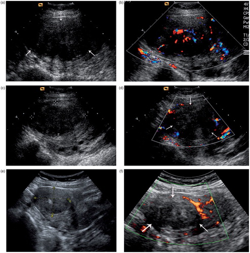
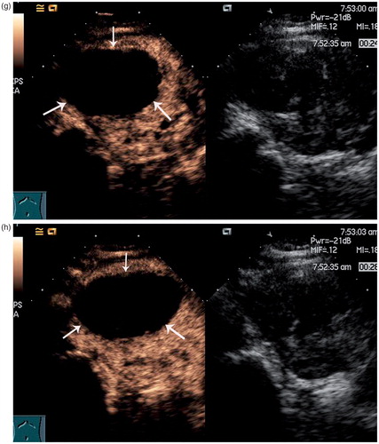
Table 1. Ablation volumes measured by two ultrasound radiologists by contrast-enhanced sonography (cm3).
According to the reliable analysis, intraclass correlation coefficient (ICC) between the two groups of ablation volumes is 0.964, and the 95% confidence interval (95%CI) is (0.913, 0.986). As for the F = 55.25, P < 0.05 from the analysis of variance, it means that there is a positive correlation between the two groups of ablation volumes. In other words, there is a good agreement between the two ultrasound radiologists’ measurements on ablation volumes obtained from contrast-enhanced sonography.
ICC between the measurements on ablation volumes from enhanced MRI
There is no contrast agent in the ablation area after the treatment of microwave ablation for uterine myomas on enhanced MRI scan (). shows the ablation volumes measured by the two MRI radiologists on enhanced MRI scan.
Figure 2. MR images in a 47-year-old woman with uterine myoma after microwave ablation therapy. (a) On contrast-enhanced MR images obtained 1 day after ablation, no enhancement is seen in the treated myoma, suggesting the complete necrosis of myoma (sagittal T1WI MR). (b) On contrast-enhanced MR images obtained 1 day after ablation, no enhancement is seen in the treated myoma, suggesting the complete necrosis of myoma (axial T1WI MR).
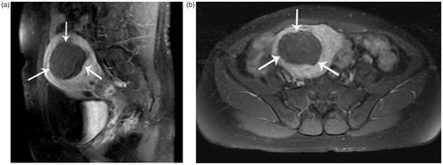
Table 2. Ablation volumes measured by two MRI radiologists on enhanced MRI scan (cm3).
According to reliable analysis, the ICC between the two groups of ablation volumes is 0.963, and the 95%CI is (0.908, 0.985). As for the F = 52.33, P < 0.05 from the analysis of variance, it means that there is a positive correlation between the two groups of ablation volumes. That is to say, there is a good agreement between the two MRI radiologists’ measurements on ablation volumes obtained from enhanced MRI.
Bland-Altman analysis of ablation volumes from contrast-enhanced sonography and enhanced MRI
Ablation volumes measured by the first ultrasound radiologist on contrast-enhanced sonography and those measured by the first MRI radiologist on enhanced MRI scan were used for Bland-Altman analysis.
The difference (D) between ablation volumes on the same subject from the two examination methods and the average (A) of ablation volumes on the same subject from the two examination methods was calculated. In our data, Correlation (D, A) = 0.05, P = 0.84, which indicates that D is independent of A. From the Bradley-Blackwood test we can see F = 0.61, P > 0.05, which indicates that the intercept and slope are 0 (α = β = 0) by doing the regression analysis to the difference (D) and the average (A). It proved that the two groups of ablation volumes obtained from contrast-enhanced sonography and enhanced MRI have the same distribution parameters, that is to say, there is no difference in the average and the standard deviation between the two groups of ablation volumes.
The difference (D) against the average (A), namely Bland-Altman graph, was further plotted, and then limits of agreement were calculated. The mean and the SD of the difference and the average of ablation volumes obtained from the two examination methods respectively are shown in . From Bland-Altman analysis, the mean difference, ablation volumes obtained from contrast-enhanced sonography minus ablation volumes obtained from enhanced MRI, is −1.22 cm3 and the SD is 4.90 cm3. Thus, limits of agreement are (−10.83 cm3, 8.39 cm3). shows that all points fall in 95% limits of agreement. Within the limits of agreement, the absolute value of the maximum difference between the two groups of ablation is 7.81 cm3.
Figure 3. Difference against average of ablation volumes from contrast-enhaced sonography and enhanced MRI (cm3), with 95% limits of agreement.
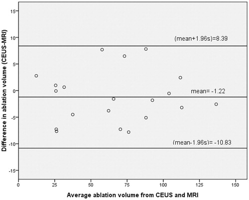
Table 3. Comparison between ablation volumes obtained from contrast-enhanced sonography and enhanced MRI (cm3).
ICC analysis between ablation volumes from contrast-enhanced sonography and enhanced MRI
Ablation volumes measured by the first ultrasound radiologist on contrast-enhanced sonography and those measured by the first MRI radiologist on enhanced MRI scan were used for ICC analysis.
The original data was plotted and the regression line was drawn. indicates that there is a linear trend between the two groups of ablation volumes obtained from contrast-enhanced sonography and enhanced MRI, so we further conducted the linear regression and correlation analysis.
Figure 4. Ablation volumes obtained from contrast-enhanced sonography and enhanced MRI scan (cm3), with regression line.
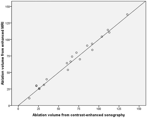
According to the reliable analysis, ICC between the two groups of ablation volumes is 0.991, and 95%CI is (0.976, 0.996). As for the F = 209.61, P < 0.05 from the analysis of variance, it means that there is a positive correlation between the two groups of ablation volumes. In other words, there is a good agreement between the two groups of ablation volumes obtained from contrast-enhanced sonography and enhanced MRI.
Discussion
US-guided percutaneous microwave ablation for uterine myomas is one of the effective treatments, with the advantages of convenient operation, shorter therapy time, minimal invasiveness, no serious complications, few adverse reactions, preserving the uterus, no influence on ovarian function, less postoperative pain and faster recovery [Citation3,Citation4]. During the treatment of microwave ablation for uterine myomas it is important to determine the therapeutic effect accurately, which means the correct judgement on the range of the ablation area. Although the greyscale ultrasound, colour Doppler ultrasound and power Doppler ultrasound play very important roles during the microwave ablation, it is inadequate to use these techniques for determining the therapeutic effect because it is difficult for greyscale ultrasound to identify necrotic tissue and residual uterine myoma, and colour Doppler ultrasound has low sensitivity to low velocity blood flow signals [Citation14,Citation15].
At present, enhanced computed tomography (CT) and enhanced MRI are considered as gold standards for assessing the therapeutic effect after hyperthermia, in which no enhancement of ablated area indicates the disappearance of the blood supply and consequent necrosis of tumour [Citation5–7]. It is widely acknowledged that the survival and development of tumour relies on tumour blood vessels. If the treatment is completely successful the lesions will show coagulation necrosis without vascular blood supply, and the enhanced CT and enhanced MRI scan will show no enhancement of ablated area. Conversely, if treatment is unsuccessful, there is still blood supply in the residual uterine myoma, and then enhancement will be seen within the uterine myomas on enhanced CT and enhanced MRI scan. Now enhanced MRI is one of the common clinical modalities for assessing the therapeutic effect of hyperthermia for tumour, including uterine myomas, because of its unparalleled soft tissue resolution and multi-section imaging technique. However, some contraindications, such as cardiac pacemaker and intrauterine contraceptive devices (IUD), limit the clinical application of MRI to some extent. In addition, if the therapeutic effects can be assessed immediately and correctly after ablation, additional ablation can be performed in time, and thus significantly improve the therapeutic effect of microwave ablation for uterine myomas. So it is necessary and important to find a new modality to assess the therapeutic effect of microwave ablation for uterine myomas.
In recent years, along with the invention of some new types of ultrasound contrast agents, such as SonoVue, the evaluation capacity of sonography for tumour vessels was greatly improved. Research has shown that contrast-enhanced sonography can accurately assess the tumour ablation area after the treatment and improve the diagnosis ability of ultrasound in detecting the residual tumour [Citation8,Citation9]. In the early stage of our study we have already used contrast-enhanced sonography to observe the ablation area after microwave ablation for uterine myomas. The purpose of this study is to explore whether contrast-enhanced sonography, which can be performed immediately after treatment and repeated within a short time, can be used as an alternative to enhanced MRI in assessing the ablated area after microwave ablation for uterine myomas.
In the analysis of measurement method comparison data, many studies give the paired t-test and simple correlation analysis of the results of the two measurement methods as an indicator of agreement. However, these two statistical analysis methods cannot take account of random errors and systematic errors at the same time. Paired t-test is sensitive to the systematic errors of measurement results, but cannot take into account the random error at the same time, while the correlation coefficient is insensitive to the system error. Consequently, the use of these two methods may mislead in the comparing the agreement of data. Although there are some limitations in the application, ICC can take into account both the random errors and systematic errors [Citation16–19]. In addition, in the analysis of the agreement of two measurement methods, Bland and Altman proposed an alternative method [Citation16–18]. The Bland-Altman method considers the impact on agreement of both random error and system error; meanwhile, making judgement by combining the statistical results with the meaning of the speciality is its unique advantage. In Bland-Altman analysis, studies reported that the two variables are equivalent only when intercept and slope are zero, and also the two variables are highly relevant at the same time [Citation20]. Therefore, in our study, we used the ICC to compare the agreement between the two ultrasound radiologists’ measurements and between the two MRI radiologists’ measurements respectively, and we used both the Bland-Altman analysis and the ICC to compare the agreement between the two groups of ablated uterine myoma volumes obtained from contrast-enhanced sonography and enhanced MRI after treatment.
Lee et al. [Citation21] suggested that the cut-off point was defined as 0.75. The agreement between the two measurement modalities can be considered as good if ICC is higher than 0.75. Our study indicates that there is a good agreement between the two ultrasound radiologists’ measurements within contrast-enhanced sonography and between the two MRI radiologists’ measurements within enhanced MRI respectively. The ablation volumes measured by the two ultrasound radiologists can be used interchangeably in clinic; so can those measured by the two MRI radiologists. Based on the results of our study, ablation volumes measured by the first ultrasound radiologist on contrast-enhanced sonography and measured by the first MRI radiologist on enhanced MRI scan were used for Bland-Altman and ICC analysis.
From the Bradley-Blackwood test, the result showed that the two groups of ablation volumes obtained from contrast-enhanced sonography and enhanced MRI have the same distribution parameters, which means no difference in the mean and the standard deviation of the two groups of ablation volumes. The mean difference of ablated volume obtained from contrast-enhanced sonography and enhanced MRI is −1.22 cm3, and the limits of agreement are (−10.83 cm3, 8.39 cm3). Among the limits of agreement, the largest absolute difference is 7.81 cm3. Considering the mean average of ablated volumes obtained from the two methods is 66.20 cm3, the magnitude of this difference, 7.81 cm3, is acceptable in clinic. So we can safely conclude that the two methods agree sufficiently well for them to be used interchangeably.
In our study, ICC between the measurements of ablated volume obtained from contrast-enhanced sonography and enhanced MRI is 0.991, higher than 0.75, and 95%CI is 0.976, 0.996. Thus we have a positive correlation between ablated volumes obtained from the two examination methods. They agree sufficiently for the one to replace the other or for using the two interchangeably in clinic.
The above analysis shows that there is a good agreement between the two groups of ablated volumes using the Bradley-Blackwood test, and the two groups of ablated volumes are highly correlated (ICC = 0.991). Therefore, we can conclude that there is equivalency between the two groups of ablated volumes obtained from enhanced MRI and contrast-enhanced sonography, and thus the two methods can be used interchangeably in clinics. In other words, contrast-enhanced sonography can be an alternative to enhanced MRI in assessing the ablated area of microwave ablation for uterine myomas. In addition, contrast-enhanced sonography is real-time and dynamic; it can be immediately performed to determine the result after the treatment, and it can clearly identify unablated areas of myomas which can be immediately re-ablated under US guidance to ensure the therapeutic effect. Contrast-enhanced sonography can be used as the preferred choice for assessing the therapeutic effect of microwave ablation for uterine myomas and subsequent follow-up as non-invasive methods in the clinic. However, since the scanning scope of contrast-enhanced ultrasound is limited, only one target area can be observed in the process of injection of contrast agent. If a full view of uterine myomas, the entire uterine, and the surrounding tissues is needed for comprehensive judgements, enhanced MRI is still indispensable.
A possible limitation of our study is that three-dimensional gadolinium-enhanced dynamic MRI could not be performed. Studies have shown that these techniques have been widely used in abdominal, chest, head and neck imaging with the advantages of short acquisition times, fewer artefacts, high temporal and spatial resolution, large coverage, uniform fat suppression, and refined signal-to-noise ratio. Three-dimensional images can be reconstructed from the original images, and so the anatomical structures of the organ and its surrounding area can be sensitively depicted in detail [Citation22–27]. Maybe this limitation could explain some of the difference in measurements between US and MRI. This issue will be paid attention in our future study.
Conclusions
Contrast-enhanced sonography and enhanced MRI can be used interchangeably in observing the ablation range of uterine myomas treated with microwave ablation. Contrast-enhanced sonography can be used as the preferred choice for assessing the therapeutic effect of microwave ablation for uterine myomas and for follow-up after treatment as non-invasive methods in clinic. Enhanced MRI can be used to determine the relationships among uterine myomas, the entire uterine, and the surrounding tissues comprehensively. In clinic, either examination method can be applied according to different purposes or a combination of the two to evaluate the therapeutic effect of microwave ablation for uterine myomas.
Declaration of interest
The authors report no declarations of interest. The authors alone are responsible for the content and writing of the paper.
References
- Vollenhoven BL, Lawrence AS, Healy DL. Uterine fibroids: A clinical review. Br J Obstet Gynaecol 1990;97:285–98
- Carlson KJ, Nichols DH, Schiff I. Indications for hysterectomy. N Engl J Med 1993;328:856–60
- Zhang J, Feng L, Zhang B, Ren J, Li Z, Hu D, et al. Ultrasound-guided percutaneous microwave ablation for symptomatic uterine fibroid treatment – A clinical study. Int J Hyperthermia 2011;27:510–16
- Zhang J, Feng L, Zhang BS, Ren JT, Li ZC, Hu DM, et al. [The study of follow up of percutaneous microwave ablation for uterine fibroids treatment]. In Chinese. Zhonghua Yi Xue Za Zhi 2011;91:48–50
- Hindley J, Gedroyc WM, Regan L, Stewart E, Tempany C, Hynyen K, et al. MRI guidance of focused ultrasound therapy of uterine fibroids: Early results. Am J Roentgenol 2004;183:1713–19
- Yoon JH, Lee EJ, Cha SS, Han SS, Choi SJ, Juhn JR, et al. Comparison of gadoxetic acid-enhanced MR imaging versus four-phase multi-detector row computed tomography in assessing tumor regression after radiofrequency ablation in subjects with hepatocellular carcinomas. J Vasc Interv Radiol 2010;21:348–56
- Liang P, Yu J, Lu M-D, Dong B-W, Yu X-L, Zhou X-D, et al. Practice guidelines for ultrasound-guided percutaneous microwave ablation for hepatic malignancy. World J Gastroenterol 2013;19:5430–8
- Wen YL, Kudo M, Zheng RQ, Ding H, Zhou P, Minami Y, et al. Characterization of hepatic tumors: Value of contrast-enhanced coded phase-inversion harmonic angio. Am J Roentgenol 2004;182:1019–26
- Zhou XD, Ren XL, Zhang J, He GB, Zheng MJ, Tian X, et al. Therapeutic response assessment of high intensity focused ultrasound therapy for uterine fibroid: Utility of contrast-enhanced ultrasonography. Eur J Radiol 2007;62:289–94
- Youk JH, Lee JM, Kim CS. Therapeutic response evaluation of malignant hepatic masses treated by interventional procedures with contrast-enhanced agent detection imaging. J Ultrasound Med 2003;22:911–20
- Liu F, Yu X, Liang P, Cheng Z, Han Z, Dong B. Contrast-enhanced ultrasound-guided microwave ablation for hepatocellular carcinoma inconspicuous on conventional ultrasound. Int J Hyperthermia 2011;27:555–62
- Wang F, Zhang J, Han ZY, Cheng ZG, Zhou HY, Feng L, et al. Imaging manifestation of conventional and contrast-enhanced ultrasonography in percutaneous microwave ablation for the treatment of uterine fibroids. Eur J Radiol 2012;9:52–6
- Orsini LF, Salardi S, Pilu G, Bovicelli L, Cacciari E. Pelvic organs in premenarcheal girls: Real-time ultrasonography. Radiology 1984;153:113–16
- Choi D, Lim HK, Kim SH, Lee WJ, Jang HJ, Lee JY, et al. Hepatocellular carcinoma treated with percutaneous radio-frequency ablation: Usefulness of power Doppler US with a microbubble contrast agent in evaluating therapeutic response – preliminary results. Radiology 2000;217:558–63
- Choi D, Lim HK, Kim SH, Lee WJ, Jang HJ, Kim H, et al. Assessment of therapeutic response in hepatocellular carcinoma treated with percutaneous radiofrequency ablation: Comparison of multiphase helical computed tomography and power Doppler ultrasonography with a microbubble contrast agent. J Ultrasound Med 2002;21:391–401
- Bland JM, Altman DG. Statistical methods for assessing agreement between two methods of clinical measurement. Lancet 1986;1:307–10
- Bland JM, Altman DG. Comparing methods of measurement: Why plotting difference against standard method is misleading. Lancet 1995;346:1085–7
- Bland JM, Altman DG. Measuring agreement in method comparison studies. Stat Methods Med Res 1999;8:135–60
- Pan X, Ni Z. [Application of intraclass correlation coefficient to reliability assessment]. In Chinese. Hua Xi Yi Ke Da Xue Xue Bao 1999;30:62–3, 67
- Fang JX, Lu Y. [Advanced Medical Statistics]. Chinese. Beijing: People’s Medical Publishing House, 2002
- Lee J, Koh D, Ong CN. Statistical evaluation of agreement between two methods for measuring a quantitative variable. Comput Biol Med 1989;19:61–70
- Lee VS, Lavelle MT, Rofsky NM, Laub G, Thomasson DM, Krinsky GA, et al. Hepatic MR imaging with a dynamic contrast-enhanced isotropic volumetric interpolated breath-hold examination: Feasibility, reproducibility, and technical quality. Radiology 2000;215:365–72
- Wetzel SG, Johnson G, Tan AG, Cha S, Knopp EA, Lee VS, et al. Three-dimensional, T1-weighted gradient-echo imaging of the brain with a volumetric interpolated examination. Am J Neuroradiol 2002;23:995–1002
- Karabulut N, Martin DR, Yang M, Tallaksen RJ. MR imaging of the chest using a contrast-enhanced breath-hold modified three-dimensional gradient-echo technique: Comparison with two-dimensional gradient-echo technique and multidetector CT. Am J Roentgenol 2002;179:1225–33
- Kataoka M, Ueda H, Koyama T, Umeoka S, Togashi K, Asato R, et al. Contrast-enhanced volumetric interpolated breath-hold examination compared with spin-echo T1-weighted imaging of head and neck tumors. Am J Roentgenol 2005;184:313–19
- Shen XY, Chai CH, Xiao WB, Wang QD. Diagnostic value of the fluoroscopic triggering 3D LAVA technique for primary liver cancer. Hepatobiliary Pancreat Dis Int 2010;9:159–63
- Grazioli L, Morana G, Kirchin MA, Schneider G. Accurate differentiation of focal nodular hyperplasia from hepatic adenoma at gadobenate dimeglumine-enhanced MR imaging: prospective study. Radiology 2005;236:166–77