
Abstract
Purpose: The emergence of thermal modalities has promoted the use of heat-sensitive phantoms for calibration, measurement, and verification purposes. However, development of durable phantoms with high precision ability to represent the temperature distribution remains a challenge. This study aims to introduce a reusable phantom that provides an accurate assessment of the heated region in various thermal modalities. Materials and methods: The phantom contains a thermochromic dye that is transparent blue at room temperature and becomes colourless after exceeding a threshold temperature. In order to determine the threshold temperature of the phantom, spectrophotometry analysis was performed. The various thermal (specific heat, thermal conductivity, melting point and latent heat of melting) and acoustic (sound speed, attenuation) properties of this phantom were measured and compared with those of the reference phantom without dye. The application of this phantom for radio-frequency and magnetic resonance guided focused ultrasound modalities was also examined. Results: The spectrophotometry analysis showed a threshold temperature of 50 ± 3 °C for this phantom. The results also demonstrated a 6 °C difference between the onset and ending temperatures of the discolouration process. Moreover, the starting temperature of colouration during cooling was found to be 4 °C lower than the ending temperature of discolouration. The sound speed, attenuation, specific heat, thermal conductivity and melting point of the heat-sensitive phantom were statistically equal to those of the reference phantom; however, the latent heat, and onset temperature of the melting of the heat-sensitive phantom were decreased by addition of the dye. Conclusions: The developed phantom is applicable for accurate evaluation of temperature variations in various thermal modalities.
Introduction
With the rapid development of thermal therapies there has been a growing interest on research concerning the influence of high temperatures on various properties of biological tissues. However, due to substantial difficulties in the preparation, handling, maintenance and standardisation of real tissues, the availability of effective phantoms with similar properties to target tissues is essential for these high temperature studies. A multitude of materials have been proposed in liquid, solid and gel (semisolid) forms; but the gel phantoms have found more popularity among researchers due to their physical similarities to both soft and hard tissues, as well as their capability to represent a wide range of thermal, electrical, acoustic and optical properties. One particular advantage of the gels is that they can easily be formed into different shapes so that realistic irradiation geometries are reproduced.
A number of gelling agents including TX-150 [Citation1], agar/agarose [Citation2–9], polyacrylamide (PAA) [Citation10–13], hydroxyethyl cellulose (HEC) [Citation14–16], gelatine [Citation17–20], gellan gum [Citation21], carrageenan [Citation22,Citation23], and sodium alginate [Citation24–26] have been suggested so far for fabrication of thermal phantoms. However, PAA hydrogels are the most popular materials for construction of thermal phantoms. These gels are transparent in optical range, enabling researchers to visualise the coagulation zone inside the phantoms by adding some heat-sensitive ingredients. The added materials may cause a large change in magnetic resonance parameters (T1 or T2) or change the transparency level of PAA phantoms upon reaching the temperature range used in hyperthermia or thermal ablation procedures [Citation27]. Bovine serum albumin (BSA) [Citation27–29], and egg white (EW) [Citation30–32] are two popular heat-sensitive agents proposed for visualisation of the heated zone inside the PAA phantoms. The main drawback of PAA-BSA and PAA-EW phantoms is the irreversibility of the coagulation process, making the phantom useless for some routine applications such as calibration of thermal devices. Moreover, the exothermal nature of acrylamide polymerisation may cause an unwanted coagulation of the heat-sensitive agent during phantom preparation. Due to these shortcomings, non-ionic surface active agents (NISAAs) have been suggested to develop heat-sensitive phantoms that have reversible reactions to the temperature changes [Citation33,Citation34]. NISAAs are a group of materials that become segregated when temperature exceeds a specific threshold level called the ‘clouding point’ (TCP), resulting in a white and opaque region in the phantom and returning to their original transparency when cooled again below the clouding point. However, similar to PAA-BSA and PAA-EW phantoms, the threshold temperature of these phantoms is inaccurate, making the researchers unable to precisely estimate the temperature profile inside the phantom. Park et al. [Citation34] evaluated the clouding and clearing points of NISAAs (the temperature that NISAAs recover their transparency) and found that there was a 17 °C difference between the starting and ending temperatures of the clouding process of a typical NISAA material. Moreover, the onset temperature of the clearing process started at about 5 °C lower than the ending temperature of the clouding process. Therefore, construction of reusable heat-sensitive phantoms for accurate estimation of temperature distribution is still a challenge.
In the present study we introduce a new heat-sensitive PAA gel containing a thermochromic dye (TCD) of type N-(2-ethoxyphenyl)-N′-(2-ethylphenyl) ethanediamine as a new phantom for hyperthermia and thermal ablation studies. TCDs are usually coloured below a specified temperature and become colourless (transparent) above this temperature level. These dyes exhibit reversible heat-sensitivity behaviour with a more accurate threshold temperature compared to that of BSA, EW and NISAAs. A number of TCDs with threshold temperatures ranging from −15 °C up to 100 °C are available. These materials are mainly composed of a dye as colour former, a weak organic acid as colour developer, and a solvent, which are microencapsulated to prevent the contents from undesired reactions. During the heating of the microcapsules the solvent alteration between the solid and liquid states causes the colour-former component to be in contact below the transition temperature (coloured state) and separated above this temperature (discoloured state). This process could be repeated for a few thousand times [Citation35].
The introduced heat-sensitive phantom could be used for more accurate visualisation of the temperature distribution during the heating and cooling stages in the high temperature experiments, evaluation of the heating and cooling rates, acoustic and electromagnetic dosimetry, and calibration of thermal devices. The low cost of preparation due to the low amount of required TCD as well as the ability to be made at flexible temperatures are the other advantages of these phantoms that make them attractive candidates for irradiation experiments including radio-frequency (RF) and magnetic resonance guided focused ultrasound (MRgFUS) modalities.
Materials and methods
Gel fabrication
For preparation of the thermal phantom, acrylamide monomer (AA; gelling agent), N, N′-methylene-bis-acrylamide (MBA) (crosslinking agent), NaCl, N,N,N′,N′-tetra-metyl-ethylene-diamine (TEMED) (catalyser), and ammonium persulfate (APS) (polymerisation primer) were purchased from Sigma-Aldrich (St Louis, MO). The TCD was obtained from Thermographic Measurements (Honiton, UK). Various types of TCDs with different colours and threshold temperatures (from −15 ° to 100 °C) are available; however, the selected dye for this study was blue at room temperature with a threshold temperature of 47° ± 2°C. The rationale for the selection of this TCD was based on the proximity of its threshold temperature to the coagulation temperature of real tissues, making the phantom desirable for simulation of organic tissues in hyperthermia studies. It is noteworthy that the proposed recipe is only valid for ablative temperature ranges (45–55 °C), and it is not applicable for mild hyperthermia application that applies a lower level of temperature (40–45 °C) [Citation36]. Therefore, in order to construct an appropriate phantom for this temperature range, a TCD with a lower threshold temperature must be used.
The composition of the fabricated heat-sensitive (HS) phantom is presented in . A reference (RE) phantom with a same recipe (but without TCD) was also made to investigate the effects of the dye on acoustic and thermal properties of the phantom. This recipe was chosen due to its self-supporting ability that provides the possibility of being used outside the container for RF and MRgFUS studies. Meanwhile, the phantom has sufficient softness to allow the penetration of the RF needle without cracking. The optimum concentration of the TCD was determined in preliminary experiments; while too much amount of dye results in a non-transparent gel, too little concentration of dye leads to low heat sensitivity and optical contrast.
Table I. Chemical composition of phantoms used in this study.
In the present study, a cylindrical mould was used (60 mL, diameter 4 cm, height 5 cm). For preparation of a typical phantom, firstly the AA, MBA, NaCl, TEMED, and TCD (for the HS phantom) were mixed in situ in a designated amount of degassed water for 15 min. Secondly, APS solution was added and the mixture was homogenised using a homogeniser (T25 Ultra-Turrax, IKA Works, Staufen, Germany) for 30 s at 5000 rpm to initiate the polymerisation. The higher rates of homogenisation may cause the formation of microbubbles inside the phantom, whereas the lower rates may result in an inhomogeneous mixture. The solution was allowed to polymerise for 24 h at room temperature before performing further experiments. After completion of polymerisation, the phantom was removed from the container and analysed by ultrasound imaging to evaluate the homogeneity of its structure. It is important to note that the acrylamide monomer is a severe neurotoxin that causes severe health hazards. Therefore, the usage of this compound requires extreme precautionary measures [Citation37].
Experimental procedures
In this study the compositions of the RE and HS phantoms were compared by Fourier transmission infra-red spectrometry (FT-IR) (Nicolet 6700, Thermo Scientific, Waltham, MA). For determination of the onset (Tod) and ending (Ted) temperatures of discolouration during heating, the spectrophotometry analysis (Dataflash 100, Datacolour International, Lawrenceville, NJ) was used. In these experiments a number of 60 HS phantom discs (4 cm diameter and 1 cm thickness) were heated in a water bath at 12 temperatures between 40° and 53 °C (five discs for each temperature). Then, by assigning one RE phantom disc as reference, the changes in the Commission Internationale de l'Eclairage Lab (CIELAB) values of the HS phantom discs including the difference in yellow/blue values (Db*), difference in darkness/lightness values (DL*), and the difference in total colour (DE*) at the different temperatures were measured. The results obtained were later analysed by ANOVA (SPSS 20, Chicago, IL) with 95% confidence interval; then, the first and last temperatures at which the CIELAB values were statistically different from the previous temperature points were considered as Tod and Ted, respectively. A similar protocol was used for measurement of the onset (Toc) and ending (Tec) temperatures of colouration during cooling.
The acoustic and thermal parameters of the HS phantom were also measured and compared with that of the RE phantom. The sound speed and the attenuation coefficient were measured by oscilloscope (GW-Instek GDS 2102, Good Will Instruments, New Taipei City, Taiwan) and ultrasound power meter (UPM-DT-10AV, Ohmic Instruments, Easton, MD), respectively. The experiments were performed at room temperature and a frequency of 1 MHz which is the typical frequency in high intensity focused ultrasound (HIFU) treatments. Thermal conductivity and specific heat capacity of the phantoms were calculated using a thermal analyser (KD2 Pro, Decagon Devices, Pullman, WA). The latent heat of melting and melting point of the phantoms were measured by differential scanning calorimetry (DSC) (DSC820 with TSO 801RO robot, Mettler-Toledo, Columbus, OH). Thermogravimetric analysis (TGA) (TGA/SDTA 851e, Mettler-Toledo) was also conducted to investigate the physical stability of the phantoms in high temperature regimes. All the above experiments were repeated for at least five times and the results were analysed by ANOVA with 95% confidence interval.
For heating of the HS phantom, the RF and MRgFUS methods were applied. The RF procedure was performed by inserting the RF electrode (RITA Medical Systems, Mountain View, CA) into the phantom with 1 cm above the bottom. The frequency and power were set to 460 MHz and 50 W, respectively. The procedure was continued for 60 s with a target temperature of 100 °C. The development of the colourless region in the phantom was recorded by digital camera during the procedure. For experimental estimation of the specific absorption rate (SAR) of electromagnetic power inside the HS phantom, the images obtained were analysed using image processing software (ImageJ 1.44 P, National Institutes of Health, Bethesda, MD). The MRgFUS experiments were also conducted using a clinical system available at the University of Malaya Medical Centre (EX-ablate, Insightec, Tirat Carmel, Israel). In these experiments the phantom was placed in a water tank and sonicated for 20 s at a frequency of 1.15 MHz with the acoustic energy and acoustic power of 2847 J and 142 W, respectively. The MR images of the HS phantom were collected at different sonication times to visualise the propagation of coagulation zone during MRgFUS treatment.
Results and discussion
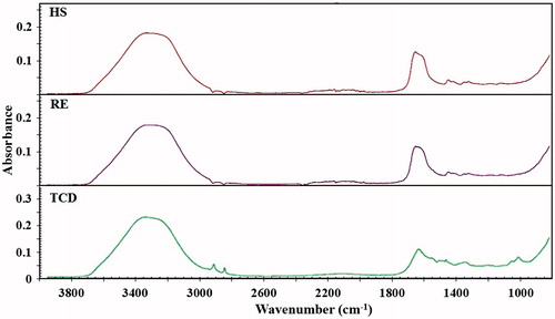
Polymerisation of acrylamide is an exothermic reaction causing a rapid temperature rise shortly after the addition of the initiator to the solution. Due to the rapid polymerisation reaction, a short time period (about 30 s) is available for mixing the APS initiator in the solution and possibly pouring the liquid into a suitable mould. Therefore, inhomogeneity of the PAA phantoms is a common issue after the completion of polymerisation. Therefore, a high-speed homogeniser was used in our experiments to rapidly mix the initiator into the mixture before polymerisation occurs. shows the ultrasound images of RE and HS phantoms that confirm the homogenous structure of both phantoms. This figure also shows that the phantoms have provided sufficient contrast to be distinguished in ultrasound images. The added TCD contains microcapsules with diameters ranging from 1 to 5 µm. As a result, the transparency of the phantom to ultrasound beam is slightly reduced due to addition of the TCD. The FT-IR spectra of the TCD and fabricated phantoms are also shown in . This figure shows that the addition of thermochromic dye has negligible impact on the absorption spectra of the phantom due to the low amount of dye used.
Figure 1. The ultrasound images of (A) the reference phantom, and (B) the heat-sensitive phantom. The bright lines at the margins are the optical reflection off the phantom surfaces.
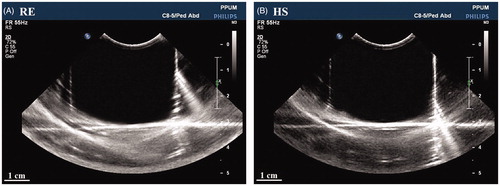
Figure 2. The FT-IR spectra of the dye, reference phantom, and heat-sensitive phantom. Due to small amount of dye used, no significant difference was detected between the FT-IR spectra of the reference and the heat-sensitive phantoms.
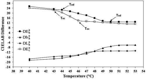
The results of spectrophotometry analysis of the HS phantom are plotted in . For determination of the threshold temperature of the HS phantom, the CIELAB values of the phantom were calculated during heating in the temperature range of 40° to 53 °C and the point at which the CIELAB values (especially DE*) become statistically constant was considered as the threshold temperature. Therefore, according to the CIELAB spectra shown in , the threshold temperature of the heat-sensitive phantoms is 50° ± 3 °C. This temperature is higher compared to the disclosed threshold temperature of the thermochromic dye (47° ± 2 °C), meaning that the threshold temperature of the TCD is increased after its addition to the phantom mixture. also shows that the temperature interval between the onset and ending temperatures of discolouration is 6 °C. Moreover, the onset temperature of colouration during cooling is 4 °C lower than the ending temperature of discolouration during heating of the HS phantom. However, the slopes of colouration are larger compared to discolouration, meaning that the colouration is completed faster than the discolouration process. Such a phenomenon occurs due to the effect of thermal history on the colouration process of the TCD, i.e. the particular colour or temperature level reached during heating. This behaviour of the TCD ink is called ‘hysteresis’. Therefore, it is colour hysteresis that describes the interval between the Ted and Toc of a TCD as a function of thermal history. In general, thermal hysteresis is a characteristic of TCDs which may slightly depend on the type of TCD used. For instance, hysteresis in phantoms containing a black-coloured TCD ink with a threshold temperature of 55 °C was measured at about 5 °C. Overall, the values measured in our study confirm that the TCD could provide a more accurate estimation of thermal profile than NISAA materials in spite of the comparable results for the hysteresis between the Toc and Ted in phantoms containing NISAA (5 °C) and TCD (4 °C).
Figure 3. The spectrophotometry spectra of the heat-sensitive phantom. Db*: The yellow/blue value; DL*: the darkness/lightness value; DE*: the total colour difference. The ‘h’ and ‘c’ subscripts represent the total colour difference during heating and cooling, respectively. Tod: the onset temperature of discolouration; Ted: the ending temperature of discolouration; Toc: the onset temperature of colouration; Tec: the ending temperature of colouration.
The thermal and acoustic properties of the RE and HS phantoms are summarised in . The data presented in this table demonstrate that both sound speed and attenuation values of the phantom are increased by addition of the dye. On the other hand, the specific heat and thermal conductivity of the HS phantom are slightly smaller compared to those of the RE phantom; although the ANOVA results do not indicate a significant difference between the sound speed, attenuation, specific heat and thermal conductivity values of the RE and HS phantoms. It should be noted that the attenuation coefficient of the phantom has a positive correlation with the applied frequency, and therefore, at frequency ranges which are regularly used in RF treatments (e.g. 460 MHz), the attenuation coefficient is drastically higher compared to the values measured at 1 MHz [Citation30]. However, due to the independency of the RF modality on the acoustic parameters, the final result of the RF experiments is not affected by alteration of the attenuation coefficient.
Table II. Acoustic and thermal properties of the reference and heat-sensitive phantoms. Data are presented as mean (SD).
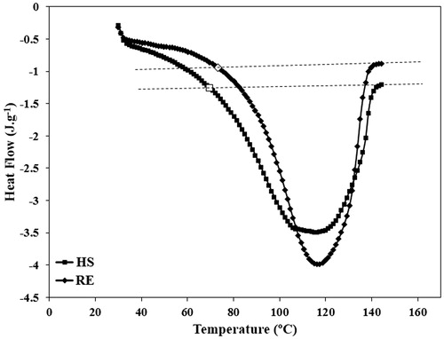
also shows that the melting point of the phantom is not affected by addition of the dye; whereas, the DSC spectra of the phantoms () illustrates a significant decrease in the onset temperature of melting (from 75 °C to 66 °C) of the HS phantom compared to the RE phantom gel. Therefore, despite the negligible influence of the TCD on the melting point of the phantom (the peak point in the DSC spectra), the transition interval between the gel and liquid phases is larger in phantoms containing TCD. Moreover, according to and ANOVA analysis, the latent heat of melting of the HS phantom is significantly lower than that of RE phantom. These facts reveal that, in spite of the similar melting points, the deformation of HS phantoms occurs at lower temperatures compared to that of RE phantoms, and thus the thermal stability of the PAA phantoms is decreased by addition of the TCD. However, this concern is not of critical importance at hyperthermia temperature ranges and need only be considered in experiments where the temperature applied is above 66 °C.
Figure 4. The DSC spectra of two typical reference and heat-sensitive phantoms. The white markers represent the onset temperatures of melting of these two typical phantoms.
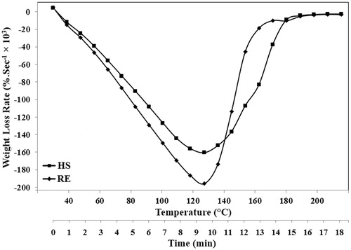
The TGA spectra of the RE and HS phantoms are also shown in . The thermal decomposition of both RE and HS phantoms occurs at similar temperature ranges which mostly correspond to water evaporation from the phantom. Although the boiling point of water is around 100 °C, evaporation of water also occurs with lower rates at milder temperatures such as room or hyperthermia temperature ranges. Therefore, due to the substantial influence of the water content on the physical properties of these phantoms, for long-term usage, water evaporation must be avoided during maintenance and thermal experiments. At higher temperatures, the rate of weight loss is increased until the temperature rises to about 120 °C. The weight loss due to water evaporation continues until the temperature reaches to nearly 200 °C, and then no significant change in the weight-loss rate is observed. However, shows that the addition of TCD may decrease the water evaporation rate, most likely due to the small increase in the viscosity of the water.
Figure 5. The TGA analysis of two typical reference and heat-sensitive phantoms.
shows the change in colour of the heated area in HS phantom gel after exposure to RF beams for 60 s. Upon exposure of the HS phantom to electromagnetic beams, a colourless region is created due to the power absorption, making the three-dimensional distribution of the temperature visible from outside. This feature of the HS phantom could be useful in many applications where the accurate three-dimensional assessment of power absorption is required.
Figure 6. (A) The colourless region developed in the heat-sensitive phantom after radiofrequency radiation for 60 s. (B) The estimated isothermal curves due to radiofrequency irradiation on X–Y and X–Z planes of the heat-sensitive phantom.
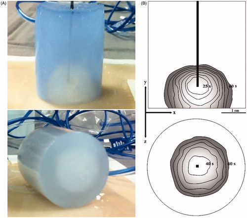
A typical application of the HS phantom for measurement of local SAR inside the phantom is demonstrated in . SAR is defined as the time rate of energy deposition per unit mass in a lossy body, and it is generally a measure of power density [Citation38,Citation39]. In this figure, the area of the colourless region as a function of irradiation time is outlined using the image processing software. As mentioned previously, the discolouration of the phantom starts at 6 °C below the threshold temperature. Based on this fact, the temperature on the boundary between the blue and colourless regions is assumed equal to the onset temperature of discolouration. This assumption could be confirmed by measurement of the colour values at the boundary using the image processing software and comparing the values obtained with those of the central area near the RF applicator that has a temperature above Ted. The colour values at the boundaries are lower (closer to dark) and gradually increase during the irradiation period, while the colour values at the central area are relatively high (close to white) and stable over the irradiation time. Therefore, the surface of the colourless region at different irradiation times coincides with the isothermal layers with temperature of 44 °C. These isothermal layers could be used for calculation of SAR on the boundary using the Pennes’ bio-heat transfer equation [Citation39,Citation40]:
where, ρ, C, and k are the density, specific heat, and thermal conductivity of the phantom, respectively. Wb, Cb, and Tb are the flow rate, specific heat, and temperature of the blood. Q is also the heat deposited in the phantom due to RF irradiation [Citation39]. It should be noted that this phantom has relatively small thermal conductivity compared to heat generation, and the time period in which thermal conductivity can be negligible is longer than the irradiation time used in this study. Moreover, there is no blood flow in the phantom. Therefore, the first and second terms on the right side are eliminated and Equation 1 is simplified as follows [Citation40]:
And thus:
where T0 is the room temperature and t is the irradiation time. Since the specific heat capacity of the HS phantom, the room temperature, Tod, and irradiation time are 3.59 kJ kg−1 K−1, 25 °C, 44 °C, and 60 s, respectively, the local SAR is estimated as 1136.8 W kg−1. A similar protocol could be used for calculation of SAR at isothermal layers with temperature of 50 °C by outlining the areas with stable colour values.
The results of phantom sonication using MRgFUS system are shown in . When the ultrasound pulses are propagated through the phantom, the MR images show a dark region with a slightly oval shape which corresponds to the heated area at the focal point (). According to , the temperature of the focal point is increased to over 50 °C after sonication for approximately 6 to 7 s; however, due to the small size and insufficient contrast at initial sonication times, the thermal lesion is not observed in MR images upon reaching the sonication time to about 8 s. After the FUS power is turned off, this region disappeared again due to the diffusion of heat through the phantom. The reversibility of colour change is an effective feature for more effective focusing of the ultrasound beams to the desired point (e.g. tumour) during MRgFUS experiments.
Figure 7. Magnetic resonance phase images of the heat-sensitive phantom during MRgFUS heating. (A–C) the temperature patterns obtained during the heating of the phantom. (D–F) development of thermal lesion due to MRgFUS treatment. (G) the final temperature pattern after sonication for approximately 19 s. (H) the temperature plot of the focal point during sonication of the phantom.
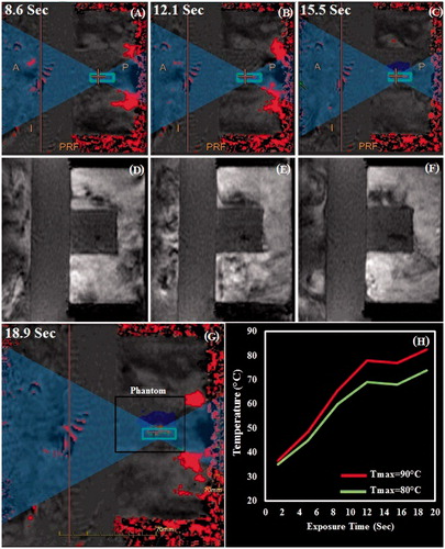
The threshold temperature of the HS phantom is close to the temperatures at which the true lesions are formed in real tissues. Therefore, the ability of HS phantom to represent thermal lesions at temperature of 50° ± 3 °C could be applied for more precise simulation of real tissues during MRgFUS experiments.
Conclusion
The thermal and acoustic properties of a polyacrylamide gel containing thermochromic dye have been described. The acoustic properties of this gel were found to be similar to those of the reference phantom, proving that the dye used may not have a significant impact on acoustic properties of the phantom. However, the thermochromic dye caused a decrease in specific heat capacity and latent heat of melting of the polyacrylamide phantom although other thermal parameters such as thermal conductivity and melting point were almost similar to that of the reference phantom. The amount of dye used for preparation of the phantom is much lower compared to other similar heat-sensitive agents such as EW, BSA, and NISAAs which causes a negligible effect on various properties of the phantom. Moreover, due to the reversibility of the colour change, the phantom could be prepared without the need to decrease the temperature continuously, while temperature control is necessary for fabrication of PAA-BSA and PAA-EW phantoms because of irreversible coagulation of the heat-sensitive agent. Another advantage of these dyes is the more precise estimation of temperature inside the phantom (error margin: 3 °C) compared to other heat-sensitive phantoms (especially PAA-EW phantoms). The phantom is reusable for several experiments and the threshold temperature could be adjusted by choosing an appropriate thermochromic dye.
The new phantom model could be used in various modalities including RF, and MRgFUS. By use of this thermochromic dye, the local SAR value inside the phantom could be precisely estimated. This experimental method of SAR evaluation has many advantages in respect of the cost requirement for measurement or observation, availability of true 3-D pattern of the local SAR, and ease of SAR estimation.
The main limitation of this phantom is the loss of transparency during the construction of sizeable phantoms. Therefore, this thermochromic dye is only suitable for construction of the phantoms with diameters smaller than 10 cm. However, the advantages of this heat-sensitive phantom make it an attractive candidate as reusable tissue-mimicking material for various high-temperature modalities.
Declaration of interest
This research was supported by Science Fund (06-01-03-SF0587) of the Ministry of Science, Technology and Innovation (MOSTI), Malaysia. The authors report no conflicts of interest. The authors alone are responsible for the content and writing of the paper.
Acknowledgements
The authors wish to thank Thermographic Measurements for providing the thermochromic dye. We would also like to extend our gratitude to Mohammed Rafiq and Maheza Irni from University of Technology Malaysia (UTM) for their support in performing acoustic characterisation tests, Mohammad Mehrali for evaluation of thermal properties, and Yus Hafizul from University of Malaya Medical Centre for conducting the MRgFUS experiments.
References
- Chou CK, Chen GW, Guy AW, Luk KH. Formulas for preparing phantom muscle tissue at various radiofrequencies. Bioelectromagnetics 1984;5:435–41
- Kato H, Ishida T. Development of an agar phantom adaptable for simulation of various tissues in the range 5–40 MHz. Phys Med Biol 1987;32:221–6
- Ito K, Furuya K, Okano Y, Hamada L. Development and characteristics of a biological tissue-equivalent phantom for microwaves. Electron Comm Jpn 2001;84:67–77
- Takimoto T, Onishi T, Saito K, Takahashi M, Uebayashi S, Ito K. Evaluation on biological tissue equivalent agar-based solid phantoms up to 10 GHz – Aiming at measurement of characteristics of antenna for UWB communications. In: Proceedings of the International Symposium on Antennas and Propagation, 2005, Seoul, South Korea, August 3–5. New York: IEEE, 2005, pp. 483–6
- Liu Z, Ahmed M, Weinstein Y, Yi M, Mahajan RL, Goldberg SN. Characterization of the RF ablation-induced ‘oven effect’: The importance of background tissue thermal conductivity on tissue heating. Int J Hyperthermia 2006;22:327–42
- Ortega R, Téllez A, Leija L, Vera A. Measurement of ultrasonic properties of muscle and blood biological phantoms. Phys Procedia 2010;3:627–34
- Siddiqi AK, Cho S. Agar-based heat-sensitive gel with linear thermal response over 65–80 °C. J Therm Anal Calorim 2013;111:1805–9
- Mylonopoulou E, Bazán-Peregrino M, Arvanitis CD, Coussios CC. A non-exothermic cell-embedding tissue-mimicking material for studies of ultrasound-induced hyperthermia and drug release. Int J Hyperthermia 2013;29:133–44
- Gasselhuber A, Dreher MR, Partanen A, Yarmolenko PS, Woods D, Wood BJ, et al. Targeted drug delivery by high intensity focused ultrasound mediated hyperthermia combined with temperature-sensitive liposomes: Computational modelling and preliminary in vivo validation. Int J Hyperthermia 2012;28:337–48
- Bini MG, Ignesti A, Millanta L, Olmi R, Rubino N, Vanni R. The polyacrylamide as a phantom material for electromagnetic hyperthermia studies. IEEE Trans Biomed Eng 1984;31:317–22
- Surowiec A, Shrivastava PN, Astrahan M, Petrovich Z. Utilization of a multilayer polyacrylamide phantom for evaluation of hyperthermia applicators. Int J Hyperthermia 1992;8:795–807
- Davidson SR, Sherar MD. Measurement of the thermal conductivity of polyacrylamide tissue-equivalent material. Int J Hyperthermia 2003;19:551–62
- Bazrafshan B, Hubner F, Farshid P, Larson MC, Vogel V, Mantele W, et al. A liver-mimicking MRI phantom for thermal ablation experiments. Med Phys 2011;38:2674–84
- Stauffer PR, Rossetto F, Prakash M, Neuman DG, Lee T. Phantom and animal tissues for modelling the electrical properties of human liver. Int J Hyperthermia 2003;19:89–101
- Ozen S, Koylu H. Phantom model of human brain tissue for cellular phone frequencies in electromagnetic field radiation absorption studies. GU J Sci 2005;18:193–200
- Prakash P, Converse MC, Mahvi DM, Webster JG. Measurement of the specific heat capacity of liver phantom. Physiol Meas 2006;27:N41–6
- Marchal C, Nadi M, Tosser AJ, Roussey C, Gaulard ML. Dielectric properties of gelatine phantoms used for simulations of biological tissues between 10 and 50 MHz. Int J Hyperthermia 1989;5:725–32
- Lazebnik M, Madsen EL, Frank GR, Hagness SC. Tissue-mimicking phantom materials for narrowband and ultrawideband microwave applications. Phys Med Biol 2005;50:4245–58
- Lindner U, Lawrentschuk N, Weersink RA, Raz O, Hlasny E, Sussman MS, et al. Construction and evaluation of an anatomically correct multi-image modality compatible phantom for prostate cancer focal ablation. J Urol 2010;184:352–7
- Yuan Y, Wyatt C, Maccarini P, Stauffer P, Craciunescu O, Macfall J, et al. A heterogeneous human tissue mimicking phantom for RF heating and MRI thermal monitoring verification. Phys Med Biol 2012;57:2021–37
- King RL, Herman BA, Maruvada S, Wear KA, Harris GR. Development of a HIFU phantom. Proc AIP Conf 2007;911:351–6
- Kato H, Yoshimura K, Kuroda M, Yoshida A, Hanamoto K, Kawasaki S, et al. Development of a phantom compatible for MRI and hyperthermia using carrageenan gel – Relationship between dielectric properties and NaCl concentration. Int J Hyperthermia 2004;20:529–38
- Yoshida A, Kato H, Kuroda M, Hanamoto K, Yoshimura K, Shibuya K, et al. Development of a phantom compatible for MRI and hyperthermia using carrageenan gel-relationship between T1 and T2 values and NaCl concentration. Int J Hyperthermia 2004;20:803–14
- Fisher JW, Rylander MN. Effective cancer laser-therapy design through the integration of nanotechnology and computational treatment planning models. In: Proceedings of SPIE, Plasmonics in Biology and Medicine V, 2008, San Jose, CA, January 21–2. Bellingham, WA: SPIE Press, 2008, p. 68690D
- Sarkar S, Gurjarpadhye AA, Rylander CG, Nichole Rylander M. Optical properties of breast tumor phantoms containing carbon nanotubes and nanohorns. J Biomed Opt 2011;16:051304–11
- Sarkar S, Zimmermann K, Leng W, Vikesland P, Zhang J, Dorn H, et al. Measurement of the thermal conductivity of carbon nanotube – Tissue phantom composites with the hot wire probe method. Ann Biomed Eng 2011;39:1745–58
- McDonald M, Lochhead S, Chopra R, Bronskill MJ. Multi-modality tissue-mimicking phantom for thermal therapy. Phys Med Biol 2004;49:2767–78
- Bu-Lin Z, Bing H, Sheng-Li K, Huang Y, Rong W, Jia L. A polyacrylamide gel phantom for radiofrequency ablation. Int J Hyperthermia 2008;24:568–76
- Choi MJ, Guntur SR, Lee KI, Paeng DG, Coleman A. A tissue mimicking polyacrylamide hydrogel phantom for visualizing thermal lesions generated by high intensity focused ultrasound. Ultrasound Med Biol 2013;39:439–48
- Takegami K, Kaneko Y, Watanabe T, Maruyama T, Matsumoto Y, Nagawa H. Polyacrylamide gel containing egg white as new model for irradiation experiments using focused ultrasound. Ultrasound Med Biol 2004;30:1419–22
- Divkovic GW, Liebler M, Braun K, Dreyer T, Huber PE, Jenne JW. Thermal properties and changes of acoustic parameters in an egg white phantom during heating and coagulation by high intensity focused ultrasound. Ultrasound Med Biol 2007;33:981–6
- Labuda CP, Church CC. Augmentation of HIFU-induced heating with fibers embedded in a phantom. Ultrasound Med Biol 2011;37:442–9
- Miyakawa M, Hoshina S. A self-supporting gel phantom used for visualization and/or measurement of the three-dimensional distribution of SAR. In: Proceedings of the IEEE EMC Symposium, 2002, Minneapolis, MN, August 19–23. New York: IEEE, 2002, pp. 671–6
- Park SK, Anjaneya Reddy Guntur SR, Lee KI, Paeng D-G, Choi MJ. Reusable ultrasonic tissue mimicking hydrogels containing nonionic surface-active agents for visualizing thermal lesions. IEEE Trans Biomed Eng 2010;57:194–202
- Kulčar R, Friškovec M, Hauptman N, Vesel A, Gunde MK. Colorimetric properties of reversible thermochromic printing inks. Dyes Pigments 2010;86:271–7
- Partanen A, Yarmolenko PS, Viitala A, Appanaboyina S, Haemmerich D, Ranjan A, et al. Mild hyperthermia with magnetic resonance-guided high-intensity focused ultrasound for applications in drug delivery. Int J Hyperthermia 2012;28:320–36
- Siddiqi AK. Development of tissue-equivalent heat-sensitive gel for the experimental verification of near infrared (NIR) laser-mediated cancer detection and therapy [MA Dissertation]. Atlanta, GA: Georgia Institute of Technology; 2009
- Kanda MY, Ballen M, Salins S, Chung-Kwang C, Balzano Q. Formulation and characterization of tissue equivalent liquids used for RF densitometry and dosimetry measurements. IEEE Trans Microw Theory Tech 2004;52:2046–56
- Paulides MM, Stauffer PR, Neufeld E, Maccarini PF, Kyriakou A, Canters RA, et al. Simulation techniques in hyperthermia treatment planning. Int J Hyperthermia 2013;29:346–57
- Arora D, Skliar M, Cooley D, Blankespoor A, Moellmer J, Roemer R. Nonlinear model predictive thermal dose control of thermal therapies: Experimental validation with phantoms. In: Proceedings of the American Control Conference, 2004, Boston, MA, June 30–July 2. New York: IEEE, 2004, pp. 1627–32