
Abstract
Focused ultrasound (FUS) is a versatile technology for non-invasive thermal therapies in oncology. Indeed, this technology has great potential for local heat-mediated drug delivery from thermosensitive liposomes (TSLs), thus improving therapeutic efficacy and reducing toxicity profiles. In the present study we evaluated the influence of FUS parameters on the release of calcein from TSLs used to model a hydrophilic drug. Quantitative calcein release from TSLs (DPPC/CHOL/DSPE-PEG2000: 90/5/5) and non-thermosensitive liposomes (NTSLs) (DPPC/CHOL/DSPE-PEG2000: 65/30/5) was measured by spectrofluorimetry after both water bath and FUS-induced in vitro heating. The heating of TSLs at 42 °C in a water bath resulted in a maximum calcein release of 45%. No additional calcein release was observed at temperatures above 42 °C. A similar percentage of calcein release was achieved when TSLs were exposed to 1 MHz sinusoidal waves at peak negative pressure of 1.5 MPa, 40% duty cycle, for 10 min (i.e. above 42 °C). No release was detected when NTSLs were heated in a water bath. For both TSLs and NTSLs, the calcein release was increased by more than 10% for acoustic pressures ranging from 1.5 MPa to 2 MPa. This additional release was attributed to the mechanical stress generated by FUS, which was sufficient to disrupt the liposomal membrane. Furthermore, analysis of cryo-TEM images showed a significant decrease in liposome size (14%) induced by the thermal effect, whereas the liposome diameter remained unaffected by the FUS-triggered non-thermal effects.
INTRODUCTION
The early use of free anticancer drugs in conventional chemotherapy revealed that their therapeutic efficacy is strongly limited by potentially life-threatening toxicities due to an undesired high drug accumulation in healthy tissues. Recent developments in nanopharmacology offer attractive features to overcome these limitations [Citation1]: (1) the ability to protect and encapsulate drugs in nanoparticles, limiting thus their unspecific accumulation in healthy tissues, and (2) the nanometer size of nanoparticles and their PEGylation, allowing a long circulatory half-life and the selective delivery of drugs to solid tumours by the enhanced permeability and retention (EPR) effect [Citation2]. Nowadays, the US Food and Drug Administration (FDA) and European Medicines Agency have clinically approved the use of drug-loaded nanoparticles such as Doxil® [Citation3] and Abraxane® [Citation4,Citation5] for cancer therapy. These treatments increase the drug therapeutic efficacy and reduce their side effects in healthy tissues [Citation1]. Nevertheless, nanoparticle accumulation in the tumour tissue does not necessarily result in higher intratumoural drug concentration [Citation6,Citation7]. Drug release from the nanoparticles is a slow phenomenon that is dependent on passive leakage. Therefore, dose-related therapeutic efficacy is controlled by this passive process. In addition, the treatments that exploit the EPR effect are not relevant for all tumour stages, since the tumour vasculature is particularly dependent on the tumour type and growth state [Citation8]. To overcome these limitations, the design of responsive nanoparticles that will release the loaded drugs in response to external stimuli in the tumour is greatly desirable [Citation9].
Among external stimuli, focused ultrasound (FUS) is a completely non-invasive technology using an external ultrasound transducer to deposit a high acoustic intensity precisely into a volume of interest within deep-situated tissues, providing thus either a strong mechanical and/or thermal stimulus [Citation10]. Ultrasound-mediated mechanical stimulus has been reported to enhance the extravasation of free drug [Citation11] and drug-loaded liposomes [Citation12] from the tumour vascular compartment into tumour interstitial tissues. Moreover, mechanical stimuli, including inertial cavitation [Citation13–15] and acoustic streaming [Citation16], serve as external triggers for targeted drug release from nanoparticles. Recent investigations support the extended use of FUS for temperature-induced local drug delivery at mild hyperthermia (i.e. 41–43 °C for 0.5–1 h) [Citation17–20]. Mild hyperthermia has been shown to improve the accumulation of drug-loaded nanoparticles [Citation21] within the heated lesion through increased perfusion and vascular permeability [Citation22,Citation23]. It also serves as an external trigger for targeted drug release from thermosensitive liposomes (TSLs) [Citation22,Citation24]. As TSLs release loaded drugs at the melting phase transition temperature (Tm) of the lipid bilayer (i.e. 41–43 °C), the exposure of tumour tissue to mild hyperthermia induces local release of the drugs only within the heated lesion [Citation24–26]. In vitro [Citation27,Citation28] and in vivo experiments [Citation17,Citation29,Citation30] with doxorubicin-loaded TSLs in association with FUS-mediated hyperthermia clearly reported an improved therapeutic efficacy of temperature-triggered drug delivery. Recently, a thermosensitive liposomal formulation, also known as ThermoDox®, has been developed that releases rapidly the loaded doxorubicin within the heated cancer lesions. This liposomal formulation contains three lipid components including 1,2-dipalmitoyl-sn-glycero-3-phosphocholine (DPPC), 1-stearoyl-2-hydroxy-sn-glycero-3-phosphocholine (MSPC) and 1,2-distearoyl-sn-glycero-3-phosphoethanolamine-N-PEG2000 (DSPE-PEG2000) in 90/10/4 molar ratio (Tm ≈ 41 °C) [Citation31,Citation32]. Some investigations reported that this formulation’s performance is superior to the Doxil formulation in releasing encapsulated doxorubicin [Citation33,Citation34]. While proven in reducing tumour growth in preclinical trials [Citation31,Citation32], ThermoDox has recently begun phase III clinical trials for hepatocarcinoma therapy. Despite its significant therapeutic benefit, 50% of its loaded doxorubicin is released within 1 h in healthy tissues [Citation35,Citation36]. As is broadly suspected, the lysolipid (i.e. MSPC) dissociation in blood from the liposome bilayer mediated by endothelial cells, erythrocytes and plasma protein interactions leads to the systemic instability of the ThermoDox formulation.
In this context we recently designed lysolipid-free thermosensitive stealth liposomes. This liposomal formulation consists of DPPC, cholesterol (CHOL) and DSPE-PEG2000 in molar ratio of 90/5/5. The present work reports the physicochemical characterisation of these TSLs and the FUS influence on the release of calcein, a model for hydrophilic drug, from these liposomes.
Materials and methods
Materials
1,2-Dipalmitoyl-sn-glycero-3-phosphocholine (DPPC, Tm = 41.5 ± 0.5 °C) was purchased from Genzyme (Liestal, Switzerland) and 1,2-distearoyl-sn-glycero-3-phosphoethanolamine-N-PEG2000 (DSPE-PEG2000) from Avanti Polar Lipid (Alabaster, AL). Cholesterol (CHOL), calcein, phosphate buffer saline (PBS) and Triton X-100 were obtained from Sigma-Aldrich (St Quentin-Fallavier, France). All chemical substances and analytical grade solvents were used without further purification. Water was purified using a RIOS/Milli-Q system from Merck-Millipore (Molsheim, France).
Liposome preparation and calcein encapsulation
Calcein was used as a model hydrophilic drug (622.53 Da) and was encapsulated into TSLs and non-thermosensitive liposomes (NTSLs). Liposomes were prepared from DPPC/CHOL/DSPE-PEG2000 using various molar ratios by the film hydration technique, followed by extrusion and ultracentrifugation to remove non-encapsulated compounds. The molar percentage of DSPE-PEG2000 was fixed at 5%, since it has been shown that this ratio leads to satisfying stealth liposomes [Citation37]. Formulations of 0, 5, 10, 15, 20 and 30 mol% CHOL were prepared. Briefly, 1.2 × 10⁻⁴ mol of total lipids was dissolved in 10 mL of chloroform in a 50 mL round-bottomed flask. The solvent was evaporated under reduced pressure for 40 min at 45 °C to form a thin lipid film. Multilamellar liposomes with or without enclosed 40 mM calcein were formed by hydration of the lipid film at 50 °C with 10 mL PBS (10 mM phosphate, 138 mM NaCl, pH 7.4). The resulting mixture was stirred vigorously followed by extrusion at 60 °C, above the DPPC Tm, under nitrogen pressure (508 psi). The number of extrusion cycles was varied between 5 and 15 for size optimisation studies, and was fixed to 10 for calcein release studies. Liposome suspensions were extruded through a stack of two 100 nm polycarbonate filters (Merck-Millipore). In order to remove non-encapsulated calcein, the liposome suspension was centrifuged twice at 150,000 g in a Beckman ultracentrifuge (Optima LE-80 K, Beckman Coulter, Brea, CA) for 4 h at 4 °C. The pellet was re-suspended in 500 µL of PBS and stored at 4 °C in brown vials until use.
Dynamic light scattering
The mean hydrodynamic diameter (dH) and polydispersity index (PDI) of the liposomes were determined using a Malvern Zetasizer Nano ZN (Malvern, UK, He-Ne laser) based on quasi-elastic light scattering. Measurements were carried out at 25 °C and intensity correlation functions were measured at a scattering angle of 173° using a wavelength of 633 nm. The dH was obtained from the Stokes-Einstein relation using the measured diffusion coefficient obtained from the fit [Citation38].
Differential scanning calorimetry
To test the liposomal phase transition temperature, films with various CHOL molar ratios (0, 5, 10, 15, 20 and 30%) made from a stock of 10 mg total lipids were hydrated with 100 μL of PBS for at least 12 h. Then, samples of approximately 5 mg were accurately weighed and analysed in aluminium pans (40 µL) by differential scanning calorimetry (DSC) (DSC7, Perkin-Elmer, Waltham, MA) in the temperature range of 20–80 °C at a rate of 5 °C/min. Calibration was performed using indium (Tonset = 156.60 °C) and n-decane (Tonset = −9.66 °C). Onset temperatures were determined and enthalpies were normalised with respect to DPPC weight in the sample.
Cholesterol-dependent calcein release
This assay was used to characterise the temperature sensitivity of the different liposome formulations as a function of CHOL molar percentage. Calcein release from liposomes with various CHOL molar ratios (0, 5, 10, 15 and 30%) was determined using a fluorescent spectrometer (LS 50B, Perkin Elmer, Waltham, MA). Excitation and emission wavelengths were 485 and 520 nm, respectively. For each liposomal formulation, samples were diluted in PBS before measurements, and fluorescence intensity was monitored online for 5 min at desired temperatures of 25°, 37° and 42 °C. Finally, liposomes in the cuvette were destroyed by adding 62.5 µL Triton X-100 (corresponding to 4% v/v final concentration) to measure the total fluorescence. Calcein efflux from liposomes at different temperatures was calculated as:
(1)
Where F was the fluorescence intensity of the liposome sample at a specific temperature, F0 the fluorescence intensity of the sample before heating and Ft the total fluorescence intensity of the liposomes after addition of Triton X-100.
In vitro FUS set-up and experimental settings
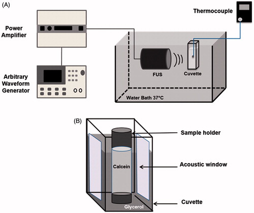
Temperature increase in the in vitro set-up was investigated as a function of the peak negative pressure (PNP) from 0 to 2 MPa, the duty cycle (DC) from 0 to 50% and the exposure time from 0 to 30 min. An arbitrary function generator (33220A, Agilent, Palo Alto, CA) was used to design the excitation signal. A tone burst centred at 1 MHz was repeated with a fixed pulse repetition frequency (PRF) of 1 kHz. This signal was then amplified using a power amplifier (ADECE, Artannes sur Indre, France) and transmitted to a 1 MHz homemade single-element transducer focused at 48 mm with a diameter of 47 mm. The size of the focal spot (i.e. at 48 mm) and the length of the focal area at −3 dB was 4 mm2 and 12 mm, respectively. FUS experiments were performed in a tank filled with degassed water and maintained at 37 °C. A dual-compartment set-up [Citation39] was positioned at the transducer focal distance (). The system consisted of a sample holder (30 mm height, 6 mm inside diameter, 8 mm outside diameter; Fischer Scientific, Illkirch, France) containing 500 µL of PBS, placed inside a glycerol-filled plastic cuvette (). The centre of the sample holder was positioned at the focal distance of the transducer. Glycerol is a good thermally absorptive medium (like tissue) that can be used to heat the sample holder by FUS [Citation28]. In fact, the heat transfer by conduction from the surrounding glycerol induces a temperature increase in the sample holder. This set-up, based on secondary heating, reproduces the in vivo scenario where ultrasound induces hyperthermia in the tumour tissue, and the liposomes accumulated in the tumour are heated by thermal conduction [Citation28]. Over the sides of the dual-compartment set-up, acoustic windows were made with 20-μm thick polyolefin heat shrink film (Rajashrink, Roissy, France) to avoid ultrasound attenuation (4% at 1 MHz) and reflection. Temperature measurements were performed using a fine wire thermocouple (Dostmann electronic, Wertheim-Reicholzheim, Germany) positioned inside the sample holder for different acoustic parameters (i.e. pressure, duty cycle or exposure time). The temperature increment was measured every 15 s for 10 min.
Figure 1. In vitro FUS set-up (A) and dual-compartment set-up (B) consisting of a sample holder placed into a glycerol-filled cuvette.
Activation protocol
For both water bath and FUS experiments, 25 µL of calcein-loaded liposomes were diluted in 500 µL of PBS and placed into the sample holder. Both NTSLs and TSLs were evaluated and each experiment was independently repeated three times. For water bath measurements, the temperature was varied from 37–49 °C. Samples were exposed to the different temperatures for 10 min. Furthermore, the calcein release kinetic was measured using a water bath at a fixed temperature of 42 °C for 30 min. Similarly, samples were exposed to FUS using different acoustic pressures ranging from 0 to 2 MPa, 40% duty cycle for an exposure time of 10 min. The calcein release kinetic was estimated under FUS conditions of 1.75 MPa, 40% duty cycle for an exposure time of 30 min. After water bath or FUS heating, samples were cooled down in an ice bath and stored at 4 °C. The calcein fluorescence was measured using a SpectraMax XPS Gemini (Molecular Devices, Sunnyvale, CA) spectrofluorimeter (λEx = 485 nm/λEm = 520 nm). Complete liposome leakage (100%) was achieved by the addition of 10 µL of Triton X-100 (4% v/v final concentration) to the liposome suspension. The percentage of calcein leakage was calculated using Equation Equation1(1) .
Cryo-transmission electron microscope sample preparation
All cryo-transmission electron microscope (cryo-TEM) experiments were conducted as described previously [Citation40]. Specimens for cryo-TEM observation were prepared using a cryoplunge cryo-fixation device (Gatan, Pleasanton, CA) in which a drop of the aqueous suspension was deposited on to glow-discharged holey-type carbon-coated grids (Ted Pella, Redding, CA). The TEM grid was then prepared by blotting the drop containing the specimen into a thin liquid layer that spanned across the holes in the support carbon film. The liquid film was vitrified by rapidly plunging the grid into liquid ethane cooled by liquid nitrogen. The vitrified specimens were mounted in a Gatan 910 specimen holder that was inserted in the microscope using a CT-3500 cryotransfer system (Gatan) and cooled with liquid nitrogen. TEM images were then obtained from specimens preserved in vitreous ice and suspended across a hole in the supporting carbon substrate. The samples were observed at −178 °C under low dose conditions (<10 e-/A2) using a JEM-1230 ‘Cryo’ microscope (JEOL, Tokyo, Japan) operated at 80 kV and equipped with a LaB6 filament. All micrographs were recorded on a Gatan 1.35 K × 1.04 K × 12 bits ES500W CCD camera. For each sample, the liposome size was measured using ImageJ software (NIH, Bethesda, MD). Data are presented as mean ± standard error of the mean (SEM) from 100 independent measurements.
Statistical analysis
Statistical analyses were performed using the non-parametric Mann-Whitney U test (StatPlus:mac, version 5.8.3.8. 2001–2009 Analyst Soft, Vancouver, BC, Canada). Significance was defined as p < 0.05 (NS, non-significant; *p < 0.05; **p < 0.01; ***p < 0.001).
Results
Preparation, characterisation and optimisation of liposomes
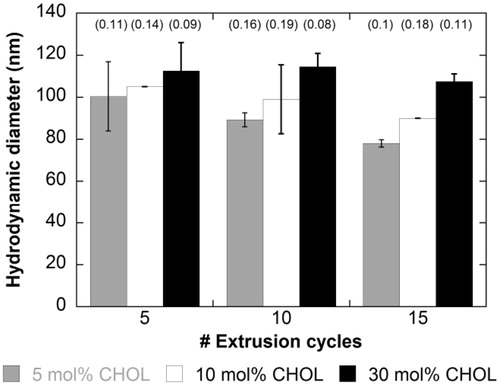
To optimise an approximate liposome size of 100 nm, formulations with 5, 10 and 30% CHOL (mol) were prepared and submitted to 5, 10 or 15 extrusion cycles. Results show that dH depended on the composition of the lipid bilayer and the number of extrusion cycles for 5 and 10% CHOL (mol) (). Indeed, dH increased as the percentage of CHOL increased for a given number of extrusion cycles, while dH decreased as the number of extrusion cycles increased at a given percentage of CHOL. However, as previously reported [Citation41], the bilayer is so rigid for 30 mol% CHOL, that beyond a certain number of extrusion cycles, liposome size could not be reduced further (). For all conditions tested, the polydispersity index remained below 0.2, thus ensuring a rather narrow size distribution. The advantage in terms of size reduction and polydispersity between 10 and 15 cycles not being obvious, we decided to fix the number of extrusion cycles to 10, yielding liposomes with a mean hydrodynamic diameter between 86 and 121 nm, and with a narrow size distribution (PDI < 0.2).
Figure 2. Liposomes size (nm) and polydispersity index (brackets) as a function of extrusion cycle number in presence of various CHOL mol% (n = 3).
Differential scanning calorimetry
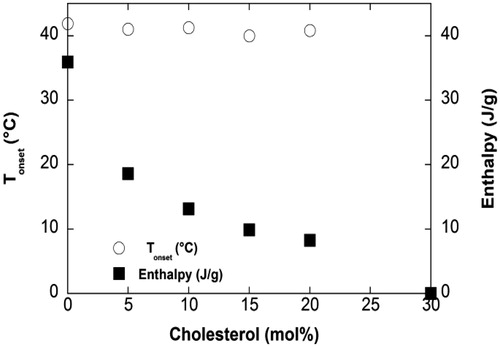
DSC analysis was performed on liposomes prepared with increasing CHOL molar ratios (0, 5, 10, 15, 20 and 30%) to assess the effect of the CHOL insertion on the enthalpy of DPPC (). CHOL-free liposomes (DPPC/DSPE-PEG2000 95:5 mol%) exhibited the typical pre-transition corresponding to the conversion of the lamellar gel phase Lβ’ into the ripple gel phase Pβ’, and the main transition related to the passage from the Pβ’ phase to the lamellar liquid-crystalline phase Lα (, inset) [Citation42]. The pre-transition occurred at Tonset 34.6 °C with an enthalpy ΔH of 2.8 J/g of DPPC, and the main phase transition at Tonset 41.9 °C with an enthalpy of ΔH of 35.9 J/g of DPPC (). The presence of both typical pre-transition and transition of DPPC reflects 5 mol% DSPE-PEG2000 and does not drastically affect thermal properties of DPPC. However, as previously reported the DSPE-PEG2000 insertion within DPPC bilayers resulted in lower enthalpy than that usually reported for pure DPPC bilayers (around 40 J/g) [Citation42]. Above 5 mol% CHOL, the pre-transition was abolished, thus proving CHOL insertion within the bilayer. In addition, as CHOL molar % increased between 5 and 20 mol%, Tonset of the main transition was slightly shifted down to about 41°C, and the enthalpy decreased to 8.3 J/g of DPPC for 20 mol% CHOL (). At 30 mol% CHOL, no thermal event could be detected, supporting that DPPC phase transition was fully abolished (). These results suggest that CHOL insertion within DPPC/DSPE-PEG2000 (5 mol%) bilayers allows fine-tuning of the temperature sensitivity of liposome formulations.
Figure 3. DSC thermograms obtained from liposomes made of DPPC/CHOL/DSPE-PEG2000 upon heating at 5 °C/min according to CHOL mol%.
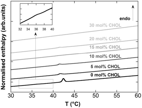
Figure 4. Tonset and corresponding normalised enthalpy obtained from liposomes upon heating at 5 °C/min according to CHOL mol%.
Calcein leakage from liposomes
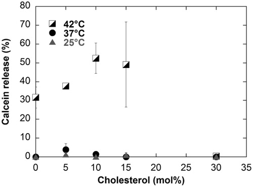
To assess liposome temperature sensitivity, a 40-mM calcein solution was encapsulated within liposomes containing a concentration range of CHOL (0 to 30 mol% CHOL). Calcein release upon water bath-induced hyperthermia was monitored using spectrofluorimetry. Before heating, a residual fluorescence signal F0 was measured corresponding to free calcein. Therefore, the percentage rise measured can only correspond to additional calcein release (). At 25 °C and 37 °C, none of the formulations exhibited substantial calcein release. At 42 °C, liposomes prepared with 0, 5, 10 and 15 mol% CHOL released 32, 38, 53, and 49% of the encapsulated calcein, respectively. No calcein release from liposomes containing 30 mol% CHOL was detected (<0.5%). In agreement with DSC experiments, these results show that liposomal formulations containing 0–15% CHOL are considered temperature-sensitive, whereas the liposomes with 30 mol% CHOL are non-temperature-sensitive.
Figure 5. Release profile of encapsulated calcein from the different liposomal systems according to CHOL mol% and heating temperature for 5 min (n = 3) using a water bath.
Ultrasound-induced hyperthermia
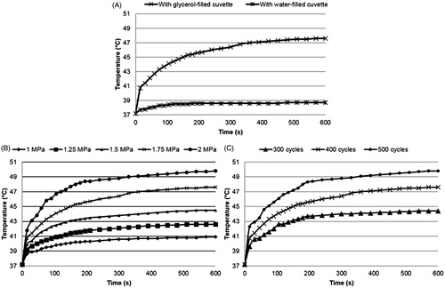
The temperature increase was measured in the sample holder as a function of the ultrasound parameters over time. For these acoustic parameters (PNP: 1.75 MPa, number of cycles: 400, PRF: 1 kHz), the use of the glycerol-filled cuvette allowed a substantial 9 °C temperature increase compared to the water-filled cuvette (). In , curves are displayed as a function of the acoustic pressure for 400 cycles and 1 kHz PRF. For each pressure, a rapid increase in temperature occurred within the first 2 min of ultrasound exposure. After 5 min insonation, the temperature inside the sample holder was quasi-stabilised. At 2 MPa, the maximum temperature at the focus was 49.8 °C after 10 min insonation. The maximum temperature dropped when the PNP decreased. Using 400 cycles and 1 kHz PRF, a PNP higher than 1 MPa was required to reach the liposome phase transition temperature of 41.5 °C, allowing calcein release from TSLs. Notice that at 1.25 MPa, the temperature of 41.5 °C was only reached in a restricted volume (i.e. on the location of the focal point). Transmitting higher acoustic pressures resulted in an increase of the volume of solution in which the temperature exceeded 41.5 °C [Citation27]. As shown in , a similar trend is observed for temperature increase as a function of the number of cycles (for 1.75 MPa PNP, 1 kHz PRF).
Figure 6. (A) Comparison of the temperature increases induced by FUS (1.75 MPa, 400 cycles, 1 kHz PRF) using glycerol-filled or water-filled cuvettes. Panels B and C show time–temperature curves as a function of the acoustic parameters using the glycerol-filled cuvette. Results are displayed as a function of the peak-negative pressure (400 cycles, 1 kHz PRF) in panel (B) and the number of cycles (1.75 MPa, 1 kHz PRF) in panel (C).
In vitro calcein release
To assess the role thermal effect has on calcein release from TSLs and NTSLs, the liposome samples were heated between 37 °C and 49 °C in a water bath for 10 min (). As expected, no temperature-dependent calcein release from NTSLs was observed. For TSLs, the calcein release increased linearly between 37° and 42 °C, reaching a plateau at temperatures above 42 °C. Even at 49 °C, the thermally induced calcein release never exceeded 45% for this liposomal formulation. Most of the calcein (i.e. 55%) was still trapped inside the liposomes in the minutes following heating.
Figure 7. Temperature dependence of liposome calcein release after 10 min water bath (WB) heating (A). Results after FUS exposure are displayed as a function of acoustic pressure in panel B for 400 cycles, repetition of 1 kHz and 10 min insonation (n = 3).
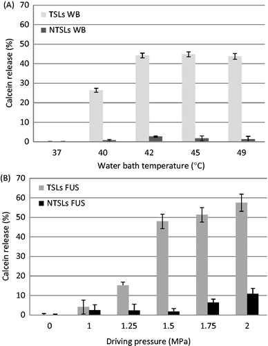
As shown in , TSLs and NTSLs were exposed to acoustic pressures ranging from 1 MPa to 2 MPa (400 cycles, 1 kHz PRF) for 10 min. For TSLs, a weak calcein release was detected for pressure amplitudes from 1 to 1.25 MPa (i.e. <15%). In these conditions the temperature inside the whole sample holder was not sufficient () to induce a significant release. At 1.5 MPa, the calcein release was 47%, a value close to the maximum leakage obtained after water bath heating. Above this pressure, the calcein release continued to slightly increase and reached 57% at 2 MPa. For NTSLs, no calcein release (<2%) was detected for pressure amplitudes below 1.5 MPa, whereas it increased to 6% and 11% at 1.75 MPa and 2 MPa, respectively.
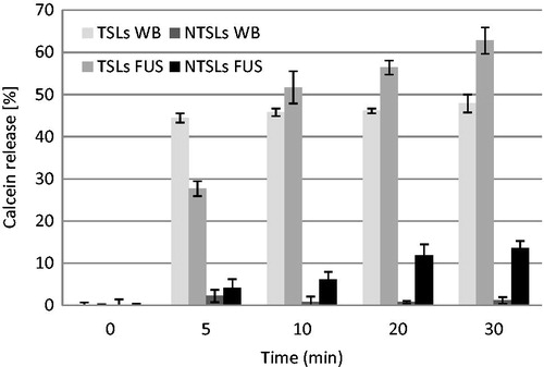
Kinetics of FUS-mediated (1.75 MPa, 400 cycles, 1 kHz PRF) calcein release from TSLs and NTSLs were compared to the release induced by water bath heating at 42 °C (). The FUS parameters were specifically selected in order to observe an additional calcein release compared to release following water bath heating () over time. Water bath heating resulted in a quick calcein release from TSLs during the first 5 min before reaching a plateau. In agreement with the previous results (), no calcein release from NTSLs was detected over time. The exposure of TSLs to FUS induced a progressive calcein release during the first 10 min. This delay corresponds to the time required to heat the sample holder containing the liposomes by FUS. However, a slow additional release up to 62% was observed after 30 min. The exposure of NTSLs to FUS heating induced a gradual calcein release, reaching 14% after 30 min insonation; 30 min later, FUS-mediated release was 15% and 13% higher than water bath heating for TSLs and NTSLs, respectively.
Figure 8. Calcein release from liposomes as a function of the exposure duration for water bath (WB) and FUS heating (n = 3).
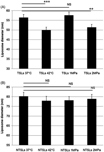
Influence of the thermal effect on liposome size
To investigate the FUS influence on the size of liposomes, firstly cryo-TEM experiments were performed on liposomes heated in a water bath at 37 °C () and 42 °C () for 10 min. Secondly, cryo-TEM experiments were carried out on liposomes exposed to FUS (400 cycles, 1 kHz PRF, 10 min) for acoustic pressures of 1 MPa () and 2 MPa (). For each experimental condition the diameter of 100 liposomes was measured and reported in for TSLs and NTSLs. The mean diameter of NTSLs is larger than TSLs because of CHOL addition within the NTSL bilayers, as mentioned above (). Furthermore, no significant difference in NTSL size was detected (p > 0.05) for the different heating conditions. As shown in , water bath heating at 42 °C or FUS exposure at 2 MPa induced a significant decrease in TSL diameter (i.e. 14%) compared to water bath heating at 37 °C. At 1 MPa, no significant difference in liposome diameter (p > 0.05) was noticed. In agreement with previous data ( and ), this result shows that the FUS-induced temperature increment was not sufficient to induce a calcein release at this acoustic pressure.
Figure 9. Cryo-TEM images of TSLs after 10 min heating in a water bath at 37 °C (A) and at 42 °C (B). Images of TSLs after FUS exposure (400 cycles, 1 kHz PRF, exposure time 10 min) at 1 MPa and at 2 MPa are displayed in panels C and D, respectively. Scale bar, 100 nm.
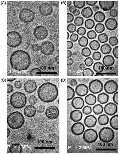
Figure 10. Mean diameters of TSLs (A) and NTSLs (B) exposed to different water bath and FUS heating for 10 min (n = 100 liposomes). Statistical analysis was performed using the nonparametric Mann-Whitney test. Significance was defined as p < 0.05 (*p < 0.05, **p < 0.01, ***p < 0.001, NS, non-significant).
Discussion
Over recent decades, FUS has emerged as a versatile technology for non-invasive thermal therapies in oncology [Citation43,Citation44]. The ultrasound waves are attenuated as they pass through tissue due to scattering and absorption of the acoustic energy. Some of this energy absorbed is converted into heat (i.e. thermal effect) [Citation45]. The exact amount depends on the ultrasound parameters and the tissue exposed. With regard to ultrasound, acoustic parameters (i.e. amplitude, length and frequency) of the propagating wave, the tissue volume heated and exposure time influence the efficacy of hyperthermia. The heating of tissue depends on its molecular composition, thermal conduction (i.e. absorption coefficient) and blood perfusion. However, the ultrasound/tissue interaction can induce other biophysical effects, such as radiation force or cavitation (i.e. mechanical effects) [Citation46]. In this in vitro study, the FUS influence on calcein-loaded thermosensitive stealth liposomes was investigated.
The liposomes were first incubated in a water bath at temperatures ranging between 37° and 49 °C. Agreeing with previous studies [Citation47], a temperature increase from 37° to 42 °C resulted in a gradual increase in calcein release from TSLs, reaching a plateau at 42 °C with a maximum release rate of 45% (). As previously reported, no additional release was observed for temperature above 42 °C. In the aqueous compartment of liposomes, calcein can display some interactions with the choline head groups of phospholipids [Citation48]. The strength of this interaction might causes less desorption and so less release from the liposomes [Citation49], thus explaining our data on . As expected, no calcein release from NTLs was detected.
Concerning FUS, an acoustic pressure of 1.5 MPa PNP induced a calcein release from TSLs as efficient as water bath heating (). In addition, no calcein release from NTSLs was observed under such acoustic pressures. These results suggest that thermal effects might play a major role in calcein release from TSLs under an acoustic pressure below 1.5 MPa. However, the exposure of TSLs to acoustic pressures above 1.5 MPa (i.e. 1.75 MPa and 2 MPa) resulted in an additional calcein release (up to 12%). Under such high pressures, a similar calcein release trend from NTSLs was observed (up to 11%). As previously reported [Citation50,Citation51], these results support that acoustic pressures above 1.5 MPa induce mechanical effects (i.e. cavitation and/or radiation forces) that disrupt the liposomal membrane of both TSLs and NTSLs, partially destabilising the calcein/phospholipid interaction and thus leading to an additional calcein release. This supplementary release increases with ultrasound beam energy and the total exposure time.
Acoustic cavitation and radiation forces are two main mechanical effects induced by high energy FUS. Cavitation is defined as the formation and the activity of gas bubbles in a medium exposed to ultrasound [Citation52]. The sustained growth of cavitation bubbles and their oscillations over several acoustic cycles is known as stable cavitation [Citation46]. Exposure to high intensity pulsed waves can lead to inertial cavitation, corresponding to a violent bubble radial expansion and immediate collapse [Citation53]. Recently, Ninomiya et al. [Citation54] reported that cavitation bubble collapse leads to an increase in local temperature, therefore enhancing TSL drug release. In addition, NTSLs show good stability at high temperature. However, as previously reported [Citation54], the collapse of cavitating bubbles or their oscillations near liposomes could induce a local mechanical stress, resulting in destabilisation of the NTSL membrane, thus enhancing its permeability and drug release. According to previous studies [Citation51], this destabilisation of NTSL membrane size is the specific signature of the mechanical effect. In addition, Schroeder et al. [Citation10] report that the exposure of liposomes to an oscillating ultrasonic field induces the formation of gas bubble nuclei in the hydrophobic region of the liposomal bilayer. These nuclei grow until they permeate the membrane through the formation of transient hydrophilic pores, resulting in the drug release. After ultrasound exposure, the liposomal membrane resumes its initial impermeable state. To date, this mechanism is not clearly understood and further investigation is still necessary.
Regarding high intensity focused ultrasound (HIFU), radiation forces can result in local particle displacement and fluid currents, also as known as acoustic streaming [Citation55]. Acoustic streaming may involve (1) bulk streaming, which produces fluid currents responsible for particle movements in the direction of the propagating ultrasound wave, and (2) micro-streaming, wherein local currents are created near cavitating bodies. In our in vitro set-up, the radiation force can steer the solution with liposomes inside the cuvette. This steering process would favour heat convection and calcein release from TSLs. According the available in vivo data [Citation18,Citation56,Citation57], the contribution of radiation force in in vivo heat convection and drug release from TSLs has not been established yet. However, we can reasonably assume that the organisation of biological tissues (i.e. extracellular matrix, vascular perfusion) and vascular network (i.e. blood flow, size of vessels) might limit the contribution of radiation force in this process. As recently described, micro-streaming can also induce shear stresses that destabilise the liposome bilayer, thus releasing the encapsulated drug. Indeed, Oerlemans et al. [Citation16] loaded hydrophilic (i.e. fluorescein) and hydrophobic (i.e. Nile Red) fluorescent dyes into NTLs and TSLs, and then measured the release kinetics under HIFU. Interestingly, the authors did not observe Nile Red release from either TSLs or NTSLs when they were exposed to increased temperatures above the lipid Tm in a water bath. Nevertheless, HIFU exposure of both liposome formulations induced Nile Red release. In this case, the release observed was attributed to non-cavitational and non-thermal effects of HIFU. The authors speculated that radiation force-mediated streaming induced liposome collisions during HIFU exposure could be responsible for the release of Nile Red from the liposome bilayer. Nevertheless, further in vivo investigations are necessary to evaluate the contribution of radiation forces in the FUS-triggered drug release from TSLs.
Cryo-TEM observations showed that the incubation of liposomes to temperatures above their Tm in a water bath induced a slight, but significant, diminution in the mean TSL diameter (i.e. 14%), while the NTSL size remained unchanged. A similar decrease in the mean TSL diameter was observed after their exposure to acoustic pressures inducing temperature elevations above their Tm. This decrease in TSL size is the specific signature of the thermal effect [Citation58]. Moreover, Leirer et al. [Citation58] reported that at temperatures above their Tm, TSLs exhibit budding or pearl formation. These buds remained connected to the parent liposomes, presumably by a small neck. The cooling of these TSLs to temperatures below their Tm led to complete rupture and fission of this neck, while the parent liposome remained intact with a decreased size. Although both thermal and mechanical actions are responsible for calcein release from TSLs ( and ) at high acoustic pressures (i.e. above 1.5 MPa), the mechanical effects do not influence liposome size. Indeed, when exposed to an acoustic pressure of 2 MPa, the decrease in TSL size was similar to that induced by the thermal effect alone (i.e. water bath at 42 °C). Moreover, increasing the acoustic pressure does not influence the mean NTSL diameter. According to previous studies [Citation59], these results indicated that the liposome diameter remained unaffected by the mechanical effect induced by high acoustic pressures. In fact, Shroeder et al. [Citation10] suggested that FUS-mediated mechanical effects induced the release of encapsulated compound through the formation of transient pores in the liposomal membrane, without any change in liposome size distribution [Citation59].
Compared to other in vitro studies using a set-up based on a water-filled tank [Citation16,Citation50,Citation54], our in vitro set-up approaches the in vivo scenario [Citation28] and requires much lower ultrasound energy to induce a significant temperature increase (). Moreover, due to high-energy tissue absorption, ultrasonic energy inducing the desired temperature elevation in vivo is usually lower than the energy required in vitro [Citation44]. Therefore, the use of higher ultrasonic energy in vitro leads to an overestimation of the role of the mechanical effects in vivo.
Conclusions
In summary, the phase transition temperature of liposomes (i.e. 41.5 °C) can be reached in vitro while using FUS parameters of 1.25 MPa, 400 cycles and 1 kHz PRF. FUS triggers the calcein release from thermosensitive stealth liposomes through thermal effect for acoustic pressures below 1.5 MPa. However, at high acoustic pressures (i.e. above 1.5 MPa), FUS induces mechanical effects which contribute to an additional calcein release from TSLs. Our findings suggest that the thermal effect is the prevailing mechanism inducing calcein release from TSLs when exposed to FUS. Cryo-electron microscopy observations demonstrate that the thermal effect results in a decrease in the liposome diameter, whereas mechanical stress does not alter liposome size. This observation supports the hypothesis that mechanical effects induced the formation of transient pores in the liposome membrane. Further research is necessary to clearly understand how the mechanical stress affects the liposome membrane.
Acknowledgements
The authors thank Michalakis Averkiou (University of Cyprus) for providing FUS transducer, Mario Ries and Roel Deckers (UMC Utrecht), Justin Tessié (IPBS-CNRS UMR 5089, Toulouse) for fruitful discussions, Sylvie Attucci (Inserm U-1100, Tours) for her technical assistance in spectrofluorimetry, Jacqueline Butterworth for proofreading the manuscript.
Declaration of interest
This research work was funded by the Agence Nationale pour la Recherche (ANR-10-TecSan-007), the European Commission FP7 Program SONODRUGS (NMP4-LA-2008-213706) and FEDER PRESAGE (3431 – 35438). Institut Galien Paris-Sud is a member of the Laboratory of Excellence LERMIT supported by a grant from ANR (ANR-10-LABX-33). The authors alone are responsible for the content and writing of the paper.
References
- Jain RK, Stylianopoulos T. Delivering nanomedicine to solid tumors. Nat Rev Clin Oncol 2010;7:653–64
- Bertrand N, Wu J, Xu X, Kamaly N, Farokhzad OC. Cancer nanotechnology: The impact of passive and active targeting in the era of modern cancer biology. Adv Drug Deliv Rev 2014;66:2–25
- Barenholz Y. Doxil® – The first FDA-approved nano-drug: Lessons learned. J Control Release 2012;160:117–34
- Gradishar WJ, Tjulandin S, Davidson N, Shaw H, Desai N, Bhar P, et al. Phase III trial of nanoparticle albumin-bound paclitaxel compared with polyethylated castor oil-based paclitaxel in women with breast cancer. J Clin Oncol 2005;23:7794–803
- Gaitanis A, Staal S. Liposomal doxorubicin and nab-paclitaxel: Nanoparticle cancer chemotherapy in current clinical use. Methods Mol Biol 2010;624:385–92
- Al-Jamal WT, Al-Ahmady ZS, Kostarelos K. Pharmacokinetics and tissue distribution of temperature-sensitive liposomal doxorubicin in tumor-bearing mice triggered with mild hyperthermia. Biomaterials 2012;33:4608–17
- Al-Ahmady ZS, Al-Jamal WT, Bossche JV, Bui TT, Drake AF, Mason AJ, et al. Lipid-peptide vesicle nanoscale hybrids for triggered drug release by mild hyperthermia in vitro and in vivo. ACS Nano 2012;6:9335–46
- Blanco E, Hsiao A, Mann AP, Landry MG, Meric-Bernstam F, Ferrari M. Nanomedicine in cancer therapy: Innovative trends and prospects. Cancer Sci 2011;102:1247–52
- Yan Y, Such GK, Johnston AP, Best JP, Caruso F. Engineering particles for therapeutic delivery: Prospects and challenges. ACS Nano 2012;6:3663–9
- Schroeder A, Kost J, Barenholz Y. Ultrasound, liposomes, and drug delivery: Principles for using ultrasound to control the release of drugs from liposomes. Chem Phys Lipids 2009;162:1–16
- Khaibullina A, Jang BS, Sun H, Le N, Yu S, Frenkel V, et al. Pulsed high-intensity focused ultrasound enhances uptake of radiolabeled monoclonal antibody to human epidermoid tumor in nude mice. J Nucl Med 2008;49:295–302
- Li L, ten Hagen TL, Bolkestein M, Gasselhuber A, Yatvin J, van Rhoon GC, et al. Improved intratumoral nanoparticle extravasation and penetration by mild hyperthermia. J Control Release 2013;167:130–7
- Somaglino L, Bouchoux G, Mestas JL, Lafon C. Validation of an acoustic cavitation dose with hydroxyl radical production generated by inertial cavitation in pulsed mode: Application to in vitro drug release from liposomes. Ultrason Sonochem 2011;18:577–88
- Lafon C, Somaglino L, Bouchoux G, Mari JM, Chesnais S, Ngo J, et al. Feasibility study of cavitation-induced liposomal doxorubicin release in an AT2 Dunning rat tumor model. J Drug Target 2012;20:691–702
- Evjen TJ, Hagtvet E, Moussatov A, Rognvaldsson S, Mestas JL, Fowler RA, et al. In vivo monitoring of liposomal release in tumours following ultrasound stimulation. Eur J Pharm Biopharm 2013;84:526–31
- Oerlemans C, Deckers R, Storm G, Hennink WE, Nijsen JF. Evidence for a new mechanism behind HIFU-triggered release from liposomes. J Control Release 2013;168:327–33
- de Smet M, Heijman E, Langereis S, Hijnen NM, Grull H. Magnetic resonance imaging of high intensity focused ultrasound mediated drug delivery from temperature-sensitive liposomes: An in vivo proof-of-concept study. J Control Release 2011;150:102–10
- Ranjan A, Jacobs GC, Woods DL, Negussie AH, Partanen A, Yarmolenko PS, et al. Image-guided drug delivery with magnetic resonance guided high intensity focused ultrasound and temperature sensitive liposomes in a rabbit Vx2 tumor model. J Control Release 2012;158:487–94
- Negussie AH, Yarmolenko PS, Partanen A, Ranjan A, Jacobs G, Woods D, et al. Formulation and characterisation of magnetic resonance imageable thermally sensitive liposomes for use with magnetic resonance-guided high intensity focused ultrasound. Int J Hyperthermia 2011;27:140–55
- Staruch RM, Ganguly M, Tannock IF, Hynynen K, Chopra R. Enhanced drug delivery in rabbit VX2 tumours using thermosensitive liposomes and MRI-controlled focused ultrasound hyperthermia. Int J Hyperthermia 2012;28:776–87
- Matteucci ML, Anyarambhatla G, Rosner G, Azuma C, Fisher PE, Dewhirst MW, et al. Hyperthermia increases accumulation of technetium-99m-labeled liposomes in feline sarcomas. Clin Cancer Res 2000;6:3748–55
- Kong G, Dewhirst MW. Hyperthermia and liposomes. Int J Hyperthermia 1999;15:345–70
- Kong G, Braun RD, Dewhirst MW. Characterization of the effect of hyperthermia on nanoparticle extravasation from tumor vasculature. Cancer Res 2001;61:3027–32
- Yatvin MB, Weinstein JN, Dennis WH, Blumenthal R. Design of liposomes for enhanced local release of drugs by hyperthermia. Science 1978;202(4374):1290–3
- Needham D, Anyarambhatla G, Kong G, Dewhirst MW. A new temperature-sensitive liposome for use with mild hyperthermia: Characterization and testing in a human tumor xenograft model. Cancer Res 2000;60:1197–201
- Mackay JA, Chilkoti A. Temperature sensitive peptides: Engineering hyperthermia-directed therapeutics. Int J Hyperthermia 2008;24:483–95
- Escoffre JM, Novell A, de Smet M, Bouakaz A. Focused ultrasound mediated drug delivery from temperature-sensitive liposomes: In-vitro characterization and validation. Phys Med Biol 2013;58:8135–51
- Mannaris C, Efthymiou E, Meyre ME, Averkiou MA. In vitro localized release of thermosensitive liposomes with ultrasound-induced hyperthermia. Ultrasound Med Biol 2013;39:2011–20
- Kheirolomoom A, Lai CY, Tam SM, Mahakian LM, Ingham ES, Watson KD, et al. Complete regression of local cancer using temperature-sensitive liposomes combined with ultrasound-mediated hyperthermia. J Control Release 2013;172:266–73
- Park SM, Kim MS, Park SJ, Park ES, Choi KS, Kim YS, et al. Novel temperature-triggered liposome with high stability: Formulation, in vitro evaluation, and in vivo study combined with high-intensity focused ultrasound (HIFU). J Control Release 2013;170:373–9
- Chang HI, Yeh MK. Clinical development of liposome-based drugs: formulation, characterization, and therapeutic efficacy. Int J Nanomedicine 2012;7:49–60
- Shenoi MM, Shah NB, Griffin RJ, Vercellotti GM, Bischof JC. Nanoparticle preconditioning for enhanced thermal therapies in cancer. Nanomedicine (Lond) 2011;6:545–63
- Tagami T, Foltz WD, Ernsting MJ, Lee CM, Tannock IF, May JP, et al. MRI monitoring of intratumoral drug delivery and prediction of the therapeutic effect with a multifunctional thermosensitive liposome. Biomaterials 2011;32:6570–8
- Tagami T, Ernsting MJ, Li SD. Optimization of a novel and improved thermosensitive liposome formulated with DPPC and a Brij surfactant using a robust in vitro system. J Control Release 2011;154:290–7
- Chiu GN, Abraham SA, Ickenstein LM, Ng R, Karlsson G, Edwards K, et al. Encapsulation of doxorubicin into thermosensitive liposomes via complexation with the transition metal manganese. J Control Release 2005;104:271–88
- Banno B, Ickenstein LM, Chiu GN, Bally MB, Thewalt J, Brief E, et al. The functional roles of poly(ethylene glycol)-lipid and lysolipid in the drug retention and release from lysolipid-containing thermosensitive liposomes in vitro and in vivo. J Pharm Sci 2010;99:2295–308
- Li L, ten Hagen TL, Schipper D, Wijnberg TM, van Rhoon GC, Eggermont AM, et al. Triggered content release from optimized stealth thermosensitive liposomes using mild hyperthermia. J Control Release 2010;143:274–9
- Berne B, Pecora R. Dynamic light scattering: With applications to chemistry, biology, and physics. New York: Courier Dover, 2000
- Novell A, Escoffre JM, Al-Sabbagh C, Mannaris C, Fattal E, Tsapis N, et al. Role of thermal and mechanical effects on drug release from thermosensitive nanocarriers. Proc IEEE Ultrasonics Symposium, Dresden, Germany, 2012
- Gaillard C, Douliez JP. Cryo-TEM and AFM for the characterization of vesicle-like nanoparticle dispersions and self-assembled supramolecular fatty-acid-based structures: A few examples. Curr Microsc Contrib Adv Sci Technol 2012;2:912–22
- Semple SC, Chonn A, Cullis PR. Influence of cholesterol on the association of plasma proteins with liposomes. Biochemistry 1996;35:2521–5
- Carion-Taravella B, Lesieur S, Chopineau J, Lesieur P, Ollivon M. Phase behavior of mixed aqueous dispersions of dipalmitoylphosphatidylcholine and dodecyl glycosides: A differential scanning calorimetry and X-ray diffraction investigation. Langmuir 2002;18:325–35
- Dromi S, Frenkel V, Luk A, Traughber B, Angstadt M, Bur M, et al. Pulsed-high intensity focused ultrasound and low temperature-sensitive liposomes for enhanced targeted drug delivery and antitumor effect. Clin Cancer Res 2007;13:2722–7
- Patel PR, Luk A, Durrani A, Dromi S, Cuesta J, Angstadt M, et al. In vitro and in vivo evaluations of increased effective beam width for heat deposition using a split focus high intensity ultrasound (HIFU) transducer. Int J Hyperthermia 2008;24:537–49
- Fry WJ, Wulff VJ, Tucker D, Fry FJ. Physical factors involved in ultrasonically induced changes in living systems: I. Identification of non-temperature effects. J Acoust Soc Am 1950;22:867–76
- Deckers R, Moonen CT. Ultrasound triggered, image guided, local drug delivery. J Control Release 2010;148:25–33
- Han HD, Kim TW, Shin BC, Choi HS. Release of calcein from temperature-sensitive liposomes in a poly(n-isopropylacrylamide) hydrogel. Macromol Res 2005;13:54–61
- Maherani B, Arab-Tehrany E, Kheirolomoom A, Geny D, Linder M. Calcein release behavior from liposomal bilayer: Influence of physicochemical/mechanical/structural properties of lipids. Biochimie 2013;95:2018–33
- Calvagno MG, Celia C, Paolino D, Cosco D, Iannone M, Castelli F, et al. Effects of lipid composition and preparation conditions on physical-chemical properties, technological parameters and in vitro biological activity of gemcitabine-loaded liposomes. Curr Drug Deliv 2007;4:89–101
- Afadzi M, Davies Cde L, Hansen YH, Johansen T, Standal OK, Hansen R, et al. Effect of ultrasound parameters on the release of liposomal calcein. Ultrasound Med Biol 2012;38:476–86
- Evjen TJ, Nilssen EA, Fowler RA, Rognvaldsson S, Brandl M, Fossheim SL. Lipid membrane composition influences drug release from dioleoylphosphatidylethanolamine-based liposomes on exposure to ultrasound. Int J Pharm 2011;406:114–16
- Apfel RE. Acoustic cavitation: A possible consequence of biomedical uses of ultrasound. Br J Cancer Suppl 1982;5:140–6
- Yudina A, Lepetit-Coiffe M, De Smet M, Langereis S, Grull H, Moonen C. In vivo temperature controlled ultrasound-mediated intracellular delivery of cell-impermeable compounds. J Control Release 2012;161:90–7
- Ninomiya K, Kawabata S, Tashita H, Shimizu N. Ultrasound-mediated drug delivery using liposomes modified with a thermosensitive polymer. Ultrason Sonochem 2014;21:310–16
- Sarvazyan AP, Rudenko OV, Nyborg WL. Biomedical applications of radiation force of ultrasound: Historical roots and physical basis. Ultrasound Med Biol 2010;36:1379–94
- Partanen A, Yarmolenko PS, Viitala A, Appanaboyina S, Haemmerich D, Ranjan A, et al. Mild hyperthermia with magnetic resonance-guided high-intensity focused ultrasound for applications in drug delivery. Int J Hyperthermia 2012;28:320–36
- Gasselhuber A, Dreher MR, Partanen A, Yarmolenko PS, Woods D, Wood BJ, et al. Targeted drug delivery by high intensity focused ultrasound mediated hyperthermia combined with temperature-sensitive liposomes: Computational modelling and preliminary in vivo validation. Int J Hyperthermia 2012;28:337–48
- Leirer C, Wunderlich B, Myles VM, Schneider MF. Phase transition induced fission in lipid vesicles. Biophys Chem 2009;143:106–9
- Schroeder A, Avnir Y, Weisman S, Najajreh Y, Gabizon A, Talmon Y, et al. Controlling liposomal drug release with low frequency ultrasound: Mechanism and feasibility. Langmuir 2007;23:4019–25