
Abstract
Purpose: Heat stress has been shown to reduce muscle atrophy and enhance muscle regeneration. However, the role of heat stress on extracellular matrix (ECM) remodelling remains poorly understood. Here, we examined the effect of heat exposure on intramuscular fibrosis and its associated signalling in soleus and plantaris muscles after tenotomy. Materials and methods: Male Wistar rats were randomly divided into four groups: sedentary control (CON), control plus heat stress (CON+HEAT), tenotomy (TEN) for 8 days, and tenotomy for 8 days plus heat stress (TEN+HEAT). Whole body heat stress was maintained at 40.5–41.5 °C for 30 min, 24 h before and 1–6 days after tenotomy. Results: Tenotomy resulted in muscle atrophy and a substantial increase in intramuscular collagen content, which was more pronounced in soleus than in plantaris muscles, whereas laminin content remained unaffected. These effects were associated with increases in MMP-2 activity, TIMP-2, and TGF-β1 protein expressions. Heat stress, however, attenuated tenotomy-induced intramuscular collagen accumulation in soleus muscle and reduced TIMP-2 and TGF-β1 protein expressions, but had no effect on MMP-2 activity in both muscles. These alterations were concomitant with the induction of heat shock protein 72 (Hsp72). Conclusion: These data demonstrated that heat stress could reduce intramuscular fibrosis, at least in part, through decreasing TGF-β1 and TIMP-2 protein expressions of tenotomised soleus muscle. The results from this study shed light on the mechanism and suggest the potential therapeutic effect of heat stress in alleviating intramuscular fibrosis after tenotomy.
Introduction
Extracellular matrix (ECM) of skeletal muscle plays an important role in maintaining the structural integrity of muscle, force transmission, and regeneration of muscle fibre following injuries [Citation1,Citation2]. Thus, disruption of ECM can lead to impairments of muscle function and lateral transmission of force [Citation3,Citation4], as well as delayed muscle repairing processes [Citation5,Citation6].
Skeletal muscle ECM is in a dynamic state of continuous synthesis and degradation of its components in response to changes in physiological as well as mechanical challenges [Citation2]. These processes are controlled by the balance of matrix metalloproteinases (MMPs) and their inhibitors of tissue inhibitors of metalloproteinases (TIMPs) [Citation7,Citation8]. Skeletal muscle fibrosis occurs whenever there is an imbalance between MMPs and TIMPs in favour of TIMPs [Citation2,Citation8]. MMPs are a family of calcium- and zinc-dependent endopeptidase and act as a key regulator for the remodelling and degradation of ECM components in both physiological and pathological processes [Citation9]. Although several types of MMPs are identified and expressed in skeletal muscles, MMP-2 (gelatinase A) and MMP-9 (gelatinase B) are thought to be responsible for ECM remodelling of skeletal muscles [Citation9–11]. MMP-9 is associated with ECM degradation during inflammation [Citation12], MMP-2 is believed to be responsible for muscle disuse atrophy [Citation13]. Specifically, the MMP-2 activity and its mRNA as well as protein expression were up-regulated in various animal models of mechanical unloading [Citation10,Citation13,Citation14]. In addition, transgenic mice with MMP-2 promoter/enhancer reporter construction exhibited a higher MMP-2 promoter/enhancer activity compared with the wild-type mice following tenotomy [Citation13].
In addition to ECM degradation, there are various signal transduction pathways regulating collagen expression, a major structural protein in ECM of skeletal muscles. Of these, transforming growth factor (TGF)-β1 is a potent regulator of type I and type III collagen synthesis [Citation1]. TGF-β1 is a cytokine mainly produced from macrophages and fibroblasts [Citation15,Citation16] in the form of latent TGF-β1 complex, which comprises disulphide linkages between cysteine residues of latent TGF-β1 through its latency-associated proteins (LAPs) and specific cysteine residues in the latent-TGFβ-binding protein (LTBP) [Citation17]. TGF-β1 activity is controlled by the liberation of active TGF-β1 form, which, in turn, binds to its receptor and activates TGF-β1 signalling responses [Citation16]. TGF-β1 causes collagen accumulation via several mediating pathways including intracellular Smad-dependent signalling that activates the transcription of profibrotic gene such as type I collagen [Citation17]. In addition, TGF-β1 can stimulate fibroblast growth and differentiation, and convert satellite cells into myofibroblasts, leading to enhanced muscle fibrosis and delay of muscle recovery following injuries [Citation18].
To date, in spite of many attempts, there is no single effective method to completely cure muscle fibrosis. Although, several pharmacological agents such as decorin and suramin, a TGF-β1 inhibitor, have been prescribed to combat fibrosis and many connective tissue disorders [Citation19,Citation20], these drugs do not come without side effects. Therefore, non-pharmacological intervention should be considered as an alternative approach to reduce fibrosis by promoting the balance of metalloproteinase expression and reducing TGF-β1 expression. Interestingly, the protective role of heat shock protein 70 (Hsp70) against tissue fibrosis has recently been documented [Citation21–23]. In one study, Takeuchi et al. [2013] have demonstrated that an application of heat stress resulted in decreased collagen fibre area following a crush-induced muscle injury [Citation24]. Moreover, induction of Hsp70 expression was reported to be associated with the attenuation of fibrotic markers including α-smooth muscle actin (α-SMA), collagen type I, TGF-β1, and TIMP-1 expressions in cardiac muscle [Citation21]. In view of a paucity of study examining the role of heat stress on muscle ECM remodelling, it is still unclear whether heat stress could attenuate intramuscular fibrosis especially during mechanical unloading. The first purpose of this study, therefore, was to evaluate the effect and mechanism of heat treatment on collagen accumulation induced by tenotomy in rat skeletal muscles. This animal model was chosen because tendon rupture is very commonly encountered by many clinicians and surgeons following orthopaedic surgery, trauma, or degenerative musculoskeletal diseases [Citation25]. We hypothesised that heat treatment could attenuate tenotomy-induced collagen accumulation through modulating MMP-2/TIMP-2 balance and reducing TGF-β1 expression.
In addition, there is growing evidence indicating that the adaptive responses of skeletal muscles to heat stress/Hsp70 and alterations of collagen content and its metabolism are fibre type-specific, i.e. more pronounced in a slow-twitch muscle than in a fast-twitch muscle [Citation26,Citation27]. Therefore, the second objective of this study was to determine whether there was a fibre type-specific in the attenuation of tenotomy-induced collagen accumulation by heat stress.
Materials and methods
Animals
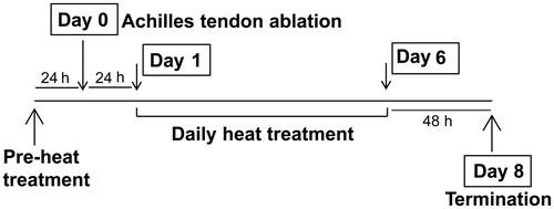
Adult male Wistar rats (10 weeks old), weighing approximately 290–310 g, were obtained from the National Laboratory Animal Centre, Salaya, Nakhon Pathom, Thailand. All animal procedures and experiments were conducted with the approval of the animal use and care guidelines established by the Ethics Committee on the Use of Experimental Animals, Faculty of Science, Mahidol University (Protocol no. MUSC56-011-273). Rats were randomly assigned to four groups (n = 8 in each group): a control group (CON), a control with heat stress group (CON+HEAT), a tenotomy without heat stress group (TEN), and a tenotomy with heat stress group (TEN+HEAT). All rats were housed at 21 °C and maintained on a 12:12 h light:dark cycle. The CON group were given standard food and water ad libitum while the CON+HEAT, TEN, and TEN+HEAT groups were pair fed to eliminate confounding factors of food intake between groups. Heat stress was applied 24 h before and after tenotomy and continued for another 5 days. Rats were terminated 48 h after the last heat exposure ().
Figure 1. Heat stress protocol.
Tenotomy
Rats were anaesthetised with a cocktail of tiletamine and zolazepam (Zoletil®; 25 mg/kg, i.p.) and xylazine (8 mg/kg, i.p.). All surgical procedures were performed under aseptic technique. The right hind limb was cleaned with Betadine® solution to prevent infection, then the skin of calf down to the Achilles tendon and calcaneous bone was incised. The Achilles tendon was carefully excised from the distal part that attached with calcaneous bone to the proximal part for 3 mm in length without disturbing nerve and blood supply. Thereafter, the skin was sutured with a surgical thread and the rats were allowed to recover before returning to their cages.
Heat treatment
Heat stress was performed as described previously by Selsby et al. [Citation28]. In brief, rats were anaesthetised with 5% isoflurane gas–oxygen mixture and the unconscious stage of the rats was maintained with 3% concentration using a SurgiVet® Classic T3™ vaporiser (VCT302; Smith medical, Waukesha, WI, USA). The core temperatures of the rats were monitored throughout the experiment by using a rectal probe (YSI401; Yellow Spring, OH, USA), which was inserted approximately 4 cm into the rectum and taped to the tail to prevent displacement. For heat treatment the rats were wrapped around with a pre-warmed thermal blanket exposing the head and tail. The core temperature, which was maintained at 40.5–41.5 °C, was continuously monitored and recorded every 5 min for 30 min. Thereafter, the thermal blanket was removed while the core temperature was kept monitored. After recovery from anaesthesia, 10–15 mL of water was given orally to the rats in order to prevent dehydration [Citation29] and the rats were returned to their cages after body temperature decreased to 37 °C. The TEN group was similarly treated but without heat exposure and the core temperature was maintained at 37 °C.
Sample collection
Rats were anaesthetised with pentobarbital sodium (75 mg/kg, i.p.), soleus and plantaris muscles were rapidly removed, and the rats were euthanised by cardiac puncture. Muscle samples were trimmed of excess connective tissue, weighed, and the entire fibre was horizontally cut into halves. The upper half was immediately frozen in liquid nitrogen and stored at −80 °C for biochemical analysis. The lower half was mounted into optimal cutting temperature (OCT) Tissue tek® (4583; Electron Microscopy Sciences, Hatfield, PA) and immersed in isopentane (M32631, Sigma, St Louis, MO) pre-cooled by liquid nitrogen. The frozen muscle samples were immediately stored in −80 °C for histological and immunohistochemical analyses.
Western blot analysis
Approximately 50 mg of muscle samples were homogenised using a glass homogeniser (Glas-Col, Terre Haute, IN) with an ice-cold buffer containing 50 mM Tris-HCl pH 7.5, 5 mM EDTA, 1 mM PMSF, and a protease inhibitor cocktail (P8340; Sigma) at a mass to buffer ratio of 20:1. Muscle homogenates were centrifuged at 10,000 g for 10 min (4 °C) to collect the supernatant. Protein concentration of supernatant was determined in triplicate by using bicinchoninic acid (BCA) assay kit (Thermo Scientific; Rockford, IL) with bovine serum albumin (BSA, Sigma) as a standard.
Under reducing conditions (except TGF-β1, type I collagen, and GAPDH, which were performed under non-reducing conditions), 1 mg/mL of cytosolic protein in sample buffer solution were denatured by heating at 60 °C for 10 min and loaded into the SDS-polyacrylamide gel. The electrophoresis was run at 60 V for 30 min on a 5% stacking gel and 110 V for 90 min on a 10–12.5% separating gel at room temperature (RT). Proteins were then transferred for 90 min at 100 V onto BioTrace™ NT nitrocellulose blotting membrane (CA27376-991; Pall, Port Washington, NY). Non-specific binding was blocked with 5% non-fat milk in Tris-buffered saline (TBS) plus Tween-20® (20 mM Tris, pH 7.5, 150 mM NaCl, and 0.1% Tween-20®) for 90 min at RT. The membrane was incubated with a primary antibody in blocking buffer overnight at 4 °C. The following primary antibodies were used: 1:1,000 anti-α-Tubulin (05-829, Millipore, Temecula, CA), 1:1,000 anti-GAPDH (2118, Cell Signaling Technology, Danvers, MA), 1:1,000 anti-Hsp72 (SPA-810, StressGen, Victoria, BC, Canada), 1:1,000 anti-TGF-β1 (ab64715, Abcam, Cambridge, MA), 1:1,000 anti-TIMP-2 (ab1828, Abcam), and 1:100 anti-type I collagen (sc397573, Santa Cruz Biotechnology, Santa Cruz, CA). Following a series of extensive washes with TBS plus Tween-20® buffer, the membrane was incubated for 90 min with 1:10,000 goat anti-mouse IgG peroxidase conjugate (31430, Thermo Scientific) or 1:5,000 goat anti-rabbit IgG conjugated horseradish peroxidase (sc2091, Santa Cruz Biotechnology) secondary antibodies. Protein bands were visualised by enhanced chemiluminescence (ECL) (34082, Thermo Scientific) and exposed to CL-XPosure film (34090, Thermo Scientific). Band intensity was measured by using Image J software version 1.44o (National Institutes of Health, Bethesda, MD).
Gelatin zymography
Approximately 50 mg of muscle samples were homogenised with RIPA buffer without EDTA (50 mM Tris-HCl pH 7.5, 150 mM NaCl, and 1% Triton X-100) at a dilution of 1:10 (w/v). The homogenates were centrifuged at 13,000 g for 10 min at 4 °C, the supernatants were collected and protein concentration determined by using BCA assay. Non-reducing proteins (2.5 mg/mL) without boiling were loaded into 8% SDS-polyacrylamide co-polymerised with 0.1% gelatin (G1890, Sigma). The gel electrophoresis was run at 60 V for 30 min on a 5% stacking gel and 125 V on an 8% separating gel at 4 °C until the dye front reached the end of the gel (about 90 min) and then continued for further 30 min. Gels were washed in a renaturing buffer (2.5% Triton X-100 in distilled water) for 30 min twice at room temperature with gentle shaking. Then gel was incubated with a developing buffer containing 50 mM Tris-HCl pH 7.5, 0.2 M NaCl, 5 mM CaCl2, and 0.02% NaN3 for 30 min at room temperature with gentle shaking before being incubated for 24 h at 37 °C. Thereafter, gel was stained with Coomassie brilliant blue R (B8647, Sigma) for 1 h and de-stained with de-staining solution (methanol:acetic acid:water, 5:7.5:87.5%) for 30 min to visualise MMP-2 and MMP-9 activities. The proteolytic activity of MMP-2 and MMP-9 were quantified using Image J software version 1.44o.
Histological analysis
Embedded muscles were placed at −20 °C for 30 min before sectioning at 10 µm thickness using a cryostat (CM 1850, Leica, Wetzlar, Germany). Muscle sections were stained with haematoxylin and eosin (H&E) and mounted with Permount (SP15, Fisher Scientific, Loughborough, UK) to visualise muscle morphology and determine the fibre cross-sectional area (CSA). The image acquisitions were taken under a light microscope (BX53, Olympus, Tokyo, Japan) with digital camera (DP73, Olympus) at 200 × magnification. Four images per muscle section were captured to determine CSA with cellSens Dimension 1.8.1 software (Olympus).
Picrosirius red was used to reveal collagen distribution. Horizontal cross-sections were made at 15 µm thickness. After being air-dried for 15 min, sections were stained with saturated picric acid solution for 1 h with gentle shaking and then washed twice with acidified water (0.5% glacial acetic acid). The sections were stained with 0.1% w/v of Direct Red 80 (365548, Sigma) in saturated picric acid solution for 5 min and dehydrated with a series of absolute ethanol before mounting. The image acquisitions were taken at 100 × magnification and at least six images per section were captured. The cellSens Dimension software 1.8.1 was used to determine the area fraction of collagen fibres, which was calculated by the staining area of collagen (red colour) normalised to the total area of muscle tissue (yellow colour) [Citation18].
Immunohistochemistry
Muscle sections of 10 µm thickness were allowed to air-dry for 15 min. Sections were rehydrated with 1X PBS (137 mM NaCl, 2.7 mM KCl, 8 mM Na2HPO4, 1.46 mM KH2PO4, pH 7.4), pre-fixed with 4% paraformaldehyde (only NF-κB staining sections) (15713; Electron Microscopy Sciences), and permeabilised with 0.5% Triton X-100 for 10 min. All of the subsequent steps were performed at RT. In order to block non-specific binding, the sections were incubated with 4% BSA for 1 h in a moist chamber. The following antibodies were applied to the sections and incubated in a moist chamber for 1 h before an overnight incubation at 4 °C: 1:25 primary rabbit anti-laminin antibody (L9393, Sigma) or 1:50 primary rabbit anti-phospho-NF-κB p65 (Ser 536) (sc33020, Santa Cruz Biotechnology). Sections were then washed with PBS and 1:1,000 Chomeo™ 488 goat anti-rabbit secondary antibody (ab60314, Abcam) was applied for 2 h in a dark moist chamber. Thereafter, sections were washed twice with PBS containing 0.01% Tween-20®. Only the laminin staining sections were post-fixed with 4% paraformaldehyde for 10 min. Sections were then mounted with VECTASHIELD® mounting medium containing DAPI (H-1200, Vector Laboratories, Burlingame, CA) and covered with glass coverslips. Immunofluorescence signal was observed under a fluorescence microscope (BX 53) with digital camera (DP 73) at 200 × magnification and at least four images per section were captured.
Statistical analysis
Data are presented as means ± standard error of the means (SEM). Normal distribution and homogeneity of variance were determined using Shapiro–Wilk test and Levene’s test, respectively. Two-way analysis of variance (ANOVA) was used to determine the differences between groups with respect to TEN and HEAT. If a significant interaction was detected, Tukey’s post hoc test was performed to determine the difference between treatment conditions with the significant level at p < 0.01 and p < 0.05. Statistical analysis was performed using SPSS version 17.0.
Results
Effects of heat stress on body weight, muscle wet weight and Hsp72 protein expression after tenotomy
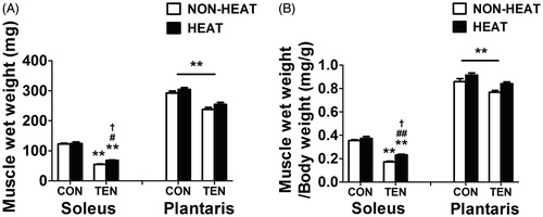
Initial body weight of rats used in this study was 296.0 ± 3.7 g. Eight days after tenotomy, the body weights of the TEN group were lower (p < 0.01) with respect to control groups (CON = 345.8 ± 2.2 g, CON + HEAT =333.7 ± 5.5 g, TEN = 316.4 ± 4.3 g, and TEN+HEAT =308.2 ± 5.2 g). Heat treatment did not have any apparent effect on the body weight of control and tenotomised rats.
Two-way ANOVA analysis revealed a significant main effect of TEN (p < 0.01) and a significant TEN × HEAT interaction (p < 0.05) on absolute soleus wet weight. Further, there were significant main effects of TEN (p < 0.01) and of HEAT (p < 0.01), and a significant TEN × HEAT interaction (p < 0.05) on the ratio of soleus wet weight to body weight. In plantaris muscle, however, there were significant main effects of TEN (p < 0.01) and of HEAT (p < 0.01) on these parameters. The absolute and ratio of soleus wet weights to body weight of the TEN group were 55.3% and 52.8%, respectively, less than that of the CON group. In the plantaris muscle, the absolute and ratio of wet weight to body weight were decreased by 18.7% and 10.5%, respectively, following tenotomy compared with control. Interestingly, however, heat treatment appeared to attenuate the tenotomy-induced losses of muscle weight in both muscles (), with a significant effect observed in soleus muscle. The absolute and ratio of muscle wet weights to body weight of the TEN+HEAT group were 24.0% and 26.1%, respectively, higher in soleus muscle and were 6.9% and 9.1%, respectively, higher in plantaris muscle compared with the TEN group.
Figure 2. Effects of heat stress and tenotomy on wet weight of soleus and plantaris muscles. (A) Absolute muscle wet weight. (B) Relative muscle wet weight normalised with body weight (n = 8 rats/group). **p < 0.01 vs. respective CON within each muscle; ##p < 0.01, #p < 0.05 vs. respective NON-HEAT within each muscle; and †p < 0.05 for TEN × HEAT interaction within each muscle.
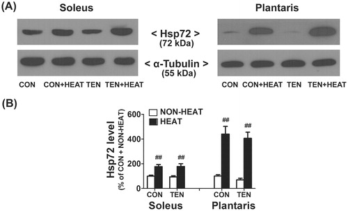
To determine whether tenotomy and whole body heat stress modulated HSP responses, the protein level of Hsp72, an inducible isoform of Hsp70, was measured as a marker of heat stress using Western blot analysis. There were no detectable differences in Hsp72 protein expression in both soleus and plantaris muscles of CON and TEN groups. Two-way ANOVA analysis indicated a significant main effect of HEAT (p < 0.01) on Hsp72 protein expression in both soleus and plantaris muscles. Compared to non-heated rats, heat exposure significantly increased of Hsp72 protein expression in both soleus and plantaris muscles of control and tenotomised rats (). Moreover, Hsp72 expression levels in the plantaris muscle were elevated by a greater magnitude than in soleus muscle after heat treatment.
Figure 3. Effects of heat stress and tenotomy on Hsp72 protein expression in soleus and plantaris muscles. (A) Representative Hsp72 protein expression using western blotting. (B) Quantified data of Hsp72. Hsp72 band density was normalised with α-tubulin (n = 8 rats/group). ##p < 0.01 vs. respective NON-HEAT within each muscle.
Effects of heat stress on muscle histology and CSA after tenotomy
illustrates the histological changes of both soleus and plantaris muscles after tenotomy without and with heat stress. In the CON and CON+HEAT groups, myofibres exhibited polygonal shape and were tightly packed to form muscle bundles with a small amount of ECM. In contrast, myofibres of the TEN group were apparently smaller in size and accompanied by enlargement of extracellular space and collagen accumulation in both muscles examined. In addition, there was an infiltration of inflammatory cells, which was more evident in the soleus muscle. Interestingly, while these morphological alterations were found in soleus muscle, the amount of collagen was relatively unaffected in the plantaris muscle after heat exposure.
Figure 4. Effects of heat stress and tenotomy on muscle morphology and fibre cross-sectional area (CSA) in soleus and plantaris muscles. (A) H&E staining of muscle cross-sections. Control (CON) and control with heat stress (CON+HEAT) muscles showing typical intact myofibres; tenotomised muscle (TEN) showing evidently variable sizes of myofibres and numerous inflammatory cells (white arrow) accumulation surrounding myofibres and extracellular space; muscle after tenotomy combined with heat stress (TEN+HEAT) illustrating few number of inflammatory cells infiltration. Scale bars = 50 µm; 200 × magnification and 1000 × magnification in insets. (B) Histogram of fibre CSA distribution (a total of 1906 fibres from CON, 1669 fibres from CON+HEAT, 1847 fibres from TEN, and 2042 fibres from TEN+HEAT of soleus muscle; 1884 fibres from CON, 1799 fibres from CON+HEAT, 2141 fibres from TEN, and 1870 fibres from TEN+HEAT of plantaris muscle). (C) Mean fibre CSA (n = 5 rats/group). **p < 0.01 vs. respective CON within each muscle.
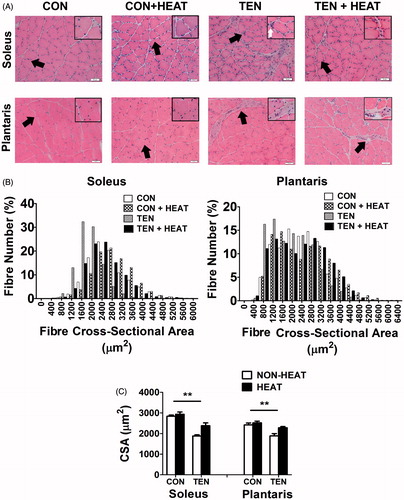
In corresponding to a decreased muscle wet weight, there was a leftward shift of myofibre area distribution curve in both muscles of the TEN group (). Two-way ANOVA analysis revealed significant main effects of TEN (p < 0.01) and of HEAT (p < 0.01, 0.05) on mean fibre CSA in both soleus and plantaris muscles. Tenotomy resulted in significantly decreased mean fibre CSA in both soleus (33.7%) and plantaris (22.1%) muscles compare to control. However, heat stress appeared to alleviate the tenotomy-induced reduction of CSA in both soleus (26.3%) and plantaris (20.9%) muscles ().
Heat stress reduces intramuscular collagen accumulation after tenotomy
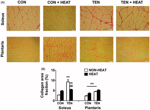
To quantify and compare the effect of heat stress on degree of fibrosis induced by tenotomy, picrosirius red staining was performed to visualise collagen distribution (). Intramuscular collagen was found to surround myofibres and muscle bundles in both soleus and plantaris muscle sections of CON and CON+HEAT groups. As expected, tenotomy markedly enhanced the accumulation of collagen surrounding nerves and blood vessels as well as endomysium in both types of muscles (). On the contrary, only a little collagen deposition was observed between the bundles of soleus muscle, but not in the plantaris muscle, of tenotomised rats after heat stress. Two-way ANOVA analysis indicated a significant main effect of TEN (p < 0.01) and a significant TEN × HEAT interaction (p < 0.01) on collagen area fraction in soleus muscle. There were significant main effects of TEN (p < 0.01) and of HEAT (p < 0.01) on collagen deposition in the plantaris muscle. Tenotomy resulted in significantly increased area fraction of collagen fibres in both soleus and plantaris muscles of TEN and TEN+HEAT groups compared to CON group. Heat treatment, however, attenuated the tenotomy-induced increased area fraction of collagen only in soleus muscle. Such a change was not observed in the plantaris muscle ().
Figure 5. Effects of heat stress and tenotomy on intramuscular collagen accumulation in soleus and plantrais muscles. (A) Picrosirius red-stained collagen fibres of control (CON), control with heat stress (CON+HEAT), tenotomy (TEN), and tenotomy combined with heat stress (TEN+HEAT), muscle fibres were stained with yellow, whilst collagen fibres were stained red. Scale bars = 100 µm. (B) Percentage of collagen area fraction (n = 4 rats/group). **p < 0.01 vs. respective CON within each muscle; ##p < 0.01 vs. respective NON-HEAT within each muscle; and ††p < 0.01 for TEN × HEAT interaction within each muscle.
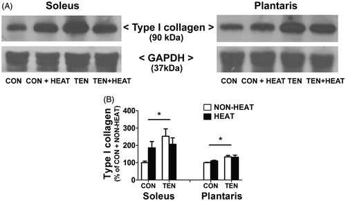
For more quantitative measurement, the amount of type I collagen, a major ECM component in skeletal muscle, was analysed by western blot analysis. Two-way ANOVA analysis revealed a significant main effect of TEN (p < 0.05), but there was no statistical interaction between TEN × HEAT interaction (p = 0.06, 0.41 in soleus and plantaris muscles, respectively) on type I collagen expression in both muscles. Tenotomy significantly increased (p < 0.05) type I collagen protein expression in both soleus and plantaris muscles compared to control. However, heat stress tended to attenuate this increase only in the soleus muscle, but not in plantaris muscle (). There was no significant difference in the extent of type I collagen in soleus muscle of control and tenotomised rats after heat treatment.
Figure 6. Effects of heat stress and tenotomy on type I collagen protein expression in soleus and plantaris muscles. (A) Representative type I collagen protein expression using western blotting. (B) Quantified data of type I collagen. Type I collagen band density was normalised with GAPDH (n = 5–6 rats/group). *p < 0.05 vs. respective CON within each muscle.
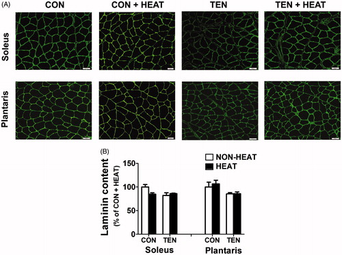
To further examine whether tenotomy and heat stress altered other ECM components, immunohistochemistry staining was performed to determine the laminin content, a major non-collagenous tissue of basement membrane responsible for the maintenance of membrane integrity. A two-way ANOVA analysis showed neither tenotomy nor heat stress affected the laminin contents in both soleus and plantaris muscles ().
Figure 7. Effects of heat stress and tenotomy on laminin content in soleus and plantrais muscles. (A) Immunofluorescence staining of laminin of control (CON), control combined with heat stress (CON+HEAT), tenotomy (TEN), and tenotomy combined with heat stress (TEN+HEAT). Scale bars = 50 µm. (B) Fluorescence intensity of laminin (n = 3–4 rats/group).
Heat stress modulates MMP-2, TIMP-2, and TGF-β1 expression after tenotomy
To determine the mechanism by which heat stress modulated intramuscular collagen turnover after tenotomy, the MMPs activities and TIMPs as well as TGF-β1 protein expression were examined.
In the present study, MMP-9 activity was not detectable in any samples in either soleus or plantaris muscles. However, three MMP-2 isoforms (i.e. pro, intermediate, and active) were identified in the gels (), except the active form of MMP-2 in the plantaris muscle. Two-way ANOVA analysis indicated a significant main effect of TEN (p < 0.01) on active and pro-MMP-2 activities in both muscles. Densitometric analysis revealed that tenotomy significantly increased the activity level of active MMP-2 (62 kDa) in soleus muscle. Interestingly, heat treatment had no additional effect on tenotomy-induced increased active MMP-2 activity. Similar results were observed for the activity of pro-MMP-2 (72 kDa) in both soleus and plantaris muscles of TEN and TEN+HEAT groups ().
Figure 8. Effects of heat stress and tenotomy on MMP-2 activities and TIMP-2 protein expression in soleus and plantaris muscles. (A) Representative MMP-9 and MMP-2 activities evaluated by gelatin zymography which determines the ability of MMPs to degrade gelatin substrate. (B) Quantified data of active form of MMP-2 and pro (latent) form of MMP-2 band density. (C) Representative TIMP-2 protein expression evaluated by western blotting. (D) Quantified data of TIMP-2. TIMP-2 band density was normalised with α-tubulin (n = 8 rats/group). **p < 0.01 vs. respective CON within each muscle, and †p < 0.05 for TEN × HEAT interaction within each muscle.
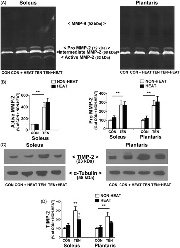
Furthermore, two-way ANOVA analysis revealed a significant main effect of TEN (p < 0.01) and a significant TEN × HEAT interaction (p < 0.05) on TIMP-2 protein expression, an endogenous inhibitor of MMP-2, only in soleus muscle. There was also a significant main effect of TEN (p < 0.01) on TIMP-2 protein expression in plantaris muscle. These data indicate that tenotomy significantly increased the expression of TIMP-2 protein in both soleus and plantaris muscles. However, heat stress attenuated this tenotomy-induced increased TIMP-2 protein expression was evident in soleus muscle ().
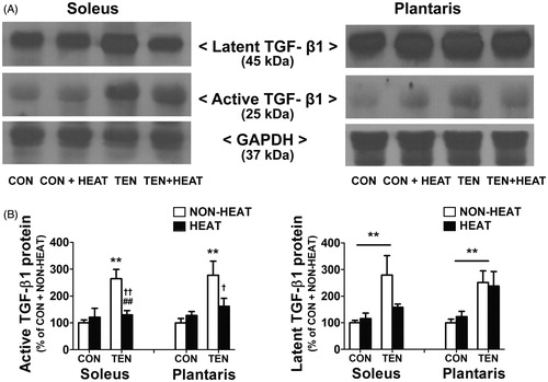
Two-way ANOVA indicated significant main effects of TEN (p < 0.05) and of HEAT (p < 0.01), and a significant TEN × HEAT interaction (p < 0.01) on the active TGF-β1 (25 kDa) protein expression in soleus muscle. In plantaris muscle, however, there was a significant main effect of TEN (p < 0.01) and a significant TEN × HEAT interaction (p < 0.05) on such parameter. Compared to CON group, the active TGF-β1 protein expression was significantly elevated (p < 0.01) in both soleus and plantaris muscles of TEN group. These increases were reversed by heat treatment (). Similar results were obtained for the protein expression of latent TGF-β1, except that no significant effect of heat stress was observed on tenotomised plantaris muscle. Thus, the expression level of latent TGF-β1 were increased 2.8 - and 2.5-fold, respectively, in tenotomised soleus and plantaris muscles compared to CON group, and were increased 1.4- and 1.9-fold, respectively, in TEN+HEAT group compared to the CON+HEAT group ().
Figure 9. Effects of heat stress and tenotomy on TGF-β1 protein expression in soleus and plantaris muscles. (A) Representative active (25 kDa) and latent (45 kDa) TGF-β1 protein expressions using western blotting. (B) Quantified data of active TGF-β1 and latent TGF-β1. TGF-β1 band density was normalised with GAPDH (n = 6–8 rats/group). **p < 0.01 vs. respective CON within each muscle; ##p < 0.01 vs. respective NON-HEAT within each muscle; and ††p < 0.01 and †p < 0.05 for TEN × HEAT interaction within each muscle.
Heat stress attenuates NF-κB activation after tenotomy in soleus muscle
NF-κB signalling is well known to play a key role in muscle disuse atrophy and inflammation [Citation30]. Generally, NF-κB, mainly the p50/p65 heterodimer, resides in the cytoplasm. When activated, NF-κB is translocated to the nucleus [Citation30] and subsequently regulates gene transcription of several inflammatory cytokines and profibrogenic activators including TGF-β1 [Citation31]. To determine whether heat stress may influence intramuscular fibrosis induced by tenotomy via the regulation of NF-κB, the level of phospho-NF-κB p65 in the nuclei was determined using immunohistochemistry. Two-way ANOVA analysis revealed a significant main effect of TEN (p < 0.01) on the level of phospho-NF-κB p65 in both muscles. The number of phospho-NF-κB p65 positive nuclei was up-regulated in both soleus and plantaris muscles following tenotomy compared with control. Although there was no statistical TEN × HEAT interaction (p = 0.16, 0.21 in soleus and plantaris muscles, respectively), heat exposure appeared to attenuate this increase in the soleus, but not in the plantaris muscles ().
Figure 10. Effects of heat stress and tenotomy on NF-κB translocation in soleus and plantrais muscles. (A) Immunofluorescence staining of phospho-NF-KB p65 (green) and DAPI (blue). Left panels, phospho-NF-κB p65 staining; middle panels, DAPI; right panels, merged image of phospho-NF-KB p65 and DAPI. The presence of NF-κB in the nucleus is indicated by arrows. Scale bars = 50 µm. (B) The number of NF-κB positive nuclei was normalised with total nuclei (n = 3–4 rats/group). **p < 0.01 vs. respective CON within each muscle.

Discussion
This study aimed to investigate the effect of heat stress on intramuscular fibrosis following tenotomy. Herein, we report for the first time that heat exposure reduced fibrosis induced by tenotomy in soleus but not in plantaris muscles and the effect was associated with suppression of TIMP-2 and TGF-β1 protein expressions, as well as an induction of Hsp72.
Tenotomy induces muscle atrophy and intramuscular fibrosis
As expected, transections of Achilles tendon for 8 days induced muscle atrophy in both soleus and plantaris muscles. However, the muscle loss was greater in a predominantly slow soleus muscle than in a predominantly fast plantaris muscle. The results were in line with the previous reports demonstrating that the antigravity muscle primarily composed of slow twitch fibres was more susceptible to atrophy after unloading compared to fast twitch muscles [Citation25,Citation32]. Furthermore, our finding showed that tenotomy induced a marked increase in intramuscular collagen accumulation measured by picrosirius red staining in the perimysium and endomysium of both muscles. Eight days after tenotomy, the collagen area fraction was more prominent in soleus (2.3-fold) than in plantaris muscles (0.9-fold) compared with the control. In parallel with this finding, type I collagen protein expression was highly increased (2.5-fold) in soleus muscle, but moderately increased (1.3-fold) in plantaris muscle following tenotomy. These results were in agreement with previous studies [Citation5,Citation25]. Jozsa et al. [Citation5], using scanning electron microscopy, found that tenotomy or immobilisation resulted in an increase of collagen content in rat skeletal muscles with a greater extent in soleus than in gastrocnemius muscles. Cross-section of supraspinatus muscle following rotator cuff tears also showed increased expressions of collagen type I and III [Citation33]. Moreover, microarray gene analysis displayed a differential response in the transcriptional regulation of collagen encoding gene (i.e. COL3A) in which the highest expression was observed in red anterior latissimus dorsi, followed by white posterior latissimus dorsi and mixed-phenotype biceps femoris muscles, respectively [Citation34]. Collectively, these data indicate that the intramuscular fibrosis following tenotomy is fibre type-specific.
Since the basement membrane associated with endomysium layer plays a crucial role in myofibre protection, we next determined whether tenotomy would affect membrane structural integrity. Surprisingly, immunofluorescence staining of the major component of the basement membrane of tenotomised soleus and plantaris muscles revealed no alterations of laminin content compared with the control muscles. This finding was in contrast with that of Zhang et al. [2013], who found a significant degeneration of laminin 2 weeks following tenotomy [Citation3]. The possible reason for this discrepancy may stem from the difference in time-scale of the study, i.e. we observed the effect only at 8 days after tenotomy.
Tenotomy enhances MMP-2 activity and increases TIMP-2 as well as TGF-β1 protein expressions in slow and fast muscles
Skeletal muscle ECM turnover or remodelling is regulated by a delicate balance between collagen synthesis and its degradation [Citation1]. With regard to ECM degradation, MMP-2 and MMP-9 are thought to play a crucial role in muscle adaptation due to altered uses and/or injuries [Citation10,Citation12]. Our finding that the MMP-2 activity was dramatically increased in both soleus and plantaris muscles 8 days after tenotomy corroborated the previous reports [Citation10,Citation13]. In contrast to MMP-2, MMP-9 activity was not detectable in all muscle samples in our study. This is not surprising, however, given that MMP-9 activity was increased only under extreme inflammation, chronic denervation, or satellite cells activation [Citation12,Citation27]. Thus, it is possible that tenotomy induced inflammation to a lesser extent [Citation35] compared with other muscle pathologies, i.e. Duchenne muscular dystrophy [Citation12].
The proteolytic activity of MMP-2 is tightly regulated by its endogenous inhibitors. Although many TIMPs have been identified, previous studies demonstrated that TIMP-2 expression was induced in response to muscle damage in both slow and fast fibres [Citation27,Citation36]. In this study we found that MMP-2 activity and TIMP-2 protein expression were increased, although at different magnitudes, following tenotomy in both muscles. TIMP-2 has a unique biological characteristic not only in MMP-2 inhibition, but also in selectively interacting with MMP-14 (MT1-MMP) to activate MMP-2 at cell surface in the presence of low concentrations of TIMP-2 [Citation8]. Since TIMP-2 is capable of inhibiting MMP-2 activation possibly through a coordination of Zn2+ at the active site of MMP-2 [Citation7], therefore, up-regulation of TIMP-2 production or low MMP-2/TIMP-2 ratio observed 8 days after tenotomy may contribute to decreased MMP-2 activity and eventually lead to the increased collagen deposition in the present study.
TGF-β1 is considered to be a potent regulator of fibrosis in various tissues including skeletal muscle [Citation37]. Its level was up-regulated in several animal models of muscle wasting and/or injuries [Citation38]. Our finding that both active and latent forms of TGF-β1 protein levels were significantly increased following tenotomy in both soleus and plantaris muscles similarly to those of others [Citation37,Citation38] and support our hypothesis. Recent studies have also demonstrated that an over-expression of TGF-β1 in mice resulted in fibrosis in both skeletal and cardiac muscles [Citation18,Citation39]. More importantly, TGF-β1 has been reported to increase the expression of TIMP-2 [Citation39,Citation40]. Taken together, these synergistic increases in TGF-β1 and TIMP-2 observed could promote the development of intramuscular fibrosis in both muscles 8 days following tenotomy.
Heat stress alleviates tenotomy-induced muscle atrophy and intramuscular fibrosis
HSPs have been shown to confer a protection against muscle damage and prevent skeletal muscle atrophy following injury and/or mechanical unloading [Citation28,Citation41]. Consistent with the previous findings [Citation28], we confirmed that heat stress could preserve muscle mass more in a slow-twitch muscle than in a fast-twitch muscle 8 days following tenotomy. The reason for this phenomenon appears to be related to a differential regulation of Hsp72 expression [Citation26,Citation28]. Under normal conditions the basal Hsp72 level was highly expressed in soleus muscle compared to that in plantaris muscle, supporting previous findings [Citation42]. This may, in part, reflect its antigravity function [Citation42]. In addition, Oishi et al. [Citation26,Citation43] found that Hsp72 was rapidly increased in a slow-twitch fibre more than in a fast-twitch fibre after heat stress. The effect may be attributed to the high rate of protein turnover observed in slow muscle [Citation44].
Another major finding in the present study was the demonstration that heat stress attenuated the collagen accumulation of tenotomised muscles only in the soleus but not in the plantaris muscles. Accumulating evidence indicates that several inflammatory cells, particularly macrophages, have been implicated in the pathogenesis of fibrosis in many tissues [Citation33,Citation35,Citation45]. Since tenotomy resulted in an infiltration of several inflammatory cells around small muscle fibres and extracellular spaces compared to control muscles () and that these alterations were associated with collagen deposition in the muscle studied, it is possible that heat stress could reduce the excessive collagen accumulation, at least in part, through modulating the inflammatory process. This hypothesis is supported by a recent study showing that direct injection of recombinant Hsp70 protein into Tibialis anterior muscle of Hsp70 knockout mice after injury hastened the inflammatory response by increasing inflammatory cell infiltration at the site of injury, thereby facilitating the repair process [Citation46]. Conversely, ablation of Hsp70 in mice caused an impairment of myofibre regeneration, a smaller CSA of regenerating fibres, and a wider extracellular space after a 10-day reloading period compared with those of wild-type mice [Citation46]. Altogether, these data suggest that heat stress could facilitate the inflammatory process and subsequently promote ECM remodelling in skeletal muscle after tenotomy. However, further studies are required to better understand this immune-modulating effect of heat stress on muscle fibrosis.
In addition, heat stress has been reported to attenuate several fibrotic markers, including, α-smooth muscle actin (α-SMA), type I collagen, TGF-β1, and TIMP expression in many animal models of fibrosis [Citation21,Citation22]. Thus, it is likely that heat exposure could alleviate tenotomy-induced intramuscular fibrosis by reducing the activation of TGF-β1 and TIMP-2. The molecular mechanism whereby heat stress suppresses TGF-β1 expression may involve multiple steps ranging from the regulation of synthesis or releasing of the pro-TGF-β1 form to the activation of pro-TGF-β1 to its active form. TGF-β1 is typically synthesised by macrophages and fibroblasts [Citation15,Citation16] in the form of latent TGF-β1 complex. TGF-β1 activity is controlled by the liberation of active TGF-β1 form, which, in turn, binds to its receptor and turns on TGF-β1 signalling responses [Citation16,Citation47]. In this study, however, we found that both the latent and active forms of TGF-β1 protein levels of tenotomised muscles were remarkably decreased to control level following heat treatment. This finding points to the transcriptional control of TGF-β1 by heat exposure. To probe this possibility, we further investigated whether NF-κB, a transcriptional factor of pro-TGF-β1 [Citation31], was inhibited in tenotomised muscles following heat exposure. Interestingly, our immunofluorescence staining showed that the number of phospho-NF-κB p65 positive nuclei was up-regulated after tenotomy compared to control muscles, and this increase was attenuated in soleus muscle by heat stress. Consistent with this finding, Senf et al. [Citation41] directly demonstrated that over-expression of Hsp70 could abolish NF-κB activation in rat soleus muscle following immobilisation. These data suggest that heat stress may reduce TGF-β1 protein expression possibly through the effect on the NF-κB pathway.
Lastly, we found that whole body heat stress reduced the tenotomy-induced increased TIMP-2 protein expression to nearly control level, while it did not affect the MMP-2 activity in either soleus or plantaris muscles. This suggests that a reduction of TIMP-2 production may give rise to the increased MMP-2 activation, thereby leading to reduced intramuscular collagen deposition in tenotomised soleus muscle. Nevertheless, a lack of such effect on intramuscular collagen in the plantaris muscle was unexpected. Clearly, a further study is required to clarify this differential response.
There are several limitations in the present study. Firstly, although heat stress-induced Hsp72 protein expression was associated with the extent of intramuscular collagen accumulation, other HSPs were also up-regulated after heat stress [Citation26,Citation48]. Therefore, the potential therapeutic effect of these HSPs could not be ruled out. Secondly, the animals in the control without heat stress group had no experience of daily isoflurane administration and this may affect our results especially on HSP expression. However, previous study clearly demonstrated that this anaesthesia had no affected on Hsp70 expression [Citation49].
Conclusion
We demonstrated that whole body heat stress attenuated tenotomy-induced intramuscular fibrosis by improving ECM remodelling through modulating the MMP-2/TIPM-2 ratio and decreasing the TGF-β1 protein expression and that this was associated with the induction of Hsp72 expression. The results from this study provide an important mechanistic insight into the benefits of heat stress on the muscle ECM remodelling after tenotomy.
Acknowledgements
Special thanks are given to Chumpol Pholpramool for reading this manuscript. We also thank the Olympus Bioimaging Center, Faculty of Science, Mahidol University for technical support.
Declaration of interest
The authors thank the Office of the Higher Education Commission, Thailand for providing a grant support via the Strategic Scholarships for Frontier Research Network for the Joint PhD Program Thai Doctoral degree for this research. The authors alone are responsible for the content and writing of the paper.
References
- Davis ME, Gumucio JP, Sugg KB, Bedi A, Mendias CL. MMP inhibition as a potential method to augment the healing of skeletal muscle and tendon extracellular matrix. J Appl Physiol 2013;115:884–91
- Gillies AR, Lieber RL. Structure and function of the skeletal muscle extracellular matrix. Muscle Nerve 2011;44:318–31
- Zhang Q, Joshi SK, Manzano G, Lovett DH, Kim HT, Liu X. Muscle extracellular matrix degradation and contractibility following tendon rupture and disuse. Muscles Ligaments Tendons J 2013;3:35–41
- Zhang C, Gao Y. Effects of aging on the lateral transmission of force in rat skeletal muscle. J Biomech 2014;47:944–8
- Jozsa L, Kannus P, Thoring J, Reffy A, Jarvinen M, Kvist M. The effect of tenotomy and immobilisation on intramuscular connective tissue. A morphometric and microscopic study in rat calf muscles. J Bone Joint Surg Br 1990;72:293–7
- Slimani L, Micol D, Amat J, Delcros G, Meunier B, Taillandier D, et al. The worsening of tibialis anterior muscle atrophy during recovery post-immobilization correlates with enhanced connective tissue area, proteolysis, and apoptosis. Am J Physiol Endocrinol Metab 2012;303:E1335–47
- Kandasamy AD, Chow AK, Ali MAM, Schulz R. Matrix metalloproteinase-2 and myocardial oxidative stress injury: Beyond the matrix. Cardiovasc Res 2009;85:413–23
- Brew K, Dinakarpandian D, Nagase H. Tissue inhibitors of metalloproteinases: Evolution, structure and function. Biochim Biophys Acta 2000;1477:267–83
- Alameddine HS. Matrix metalloproteinases in skeletal muscles: Friends or foes? Neurobiol Dis 2012;48:508–18
- Giannelli G, De Marzo A, Marinosci F, Antonaci S. Matrix metalloproteinase imbalance in muscle disuse atrophy. Histol Histopathol 2005;20:99–106
- Zimowska M, Brzoska E, Swierczynska M, Streminska W, Moraczewski J. Distinct patterns of MMP-9 and MMP-2 activity in slow and fast twitch skeletal muscle regeneration in vivo. Int J Dev Biol 2008;52:307–14
- Kherif S, Lafuma C, Dehaupas M, Lachkar S, Fournier JG, Verdiere-Sahuque M, et al. Expression of matrix metalloproteinases 2 and 9 in regenerating skeletal muscle: A study in experimentally injured and mdx muscles. Dev Biol 1999;205:158–70
- Skittone LK, Liu X, Tseng A, Kim HT. Matrix metalloproteinase-2 expression and promoter/enhancer activity in skeletal muscle atrophy. J Orthop Res 2008;26:357–63
- Bialek P, Morris C, Parkington J, St Andre M, Owens J, Yaworsky P, et al. Distinct protein degradation profiles are induced by different disuse models of skeletal muscle atrophy. Physiol Genomics 2011;43:1075–86
- Wahl SM, McCartney-Francis N, Allen JB, Dougherty EB, Dougherty SF. Macrophage production of TGF-beta and regulation by TGF-beta. Ann N Y Acad Sci 1990;593:188–96
- Lawrence DA, Pircher R, Kryceve-Martinerie C, Jullien P. Normal embryo fibroblasts release transforming growth factors in a latent form. J Cell Physiol 1984;121:184–8
- Leask A, Abraham DJ. TGF-beta signaling and the fibrotic response. FASEB J 2004;18:816–27
- Narola J, Pandey SN, Glick A, Chen YW. Conditional expression of TGF-beta1 in skeletal muscles causes endomysial fibrosis and myofibers atrophy. PLoS One 2013;8:e79356
- Zanotti S, Gibertini S, Savadori P, Mantegazza R, Mora M. Duchenne muscular dystrophy fibroblast nodules: A cell-based assay for screening anti-fibrotic agents. Cell Tissue Res 2013;352:659–70
- Taniguti AP, Pertille A, Matsumura CY, Santo Neto H, Marques MJ. Prevention of muscle fibrosis and myonecrosis in mdx mice by suramin, a TGF-beta1 blocker. Muscle Nerve 2011;43:82–7
- Wakisaka O, Takahashi N, Shinohara T, Ooie T, Nakagawa M, Yonemochi H, et al. Hyperthermia treatment prevents angiotensin II-mediated atrial fibrosis and fibrillation via induction of heat-shock protein 72. J Mol Cell Cardiol 2007;43:616–26
- Hagiwara S, Iwasaka H, Matsumoto S, Noguchi T, Yoshioka H. Association between heat stress protein 70 induction and decreased pulmonary fibrosis in an animal model of acute lung injury. Lung 2007;185:287–93
- Takahashi KA, Tonomura H, Arai Y, Terauchi R, Honjo K, Hiraoka N, et al. Hyperthermia for the treatment of articular cartilage with osteoarthritis. Int J Hyperthermia 2009;25:661–7
- Takeuchi K, Hatade T, Wakamiya S, Fujita N, Arakawa T, Miki A. Heat stress promotes skeletal muscle regeneration after crush injury in rats. Acta Histochem 2013;116:327–34
- Jamali AA, Afshar P, Abrams RA, Lieber RL. Skeletal muscle response to tenotomy. Muscle Nerve 2000;23:851–62
- Oishi Y, Taniguchi K, Matsumoto H, Ishihara A, Ohira Y, Roy RR. Muscle type-specific response of HSP60, HSP72, and HSC73 during recovery after elevation of muscle temperature. J Appl Physiol 2002;92:1097–103
- Koskinen SO, Ahtikoski AM, Komulainen J, Hesselink MK, Drost MR, Takala TE. Short-term effects of forced eccentric contractions on collagen synthesis and degradation in rat skeletal muscle. Pflugers Arch 2002;444:59–72
- Selsby JT, Dodd SL. Heat treatment reduces oxidative stress and protects muscle mass during immobilization. Am J Physiol Regul Integr Comp Physiol 2005;289:R134–9
- Tolson JK, Roberts SM. Manipulating heat shock protein expression in laboratory animals. Methods 2005;35:149–57
- Jackman RW, Cornwell EW, Wu CL, Kandarian SC. Nuclear factor-kappaB signalling and transcriptional regulation in skeletal muscle atrophy. Exp Physiol 2013;98:19–24
- Bowie A, O'Neill LA. Oxidative stress and nuclear factor-kappaB activation: A reassessment of the evidence in the light of recent discoveries. Biochem Pharmacol 2000;59:13–23
- Yimlamai T, Dodd SL, Borst SE, Park S. Clenbuterol induces muscle-specific attenuation of atrophy through effects on the ubiquitin-proteasome pathway. J Appl Physiol 2005;99:71–80
- Gumucio JP, Davis ME, Bradley JR, Stafford PL, Schiffman CJ, Lynch EB, et al. Rotator cuff tear reduces muscle fiber specific force production and induces macrophage accumulation and autophagy. J Orthop Res 2012;30:1963–70
- Nierobisz LS, Sporer KR, Strasburg GM, Reed KM, Velleman SG, Ashwell CM, et al. Differential expression of genes characterizing myofibre phenotype. Anim Genet 2012;43:298–308
- Baker JH. Segmental necrosis in tenotomized muscle fibers. Muscle Nerve 1983;6:29–39
- Koskinen SO, Wang W, Ahtikoski AM, Kjaer M, Han XY, Komulainen J, et al. Acute exercise induced changes in rat skeletal muscle mRNAs and proteins regulating type IV collagen content. Am J Physiol Regul Integr Comp Physiol 2001;280:R1292–300
- Gosselin LE, Williams JE, Deering M, Brazeau D, Koury S, Martinez DA. Localization and early time course of TGF-beta 1 mRNA expression in dystrophic muscle. Muscle Nerve 2004;30:645–53
- Burks TN, Cohn RD. Role of TGF-beta signaling in inherited and acquired myopathies. Skelet Muscle 2011;1:19
- Seeland U, Haeuseler C, Hinrichs R, Rosenkranz S, Pfitzner T, Scharffetter-Kochanek K, et al. Myocardial fibrosis in transforming growth factor-beta(1) (TGF-beta(1)) transgenic mice is associated with inhibition of interstitial collagenase. Eur J Clin Invest 2002;32:295–303
- Edwards DR, Murphy G, Reynolds JJ, Whitham SE, Docherty AJ, Angel P, et al. Transforming growth factor beta modulates the expression of collagenase and metalloproteinase inhibitor. EMBO J 1987;6:1899–904
- Senf SM, Dodd SL, McClung JM, Judge AR. Hsp70 overexpression inhibits NF-kappaB and Foxo3a transcriptional activities and prevents skeletal muscle atrophy. FASEB J 2008;22:3836–45
- Locke M, Noble EG, Atkinson BG. Inducible isoform of Hsp70 is constitutively expressed in a muscle fiber type specific pattern. Am J Physiol 1991;261:C774–9
- Oishi Y, Taniguchi K, Matsumoto H, Ishihara A, Ohira Y, Roy RR. Differential responses of HSPs to heat stress in slow and fast regions of rat gastrocnemius muscle. Muscle Nerve 2003;28:587–94
- Booth FW, Thomason DB. Molecular and cellular adaptation of muscle in response to exercise: Perspectives of various models. Physiol Rev 1991;71:541–85
- Spencer M, Yao-Borengasser A, Unal R, Rasouli N, Gurley CM, Zhu B, et al. Adipose tissue macrophages in insulin-resistant subjects are associated with collagen VI and fibrosis and demonstrate alternative activation. Am J Physiol Endocrinol Metab 2010;299:E1016–27
- Senf SM, Howard TM, Ahn B, Ferreira LF, Judge AR. Loss of the inducible Hsp70 delays the inflammatory response to skeletal muscle injury and severely impairs muscle regeneration. PLoS One 2013;8:e62687
- Dubois CM, Laprise MH, Blanchette F, Gentry LE, Leduc R. Processing of transforming growth factor beta 1 precursor by human furin convertase. J Biol Chem 1995;270:10618–24
- Oguro A, Sakurai T, Okuno M, Nagata K, Atomi Y. The change of Hsp47, collagen specific molecular chaperone, expression in rat skeletal muscle may regulate collagen production with gravitational conditions. Biol Sci Space 2004;18:150–1
- Yamasaki A, Takahashi T, Suzuki T, Fujiwara T, Hirakawa M, Ohmori E, et al. Differential effects of isoflurane and halothane on the induction of heat shock proteins. Biochem Pharmacol 2001;62:375–82