
Abstract
Purpose: A variety of medications and procedures are available for the treatment of warts, but it appeared the treatment response in systemic lupus erythematosus (SLE) patients is poor. It is necessary to investigate the feasibility, safety and efficacy of local thermotherapy for extensive viral warts. Materials and methods: A SLE patient on systemic steroid developed extensive viral warts on both her hands and feet for months. She had a high score of SLE Disease Activity Index (SLEDAI), up to 30, and was extensively treated with high and prolonged dosage of corticosteroid and intermittent use of cyclophosphamide. We applied local hyperthermia at 44 °C on a target lesion for 30 min on days 1, 2, 3, 17, 18, a protocol which has been successfully used in treating viral warts. There was no sign of clinical response in a 3-month follow-up. Then we treated the patient on a once-a-week protocol. Result: All the lesions cleared in ten weeks and there was no sign of recurrence. Conclusion: This observation suggests that more intensive local hyperthermia is required for clearing viral warts in SLE.
Introduction
Viral wart is a common skin condition caused by human papillomavirus (HPV) infection. Typical warts appear as hyperkeratotic papules or plaques that most often inflict hands (verruca vulgaris) and feet (verruca plantaris). Cutaneous warts are mainly associated with HPV type 1/2/27/57, among a hundred-plus HPV types [Citation1]. The choice of treatment for viral warts can be immunomodulatory, ablative or cytotoxic, such as repeated topical application of imiquimod, trichloroacetic acid and podophyllin, cryotherapy, laser ablation, and surgery. Susceptibility and persistence of HPV infection may be increased in patients with lupus, which is mainly caused by failure to induce an effective cellular immune response [Citation2].
Regional hyperthermia has been used to treat infectious diseases and cancers, but the efficacy was variable [Citation3]. Also, mild local hyperthermia has been used in treating verruca plantaris, verruca vulgaris and condyloma acuminata. The remarkable advantage of the method was that the patients tolerated the procedure well and complications were negligible, major complications included heat-induced swelling, bullae formation and post-treatment hyperpigmentation. Choice of treatment for patients with extensive warts and associated with immunosuppressed conditions should be more cautious, for the possibility of treatment-induced secondary infection, prolonged healing time and even the exacerbation of complicated diseases. We recently successfully treated a series of patients with extensive genital warts complicated with diabetes mellitus or pregnancy, conditions that are associated with suppression of cellular immunity [Citation4,Citation5]. Here we diagnosed a SLE patient with extensive warts. We treated the warts by local hyperthermia with a prolonged course. The trial was approved by the Ethics Committee of China Medical University.
Case presentation
A 33-year-old female was referred to our outpatient clinic in January 10, 2013. She noticed some rough warty lesions on her feet and fingers over the past months, which grew in size and number gradually. She was advised by her community doctor to take transfer factor capsules (6 mg, twice a day) for 3 months but to no avail. Physical examination revealed extensive fleshy, skin-coloured or dark-grey papules or coalesced plaques in the arch, sole, heel, toe, toe gap, and fingers. Size of the lesions ranged approximately 0.2 × 0.2 cm to 3.0 × 3.0 cm (). HPV type 2 was identified from the scrapes of a lesion on the sole of her right foot by polymerase chain reaction (PCR) (). In the study we used a patented hyperthermia device with a light source from a tungsten-halogen lamp (Patent No. ZL 200720185403.3, China Medical University, China), most wavelengths (>90%) were from 760 to 2300 nm, with a peak wavelength at 1200 nm (data supplied by the lamp supplier). For this patient we chose a confluent plaque on her right sole as the target lesion. The target lesion was 2 × 2 × 0.5 cm in size and subjected to local hyperthermia at 44 °C for 30 min/day on days 1, 2, 3, 17, 18, as described [Citation6,Citation7].In a 3-month follow-up, there was no obvious decrease in the size of the lesions. Then we treated the patient on a once-a-week protocol at 44 °C for 30 min/day on the same target lesion for ten weeks when all the warts, either targeted or non-targeted, disappeared completely ( ). During the regression of the lesions she felt intense pruritus on all the lesions (both target and un-targeted lesions). The patient was free of the warts at 1-year follow-up.
Figure 1. Extensive viral warts in a 33-year-old woman with SLE. Warty lesions before treatment (a, b, c), complete clearance of lesions ten weeks after intensive hyperthermia (d, e, f ). The targeted site is marked with a blue arrow.
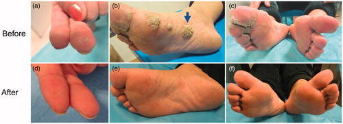
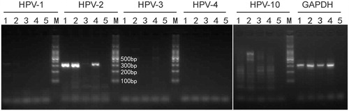
Figure 2. DNA was extracted from the scrapes of a lesion on the sole of her right foot. DNA segments specific for HPV type 2. It was identified by PCR which included HPV type 1, 2, 3, 4, 10 specific primers. The electrophoresis strip 1 represents the target lesion before treatment. Strip 2 represents the target lesion which was not noticeably improved after 2 months of treatment. Strip 3 represents the target lesion when all lesions were noticeably improved. Strip 4 represents the lesions except the target lesion when all lesions were noticeably improved. Strip 5 represents H2O.
The patient was diagnosed as SLE seven years ago. At the time of diagnosis, she had malar rash, pericardial and pleural effusion. Laboratory tests showed positive anti-nuclear, anti-Smith and anti-dsDNA antibodies. In May 2011 she was hospitalised due to leg oedema and headache, and diagnosed as relapsing SLE with a SLE Disease Activity Index (SLEDAI) score of 30. Cyclophosphamide pulse therapy, with intravenous injection of 0.6 g and 0.4 g on the first and second day in the week, was given on a 2-week basis for four cycles, on a monthly basis for the following 4 months, then on a 2-month basis for half a year. She also received concomitant intravenous pulsed methylprednisolone at an initial dosage of 500 mg and gradually maintained at 10 mg/day over a 6-month period. This was when the warty lesions started to appear.
Discussion
As compared to the general population, SLE patients display a higher prevalence of HPV infection [Citation8]. Our case had a high score of SLEDAI up to 30. She was extensively treated with high and prolonged dosage of corticosteroid and intermittent use of cyclophosphomide. Korkmaz et al. [Citation8] stated that the high prevalence of cutaneous warts in patients with LE is probably due to defects in some immune mechanisms, independently of immunosuppressive drugs. Johansson et al. [Citation9] suggested that the corticosteroid and antimalarial drugs used to treat SLE did not influence the frequency of warts. However, Klumb et al. [Citation10] demonstrated that SLE patients have a higher risk of presenting HPV infection, which is associated with the use of immunosuppressors.
Types of HPV that inflicted the skin may affect the natural course and treatment response. About 80–90% of HPV infection may clear in a couple of years, though a minor group of patients may have persistent infection and some even got malignant transformation. A study by Bruggink et al. observed that plantar warts induced by HPV type 1 were more prone to self-healing, and more responsive to cryotherapy and 40% topical salicylic acid painting, as compared to those induced by HPV type 2/27/57 [Citation1]. Our case harboured HPV 2, a factor that might have contributed to the protracted course of the disease.
Conclusion
There have been several reports of local hyperthermia being effective in the treatment of viral warts [Citation7,Citation11,Citation12]. This case study strongly suggests that local hyperthermia could help the body to build antiviral immunity. Local hyperthermia could promote migrational maturation of Langerhans cells [Citation13], stimulate cytotoxic and apoptotic effects [Citation14], and induce production of endogenous interferon in viral warts [Citation15].
Acknowledgement
Written informed consent was obtained from the patient for submission and publication of this case report and accompanying images. Yi Ren and Wei Huo contributed equally to this work.
Declaration of interest
This work was supported by the Public Welfare Programme, Ministry of Health, China (grant number 201202013); and the Innovative Research Team in Universities, Liaoning Bureau of Education (grant number LT2012012). The authors alone are responsible for the content and writing of the paper.
References
- Bruggink SC, Gussekloo J, de Koning MN, Feltkamp MC, Bavinck JN, Quint WG, et al. HPV type in plantar warts influences natural course and treatment response: secondary analysis of a randomised controlled trial. J Clin Virol 2013;57:227–32
- Yu SL, Chan PK, Wong CK, Szeto CC, Ho SC, So K, et al. Antagonist-mediated down-regulation of Toll-like receptors increases the prevalence of human papillomavirus infection in systemic lupus erythematosus. Arthritis Res Ther 2012;14:R80
- Ishikawa T, Kokura S, Sakamoto N, Ando T, Imamoto E, Hattori T, et al. Phase II trial of combined regional hyperthermia and gemcitabine for locally advanced or metastatic pancreatic cancer. Int J Hyperthermia 2012;28:597–604
- Huo W, Li GH, Qi RQ, Zhang L, Yan XX, Chen HD, et al. Clinical and immunologic results of local hyperthermia at 44 °C for extensive genital warts in patients with diabetes mellitus. Int J Hyperthermia 2013;29:17–20
- Huo W, Di ZH, Xiao BH, Qi RQ, Weiland M, Gao XH. Clearance of genital warts in pregnant women by mild local hyperthermia: A pilot report. Dermatol Ther 2014;27:109–12
- Kirnbauer R, Lenz P. Human Papillomaviruses. In: Bologia JL, Jorizzo JL, Schaffer JV, editors Dermatology (Third Edition). Volume 2. Elsevier Limited, 2012. pp 1303–19
- Huo W, Gao XH, Sun XP, Qi RQ, Hong Y, McHepange UO, et al. Local hyperthermia at 44 °C for the treatment of plantar warts: A randomized, patient-blinded, placebo-controlled trial. J Infect Dis 2010;201:1169–72
- Korkmaz C, Urer SM. Cutaneous warts in patients with lupus erythematosus. Rheumatol Int 2004;24:137–40
- Johansson E, Pyrhonen S, Rostila T. Warts and wart virus antibodies in patients with systemic lupus erythematosus. Br Med J 1977;1:74–6
- Klumb EM, Pinto AC, Jesus GR, Araujo M, Jr Jascone L, Gayer CR, et al. Are women with lupus at higher risk of HPV infection? Lupus 2010;19:1485–91
- Ma Y, Huo W, Hong YX, Chen HD, Gao XH. Successful clearance of facial common warts by local hyperthermia: Report of two cases. Dermatol Ther 2012;25:386–8
- Li X, Zhang C, Hong Y, Zhang D, Wei H, Chen HD, et al. Local hyperthermia treatment of extensive viral warts in Darier disease: A case report and literature review. Int J Hyperthermia 2012;28:451–5
- Ginhoux F, Liu K, Helft J, Bogunovic M, Greter M, Hashimoto D, et al. The origin and development of nonlymphoid tissue CD103+ DCs. J Exp Med 2009;206:3115–30
- Wang X, Gao XH, Li X, Hong Y, Qi R, Chen HD, et al. Local hyperthermia induces apoptosis of keratinocytes in both normal skin and condyloma acuminata via different pathways. Apoptosis 2009;14:721–8
- Zhu LL, Gao XH, Qi R, Hong Y, Li X, Wang X, et al. Local hyperthermia could induce antiviral activity by endogenous interferon-dependent pathway in condyloma acuminata. Antiviral Res 2010;88:187–92