
Abstract
Purpose: The aim of this paper was to introduce a new mechanism of thermal sensitivity in nanocarriers that results in a relatively low drug release at physiological temperature and rapid release of the encapsulated drug at hyperthermia and thermal ablation temperature range (40–60 °C). Materials and methods: The nanocarriers were synthesised by coating mesoporous silica nanoparticles with a thin layer of polyacrylamide. The low gelation temperature of the protective shell provides preferred routes for drug diffusion when the nanocarriers are heated within the hyperthermia temperature range. In order to determine the gelation point of polyacrylamide shell, differential scanning calorimetry was used. Various chemical, morphological, thermal, as well as drug loading capacities of these nanocarriers were characterised and their drug release behaviour was examined using magnetic resonance -guided focused ultrasound (MRgFUS). Results: Drug release measurements at different temperatures using doxorubicin showed 11.5 ± 2.4% leakage in aqueous solution at 37 °C after 30 min, while this value was significantly increased to 67.6 ± 2.5% at 60 °C. A 39.2 ± 2.2% release of doxorubicin was also obtained due to the sonication of drug-loaded nanoparticles for 5 × 20 s using MRgFUS. Conclusion: The nanocarriers developed do not exhibit a sharp transition temperature. However, a relatively high loading efficiency as well as rapid drug release at thermal ablation temperature range makes these nanostructures promising candidates for application as adjuvants to various thermal modalities such as radiofrequency and high intensity focused ultrasound.
Introduction
Utilisation of thermosensitive carriers is an effective strategy to protect healthy tissues from the negative effects of chemotherapeutic drugs as well as to obtain a synergistic effect of hyperthermia and chemotherapy [Citation1]. These carriers transfer the chemotherapeutic drugs through the body and release them specifically in the desired region which is simultaneously heated by a thermal modality [Citation1,Citation2]. In addition to site-specific targeting and reduced side effects, encapsulation of free drugs in these nanoparticles could prolong their half-life and therapeutic efficacy [Citation3].
The active release of encapsulated drug from thermosensitive nanostructures mostly relies on thermally triggered mechanisms including membrane disruption, coil-globule transition, and micellisation (or sol-gel behaviour). Membrane disruption is the most investigated mechanism which is often seen in lipid-based drug-carriers such as traditional thermosensitive liposomes (TTSLs), lysolipid-containing thermosensitive liposomes (LTSLs), polymer-modified thermosensitive liposomes, magnetoliposomes, and bubble-generating thermosensitive liposomes. For instance, drug release from lysolipid-containing thermosensitive liposomes (LTSLs) occurs by generation of stabilised defects in lipid membrane due to the micellisation of lysolipids at temperatures near 39–40 °C. These defects further provide preferred routes for rapid release of encapsulated drug at hyperthermia temperatures [Citation4–8]. The LTSL formulations that have undergone pharmaceutical development and are currently under clinical development (i.e. ThermoDox®) [Citation4], can release their contents in a few seconds after exposure to the increased temperatures and delay the tumour growth [Citation9,Citation10]. However, a number of studies have demonstrated the rapid desorption of lysolipids from LTSLs when administered in vivo, resulting in reduced thermal sensitivity and premature drug leakage at physiological conditions [Citation11–14]. There was also indirect evidence that lysolipids dissociate from LTSL upon dilution [Citation12].
The coil-globule transition is mostly seen in polymers with lower critical solution temperature (LCST) such as isopropylacrylamide-based polymers [Citation15–21]. The increased hydrophobicity of these polymers upon slight heating above physiological temperatures results in a thermo-induced conformational change from the swollen hydrophilic coils to the shrunken hydrophobic globules which provide a mechanical pressure to squeeze the encapsulated drug out of the particles [Citation16,Citation22,Citation23]. However, a relatively low mechanical strength and slow drug release due to their reliance on diffusion mechanism are two particular limitations of these phase-transitional polymers [Citation23]. Moreover, in the core-shell structures where a porous drug reservoir (e.g. mesoporous silica particles) is used, the shrinkage of polymer shell at temperatures above LCST may suppress the diffusion of encapsulated drug outward from the reservoir core [Citation24].
Poloxamers (e.g. poly(propylene oxide)-poly(ethylene oxide)-poly(propylene oxide) (PEO-PPO-PEO)) are typical examples of materials exhibiting micellisation followed by sol-gel behaviour [Citation25–27]. At micellar volumetric fractions above a specific threshold, these micelles entangle together upon heating and undergo three-dimensional and crystal-like packing, producing a hydrogel network [Citation27]. When heating is continued, phase separation and clouding due to the dissociation of the stiff gel network favours the release of entrapped drug. Although the gel-forming mechanism of poloxamers could provide an attractive solution in drug delivery, these gels may not remain stable more than several hours, and thus long-term delivery of these compounds remains a challenge. Moreover, the transition temperature of these materials is highly dependent on their concentration in aqueous solutions [Citation28].
In order to overcome the shortcomings of previously developed actuation mechanisms, a novel mechanism of drug release using thermosensitive capping agents has recently been proposed. In this mechanism a porous matrix (e.g. mesoporous silica nanoparticles) is blocked by capping agents such as double helix DNA [Citation29,Citation30], and pseudorotaxane-based nanovalves [Citation31], which rapidly dissociate in the hyperthermia temperature range and allow an ‘on–off’ drug release from the porous structure. The porous nanocore provides a high loading capacity as well as sufficient mechanical and chemical stability in a physiological environment. However, considerable drug release from these nanocarriers at physiological temperatures results in relatively high systemic toxicities and low therapeutic doses available at the target tumour. For instance, in vitro drug release ratios above 30% were observed in DNA-capped mesoporous silica nanoparticles incubated at 37 °C for 15 min [Citation29].
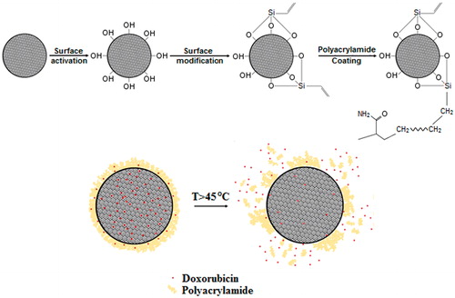
In the present research we have developed a polyacrylamide-mesoporous silica core-shell nanostructure (PMSN) as a smart carrier of anticancer drugs. Polyacrylamide (PAA) has been used previously for delivery of photosensitiser agents in photodynamic therapy of cancer [Citation32]; however, its potential as a thermosensitive capping agent for drug delivery has not been investigated. This study is an attempt to develop thermosensitive nanocarriers with a drug release mechanism based on the rapid dissociation of capping agent that provide higher loading capacity, better stability at physiological environment, and minimised drug release at body temperature. PAA possess a controllable gelation temperature that could be adjusted in the temperature range of thermal ablation by altering the cross-linker/monomer ratio. Therefore, we assumed that by irradiation of energy via the thermal ablation modalities, the PAA protective shell could be rapidly dissociated, facilitating the release of encapsulated drug. The onset gelation temperature of PAA is sufficiently higher than physiological temperature and within the therapeutic hyperthermia range, causing a minimal drug release in healthy tissues, and consequently lower systemic toxicity. To the best of our knowledge, this study is the first attempt to produce a thermoresponsive polymeric drug carrier based on the gelation of the encapsulating shell. A schema of the synthesis procedure as well as the expected mechanism of drug release from the core-shell nanoparticles is presented in .
Materials and methods
Synthesis of polymer-coated mesoporous silica nanoparticles (PMSNs)
PMSNs were synthesised using the following materials obtained from Sigma-Aldrich. Tetraethyl orthosilicate (TEOS), cetyltrimethylammonium bromide (CTAB), ammonia solution (28% wt), ethanol, hydrochloric acid aqueous solution (HCl, 37% wt) triethoxyvinylsilane (TEVS), acrylamide (AA), N,N′-methylene-bis-acrylamide (MBAA), N,N,N′,N′-tetra-metyl-ethylene-diamine (TEMED), and ammonium persulphate (APS).
The mesoporous silica nanoparticles (MSNs) were produced by modification of the Stoeber method proposed in the literature [Citation33,Citation34]. In our modified recipe, CTAB was added as a template, resulting in a mesoporous structure in silica nanoparticles. In a typical process, two types of solutions were prepared. The first solution was a mixture of 16.75 mL TEOS, 3.3 g of CTAB, and 133.25 mL ethanol. The second solution contained 3.81 mL aqueous ammonia (28% wt in water), 40.78 mL deionised water, and 105.46 mL ethanol. Under stirring, the first solution was added to the second solution and the mixture was stirred for 210 min. Then the mixture was centrifuged at 4000 rcf for 15 min and the precipitate was rinsed with ultrapure water three times followed by drying for 24 h at 80 °C. The CTAB template was later removed by incubation of the particles in a methanol solution containing 3% v/v HCl for 6 h. The MSNs were lastly obtained by another centrifuging, rinsing, and drying at 80 °C for 24 h.
In order to activate the surface of MSNs, the particles were dispersed in 100 mL of HCl solution and left stirring for 1 h at room temperature. The solution was centrifuged (4000 rcf for 15 min), washed three times in ultrapure water, and the activated mesoporous silica (AMSN) was obtained by drying the solution. Then, 1 g of AMSN was added to a beaker containing 40 mL of toluene and the mixture was dispersed using an Ultra-Turrax homogeniser (T25, Ika Works, USA) at 20,000 rpm. After 10 min, 600 µL of TEVS was slowly injected into the solution and the mixture was stirred for 24 h at room temperature. The resulting suspension was later centrifuged (4000 rcf for 15 min), washed, and dried at 80 °C for 24 h to obtain vinyl-modified MSNs (VMSNs).
PMSNs were finally prepared by the graft polymerisation method. As a typical procedure, 700 mg of VMSNs with varying amounts of AA (350, 700, and 1050 mg) were added to 50 mL of water, while the ratios of MBAA/AA and TEMED/AA in the solution were kept constant (3.2, and 16.1 mmol per mol of AA, respectively). The mixture was stirred vigorously for 30 min using Ultra-Turrax at 20,000 rpm and then, 800 µL of an aqueous solution containing 1.3% w/v APS was injected to initiate the polymerisation. After 24 h, the resulting suspension was centrifuged at 4000 rcf for 15 min, washed for three times, and dried at 50 °C for 48 h. The overall procedure for preparation of PASNs is illustrated in . It is important to note that polyacrylamide has not been reported as a toxic material; however, its monomer is a severe neurotoxin which may be present in small amounts in the product. Therefore, a stringent washing process is needed to remove the unreacted monomer from the nanoparticle composition.
Table 1. The materials needed for production of nanoparticles at different synthesis stages.
Material characterisation
The Fourier transform infrared spectroscopy (FT-IR) (Nicolet 6700, Thermo Scientific, Madison, WI) analysis was performed at wavenumber range of 600 to 4000 cm−1 to confirm the presence of hydroxyl, vinyl, and amide bonds in the nanoparticle structure. The structural morphology of the PMSNs was observed using scanning electron microscopy (SEM) (Hitachi S3400n, Tokyo, Japan), transmission electron microscopy (TEM) (Libra 120 kV, Carl Zeiss, Oberkochen, Germany), and atomic force microscopy (AFM) (AFP-200, Ambios Technology, Santa Cruz, CA). The average thickness of the polymer layer as well as the average size and volume of particles was estimated by analysis of at least 50 randomly selected nanoparticles from TEM images using ImageJ software (version 1.44 P, National Institutes of Health, Bethesda, MD).
In order to calculate the polymer:silica weight ratio inside the nanostructure, thermogravimetric analysis (TGA) (TGA/SDTA 851 e, Mettler-Toledo, OH) with scan rate of 5 °C/min was used. The gelation temperature range of the polymer shell was also determined by differential scanning calorimetry (DSC) (DSC820 with TSO 801RO robot, Mettler-Toledo) with scan rate of 5 °C/min. The TGA and DSC experiments were repeated at least five times and the results obtained were reported as mean ± standard deviation (SD) using SPSS software (SPSS 20, Armork, NY).
Drug loading experiments
For the drug encapsulation, doxorubicin was dissolved in distilled water with a concentration of 2 mg/mL. Then 3 mg of PMSNs was dispersed in 1.5 mL of this solution to adjust the drug/PMSNs weight ratio to 1. The mixture was incubated at room temperature for three different intervals (24 h, 48 h, and 72 h) in order to determine the optimum incubation time that results in the statistically maximum encapsulation value (Em). The dispersion was later centrifuged at 16,100 rcf for 60 min to separate the drug-loaded PMSNs from the supernatant. Ultraviolet-visible spectroscopy (UV-Vis) (UV-1800, Shimadzu, Kyoto, Japan) at wavelength range of 260 to 600 nm was used to determine the quantity of the drug in the aqueous supernatants. All the UV-Vis measurements were performed at room temperature and using distilled water as the baseline. An aqueous solution containing 2 mg/mL doxorubicin was prepared as the reference solution, and the quantity of the encapsulated drug was calculated by comparing the obtained UV-Vis graphs with that of the reference solution. UV-Vis analyses were all performed without dilution of the supernatants obtained. The amount of encapsulated drug within the PMSNs was calculated from the difference in concentration of the drug in the supernatant after centrifugation and its concentration in the initial doxorubicin solution. The optimum incubation time calculated in this experiment was used in further stages to encapsulate doxorubicin in the PMSNs.
Drug release experiments
In order to measure the amount of drug leakage from the polymer shell at physiological temperatures, the doxorubicin-loaded PMSNs were incubated in an aqueous solution (2 mg/mL) at 37 °C for 7 days. Then the supernatants were separated from the nanoparticles and the amounts of released drug were quantified by comparing the UV-Vis spectra of the supernatants with that of a reference solution (an aqueous solution containing doxorubicin with concentration equal to Em as a reference solution for 100% drug release).
The release behaviour at increased temperatures was also studied by preparing ten similar aqueous solutions containing 2 mg/mL of doxorubicin-loaded nanoparticles. Each solution was heated separately at a specific temperature for 30 min using a water bath. Ten different temperature values ranging from 30–75 °C (30, 37, 40, 45, 50. 55, 60, 65, 70, and 75 °C) were used in these experiments to simulate the hyperthermia and thermal ablation conditions as well as to determine the effect of temperature on the release ratio of the encapsulated drug. After heat treatments, the supernatants were separated from the PMSNs and the amounts of released drug were quantified by comparing the UV-Vis spectrum of each sample with that of the reference solution. A similar protocol was also applied (except that the tests were performed only at 75–80 °C) to calculate the ratio of drug release after sonication of nanoparticles using the magnetic resonance-guided focused ultrasound (MRgFUS) method (ExAblate, Insightec, Tirat-Carmel, Israel). For these experiments a tube containing an aqueous solution of drug-loaded nanoparticles was placed in a thermosensitive tissue-mimicking phantom which had been developed previously [Citation35]. This phantom can mimic various acoustic properties of soft tissues (e.g. attenuation and sound speed) and precisely show the heated area by a reversible colour change as well as alteration of MR parameters.
The phantom was immediately sonicated for 5 × 20 s at a frequency of 1.15 MHz with the acoustic energy and acoustic power of 5313 J and 266 W, respectively. Then the ratio of drug release due to heating by MRgFUS was measured by UV-Vis technique. All the drug loading and release experiments were repeated for at least five times and the results were analysed using ANOVA (SPSS 20) with 95% confidence interval.
Results
Material characterisation
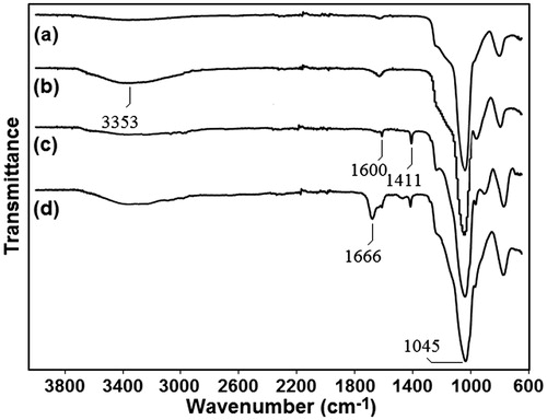
The FT-IR spectra of the MSNs, AMSNs, VMSNs, and PMSNs are shown in . The strong peak observed at 1045 cm−1 in all samples represents the Si-O-Si vibration. In the FT-IR spectrum of the AMSNs, the peak of absorbed water is stretched at 3353 cm−1. For the VMSN sample, one can clearly observe the peaks at 1411 cm−1 and 1600 cm−1 for vinyl and silane bonds, respectively. In the FT-IR spectrum of the PMSNs, the peak at 1666 cm−1 is a characteristic vibration of the amide group.
Figure 1. Schematic of the synthesis method and expected mechanism of drug release from the core-shell nanostructure. Rapid melting of protective shell provides preferred trajectories for drug release.
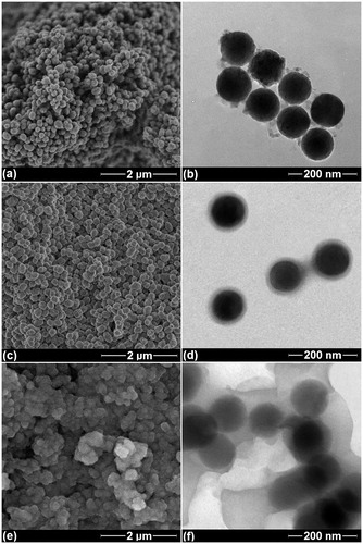
illustrates the SEM and TEM images of the PMSNs with different weight ratios of acrylamide:mesoporous silica (AA/MSN:0.5, 1.0, and 1.5). The dark spheres represent the mesoporous silica nanoparticles (MSNs) with a mean diameter of 90 ± 20 nm and the thin grey shells imply the presence of polymer, covering the MSN cores. This figure clearly demonstrates that the AA/MSN ratio of 0.5 could not provide a uniform layer around the cores (), whereas the AA/MSN ratio of 1.0 results in a uniform polymer shell with thickness of 12 ± 2 nm (). On the other hand, a particle aggregation is observed in the SEM and TEM images of the specimen with AA/MSN ratio of 1.5 () due to a relatively large amount of monomer used in this specimen. Based on the microstructures obtained, the AA/MSN ratio of 1.0 was selected to synthesise core-shell nanoparticles for further experiments.
Figure 2. The FT-IR spectra of the particles at different synthesis stages. (a) mesoporous silica (MSN), (b) activated mesoporous silica (AMSN), (c) vinyl-functionalised mesoporous silica (VMSN), and (d) polyacrylamide-coated mesoporous silica (PMSN).
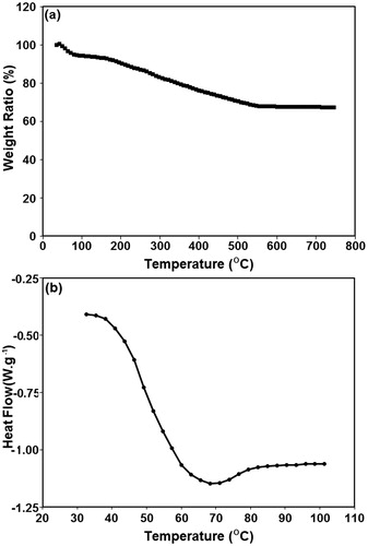
According to a typical curve obtained from TGA analysis (), a rapid weight loss occurs in PMSNs at temperatures below 100 °C that corresponds to the evaporation of water from the PAA structure. The second phase of weight loss begins at 200 °C and continues until stabilisation at the constant value of 67 ± 3% (temperatures above 630 °C). Therefore, due to the thermal stability of silica at this temperature range, the amount of weight loss (∼33 ± 3%) might represent the weight ratio of the polymer shell in the PMSN structure. The weight ratio of the polymer shell could also be calculated according to the following equation
(1)
where V, ρ, and r values represent volume, density, and radius of nanoparticles, respectively. The densities of MSNs and PAA were respectively assumed equal to 2.648 g/cm3 and 1.13 g/cm3. According to this equation, the weight ratio of the PAA shell around the MSNs is around 31% wt which is slightly lower than the value obtained from the TGA analysis. This might be due to the availability of moisture in the nanoparticles as well as the presence of small amounts of polymer inside the MSN pores.
Figure 3. Microstructural images of synthesised nanoparticles. (a, c, e) SEM, and (b, d, f) TEM images of the specimens with AA/MSN weight ratios of 0.5, 1.0, and 1.5, respectively.
The DSC analysis was also performed to determine the gelation point of the PAA coating (). Although the melting point of PAA coating is 68 ± 4 °C (the minimal point in the DSC spectra), the wide DSC peak shows that the gelation of polymer shell initiates at temperatures below 45 °C. Therefore, a partial release of encapsulated drug from the MSN pores could also occur at hyperthermia temperature range. It is important to note that this temperature range is significantly higher than the physiological temperature which may minimise the ratio of drug release at the tissues surrounding the target tumour.
Drug loading behaviour
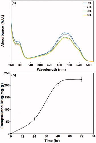
The optimal incubation time needed for encapsulation of doxorubicin in PMSN structure was determined by UV-Vis spectroscopy. The UV-Vis spectra of the supernatants after centrifugal separation of PMSNs from doxorubicin solution are illustrated in . The increase of incubation time results in reduced UV absorption intensity, and thus decreased doxorubicin concentration in the supernatant. The amounts of encapsulated doxorubicin in the PMSNs after different incubation intervals are also plotted in . With the increase of incubation time up to 48 h, the average ratio of encapsulated doxorubicin in the nanoparticles is increased (208 ± 20 mg in 1 g of PMSNs) and then, no significant change in its amount is observed. Based on this fact, incubation time of 48 h was used in all further experiments to prepare the drug-loaded nanoparticles. Moreover, an aqueous solution containing 416 µg/mL of doxorubicin (equal to the average amount of drug encapsulated in 2 mg of the PMSNs) was prepared and its UV-Vis absorbance at wavelength range of 260–600 nm was recorded as a reference for 100% drug release to quantify the ratio of drug release at the 10 different temperatures applied.
Figure 4. The thermal behaviour of core-shell nanoparticles. (a) The TGA, and (b) the DSC analysis of a typical specimen with AA/MSN weight ratio of 1.0.
Drug release behaviour
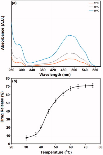
shows the absorbance intensity of three specimens heated at 37, 45, and 60 °C. The red spectrum gives an indication of the diffuse-type release of doxorubicin molecules from the PMSNs at 37 °C. The grey spectrum shows that a relatively higher amount of drug molecules could be released at 45 °C, possibly due to the partial melting of the PAA shell at this temperature. However, a more intense absorbance is established by the specimens that were heated at 60 °C. Note that the similarity of absorbance spectra at different temperatures shows the stability of encapsulated doxorubicin during the experiments. also shows the percentage of drug release at different temperatures from 30–75 °C. The percentage of drug release at physiological temperature (37 °C) was about 11.5 ± 2.4%, which is not statistically different from the drug released at 30 °C (p < 0.05). It is noteworthy that incubation of drug-loaded PMSNs at physiological temperature for 7 days resulted in a 14 ± 3% drug leakage from the polymer shell. The value obtained was not statistically different from the leakage ratios after incubation at 30 °C and 37 °C for 30 min (). Therefore, it may be concluded that the entrapped drug inside the polymer shell could be rapidly released after incubation of the drug-loaded PMSNs into the aqueous solution.
Figure 5. The amount of doxorubicin encapsulated in nanoparticles at different time intervals. (a) UV-Vis spectra of PMSNs after incubation in doxorubicin solution for 0, 24, 48, and 72 h; (b) the amount of doxorubicin encapsulated in the particles at different incubation times.
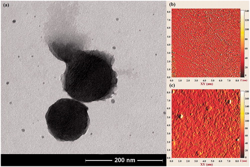
According to , the maximum ratio of drug release (67.6 ± 2.5%) could be obtained at 60 °C and then no significant increase in release ratio of the encapsulated drug is observed. The TEM image of typical PMSNs after heating at 60 °C () illustrates a partial melting of the PAA shells that produces preferred routes for release of encapsulated drug at this temperature level. The partial elimination of polymer shells from the MSN surface provides the ability to visualise the silica mesopores in this figure. The melted shell may produce relatively large aggregates on the MSN surface or detach from the MSN surface and form small polymeric spheres in aqueous solution. According to the AFM images of the nanoparticles prior to and after heating (), the partial melting of polymer shells may result in aggregation of PMSNs in aqueous solutions containing high concentrations of PMSNs.
Figure 6. The ratio of doxorubicin released at different temperatures. (a) UV-Vis spectra of S10 specimens heated at 37, 50, and 60 °C. (b) The percentage of drug release from the PMSNs after heating at various temperatures for a period of 30 min.
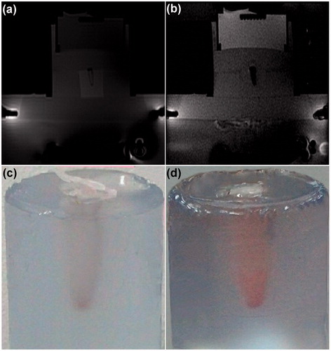
The procedure applied for sonication of drug-loaded PMSNs using MRgFUS system is shown in . When the ultrasound pulses are propagated through the thermosensitive phantom, the MR images show a dark oval region corresponding to the heated area at the focal point (). The temperature patterns of the focal region at different sonication times are also illustrated in . The region with maximum temperature was initially produced at bottom of the container (where PMSNs are precipitated) and gradually propagated through the container until all the aqueous solution reached the maximum temperature at 19 s. According to , the temperature of the focal point was increased to over 50 °C and 70 °C after sonication for approximately 4 and 16 s, respectively, producing sufficient thermal energy for gelation of the polymer shell.
Figure 7. (a) TEM image of PMSNs after heating at 60 °C. The PAA shell is partially removed because of melting at this temperature range. (b, c) AFM image of PMSNs before, and after heating at 60 °C, respectively.
The MR images of a typical thermosensitive phantom containing drug-loaded PMSN solution before and during heat treatment using focused ultrasound are shown in . The temperature rise in the surrounding thermosensitive phantom provides suitable contrast in MR images which also confirms that all of the container has been heated during the sonication. also illustrates a phantom before and after sonication using focused ultrasound. A colour change in the aqueous solution is clearly observed due to the partial leakage of red-coloured doxorubicin from the core-shell nanostructure. The measurement of drug release ratio using UV-Vis indicates that 39.2 ± 2.2% of encapsulated doxorubicin is released from PMSNs due to the sonication by focused ultrasound. This fact shows the capability of these nanoparticles for fast and effective release of encapsulated therapeutic agent when applied in a clinical setting.
Figure 8. The procedure applied for MRgFUS sonication of nanoparticles. (a–c) Magnetic resonance phase images of a typical thermosensitive phantom containing drug-loaded PMSNs during MRgFUS heating. (d–g) The temperature patterns obtained during the heating process, where higher temperatures are shown in red. (h) The temperature plot of the focal point during sonication of the phantom.
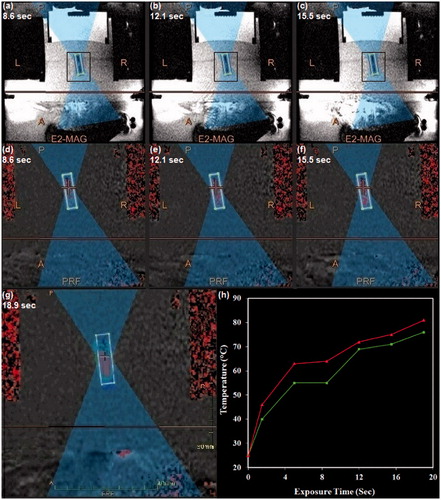
Figure 9. Influence of MRgFUS sonication on drug-loaded nanoparticles. (a, b) Magnetic resonance phase images of a typical thermosensitive phantom containing drug-loaded PMSNs before and after MRgFUS treatment, respectively. (c, d) The colour change of the aqueous solution containing drug-loaded PMSNs after MRgFUS treatment, confirming the partial release of encapsulated drug.
Discussion
The current thermosensitive drug carriers are mostly synthesised using polymers that undergo phase transition and shrinkage at temperatures above their LCST. However, the drug release from these nanoparticles is mostly based on the diffusion mechanism which may result in lower rates of drug release in comparison to lysolipid-containing thermosensitive liposomes. Moreover, the diffusion mechanism may not be successfully applied in the core-shell particles due to the suppression of encapsulated drug at temperatures above LCST.
In this research we developed a mesoporous silica-polyacrylamide core-shell nanostructure that showed thermal sensitivity in the temperature range of 40–60 °C. The nanoparticles were synthesised in an average particle size range of 102 ± 22 nm which is within the optimum particle size range (80–120 nm) for effective preferential delivery of drug to the tumours through the enhanced permeability and retention (EPR) effect [Citation36].
Doxorubicin was used as a typical guest molecule to test the encapsulation ability of the synthesised nanoparticles. The mesoporous silica in this structure acted as a drug reservoir while the polyacrylamide shell prevented the entrapped drug from rapid diffusion out of the structure. Mesoporous silica nanoparticles are known as efficient carriers of diagnostic and therapeutic agents. Among various characteristics, the biodegradability of these carriers is of particular interest and has been confirmed in a number of studies [Citation36,Citation37]. Silica nanoparticles (especially mesoporous silica) readily degrade in biological medium to form silicic acid, which is a natural trace compound in humans. The in vivo degradation of these nanoparticles is often excessively fast (<12 h) for optimal drug delivery efficacy, and thus surface modifications are needed to increase their circulation time [Citation36]. Therefore, the polyacrylamide coating not only provides a sufficient degree of thermal sensitivity in the drug carrier, this polymeric layer could also protect the silica nanoparticles from rapid degradation within biological environments.
The toxicity, biocompatibility, and biodegradation of polyacrylamide have also been evaluated in a number of studies and no obvious evidence of toxicity, but even some degree of biodegradability has been observed [Citation38]. However, more investigations are needed to confirm the safety of this polymer for application in drug delivery.
During the heating of these nanoparticles, the gelation of polymer shell at temperatures above 40 °C provided preferred routes for faster diffusion of entrapped drug outside the nanoparticles. The obtained value of drug release at physiological temperature was obviously smaller than the reported value for hollow PNIPAM hydrogel (around 60%) [Citation39] and DNA-capped mesoporous silica nanoparticles (approximately 30%) [Citation29]. The release of doxorubicin at temperatures lower than 40 °C might be due to the diffusion of the drug molecules which are entrapped in the PAA shell. By increase of the temperature above 40 °C, the ratio of drug release from the PMSNs was significantly increased due to the gradual gelation of the polymer shell. Although the maximum drug release was obtained at temperatures above the clinically applicable temperature range, the nanoparticles could release over 50% of their loading at temperatures near 50 °C which could still be sufficient for an effective targeted drug delivery.
This thermosensitive behaviour makes these nanoparticles suitable for hyperthermia and thermal ablation temperature range (45–55 °C) which is mostly applied in thermal modalities such as high intensity focused ultrasound and radiofrequency. By targeted delivery of these carriers, a significantly higher local release of the encapsulated drug could be obtained with lower systemic toxicity levels compared to the current similar products. However, in spite of a significant decrease, the amount of drug release at physiological temperatures is still challenging, which could be addressed in further studies. A wide and continuous transition range is another limitation of these nanoparticles that must be improved to obtain polymer layers with discontinuous gel-to-liquid transition temperatures. The stability tests in complete serum as well as in vivo comparative studies for both hyperthermia and thermal ablation ranges must also be conducted to determine the in vivo efficacy of the newly synthesised product.
Acknowledgements
The authors wish to thank Emad Sadeghian Nezhad for running the UV-Vis spectrometry experiments, and Yus Hafizul from University of Malaya Medical Centre for conducting the MRgFUS experiments.
Declaration of interest
This research was supported by the High-Impact Research MoE Grant, UM.C/625/1/HIR/MOE/DENT/14 from the Ministry of Higher Education Malaysia, and University of Malaya (Research Grant No. RG055/09HTM). The authors alone are responsible for the content and writing of the paper.
References
- McDaniel JR, Dewhirst MW, Chilkoti A. Actively targeting solid tumours with thermoresponsive drug delivery systems that respond to mild hyperthermia. Int J Hyperthermia 2013;29:501–10
- Shao P, Wang B, Wang Y, Li J, Zhang Y. The application of thermosensitive nanocarriers in controlled drug delivery. J Nanomater 2011;2011:389640
- Parodi A, Quattrocchi N, van de Ven AL, Chiappini C, Evangelopoulos M, Martinez JO, et al. Synthetic nanoparticles functionalized with biomimetic leukocyte membranes possess cell-like functions. Nat Nanotechnol 2013;8:61–8
- Landon CD, Park J-Y, Needham D, Dewhirst MW. Nanoscale drug delivery and hyperthermia: The materials design and preclinical and clinical testing of low temperature-sensitive liposomes used in combination with mild hyperthermia in the treatment of local cancer. Open Nanomed J 2011;3:38–64
- Gasselhuber A, Dreher MR, Rattay F, Wood BJ, Haemmerich D. Comparison of conventional chemotherapy, stealth liposomes and temperature-sensitive liposomes in a mathematical model. PLoS One 2012;7:e47453
- Ta T, Porter TM. Thermosensitive liposomes for localized delivery and triggered release of chemotherapy. J Control Release 2013;169:112–25
- Needham D, Park J-Y, Wright AM, Tong J. Materials characterization of the low temperature sensitive liposome (LTSL): Effects of the lipid composition (lysolipid and DSPE–PEG2000) on the thermal transition and release of doxorubicin. Faraday Discuss 2013;161:515–34
- Li L, ten Hagen TL, Hossann M, Süss R, van Rhoon GC, Eggermont AM, et al. Mild hyperthermia triggered doxorubicin release from optimized stealth thermosensitive liposomes improves intratumoral drug delivery and efficacy. J Control Release 2013;168:142–50
- Gasselhuber A, Dreher MR, Partanen A, Yarmolenko PS, Woods D, Wood BJ, et al. Targeted drug delivery by high intensity focused ultrasound mediated hyperthermia combined with temperature-sensitive liposomes: Computational modelling and preliminary in vivo validation. Int J Hyperthermia 2012;28:337–48
- Viglianti BL, Dewhirst MW, Boruta RJ, Park J-Y, Landon C, Fontanella AN, et al. Systemic anti-tumour effects of local thermally sensitive liposome therapy. Int J Hyperthermia 2014;30:385–92
- Chiu GN, Abraham SA, Ickenstein LM, Ng R, Karlsson G, Edwards K, et al. Encapsulation of doxorubicin into thermosensitive liposomes via complexation with the transition metal manganese. J Control Release 2005;104:271–88
- Banno B, Ickenstein LM, Chiu GN, Bally MB, Thewalt J, Brief E, et al. The functional roles of poly (ethylene glycol)-lipid and lysolipid in the drug retention and release from lysolipid-containing thermosensitive liposomes in vitro and in vivo. J Pharm Sci 2010;99:2295–308
- Ta T, Convertine AJ, Reyes CR, Stayton PS, Porter TM. Thermosensitive liposomes modified with poly (N-isopropylacrylamide-co-propylacrylic acid) copolymers for triggered release of doxorubicin. Biomacromolecules 2010;11:1915–20
- de Smet M, Langereis S, den Bosch SV, Grüll H. Temperature-sensitive liposomes for doxorubicin delivery under MRI guidance. J Control Release 2010;143:120–7
- Ruiz J-C, Burillo G, Bucio E. Interpenetrating thermo and pH stimuli-responsive Polymer networks of PAAc/PNIPAAm grafted onto PP. Macromol Mater Eng 2007;292:1176–88
- Wadajkar AS, Koppolu B, Rahimi M, Nguyen KT. Cytotoxic evaluation of N-isopropylacrylamide monomers and temperature-sensitive poly (N-isopropylacrylamide) nanoparticles. J Nanopart Res 2009;11:1375–82
- Yan R, Zhang M, Zhang W, Liu S. Temperature dependent synthesis of micro-and meso-porous silica employing the thermo-responsive polymer of poly (N-isopropylacrylamide) as structure-directing agent. J Sol-Gel Sci Technol 2011;59:315–26
- Yang X, Lee HY, Kim JC. Effect of hydrophobic comonomer content on assembling of poly (N-isopropylacrylamide) and thermal properties. J Appl Polym Sci 2011;120:2346–53
- Ward MA, Georgiou TK. Thermoresponsive polymers for biomedical applications. Polymers 2011;3:1215–42
- Scherzinger C, Schwarz A, Bardow A, Leonhard K, Richtering W. Cononsolvency of poly-N-isopropyl acryl amide (PNIPAM): Microgels versus linear chains and macrogels. Curr Opin Colloid Interface Sci 2014;19:84–94
- Deshmukh SA, Kamath G, Suthar KJ, Mancini DC, Sankaranarayanan SK. Non-equilibrium effects evidenced by vibrational spectra during the coil-to-globule transition in poly (N-isopropylacrylamide) subjected to an ultrafast heating–cooling cycle. Soft matter 2014;10:1462–80
- Li P, Xu R, Wang W, Li X, Xu Z, Yeung KW, et al. Thermosensitive poly (N-isopropylacrylamide-co-glycidyl methacrylate) microgels for controlled drug release. Colloids Surf B Biointerfaces 2013;101:251–5
- Bekhradnia S, Zhu K, Knudsen K, Sande S, Nyström B. Structure, swelling, and drug release of thermoresponsive poly(amidoamine) dendrimer–poly(N-isopropylacrylamide) hydrogels. J Mater Sci 2014;49:6102–10
- Yang Y, Yan X, Cui Y, He Q, Li D, Wang A, et al. Preparation of polymer-coated mesoporous silica nanoparticles used for cellular imaging by a ‘graft-from’ method. J Mater Chem 2008;18:5731–7
- Chiappetta DA, Sosnik A. Poly (ethylene oxide)-poly (propylene oxide) block copolymer micelles as drug delivery agents: Improved hydrosolubility, stability and bioavailability of drugs. Eur J Pharm Biopharm 2007;66:303–17
- Chen S, Li Y, Guo C, Wang J, Ma J, Liang X, et al. Temperature-responsive magnetite/PEO-PPO-PEO block copolymer nanoparticles for controlled drug targeting delivery. Langmuir 2007;23:12669–76
- Yapar EA, Ýnal Ö. Poly (ethylene oxide)–poly (propylene oxide)-based copolymers for transdermal drug delivery: An overview. Trop J Pharm Res 2013;11:855–66
- Jeong B, Gutowska A. Lessons from nature: Stimuli-responsive polymers and their biomedical applications. Trends Biotechnol 2002;20:305–11
- Ruiz-Hernandez E, Baeza A, Vallet-Regí MA. Smart drug delivery through DNA/magnetic nanoparticle gates. ACS Nano 2011;5:1259–66
- Derfus AM, von Maltzahn G, Harris TJ, Duza T, Vecchio KS, Ruoslahti E, et al. Remotely triggered release from magnetic nanoparticles. Adv Mater 2007;19:3932–6
- Thomas CR, Ferris DP, Lee J-H, Choi E, Cho MH, Kim ES, et al. Noninvasive remote-controlled release of drug molecules in vitro using magnetic actuation of mechanized nanoparticles. J Am Chem Soc 2010;132:10623–5
- Kuruppuarachchi M, Savoie H, Lowry A, Alonso C, Boyle RW. Polyacrylamide nanoparticles as a delivery system in photodynamic therapy. Mol Pharm 2011;8:920–31
- Rossi LM, Shi L, Quina FH, Rosenzweig Z. Stöber synthesis of monodispersed luminescent silica nanoparticles for bioanalytical assays. Langmuir 2005;21:4277–80
- Shin Y, Lee D, Lee K, Ahn KH, Kim B. Surface properties of silica nanoparticles modified with polymers for polymer nanocomposite applications. J Ind Eng Chem 2008;14:515–19
- Dabbagh A, Abdullah BJJ, Abu Kasim NH, Ramasindarum C. Reusable heat-sensitive phantom for precise estimation of thermal profile in hyperthermia application. Int J Hyperthermia 2013;30:66–74
- Hon NK, Shaposhnik Z, Diebold ED, Tamanoi F, Jalali B. Tailoring the biodegradability of porous silicon nanoparticles. J Biomed Mater Res A 2012;100:3416–21
- Rosenholm JM, Mamaeva V, Sahlgren C, Lindén M. Nanoparticles in targeted cancer therapy: Mesoporous silica nanoparticles entering preclinical development stage. Nanomedicine 2012;7:111–20
- Caulfield MJ, Qiao GG, Solomon DH. Some aspects of the properties and degradation of polyacrylamides. Chem Rev 2002;102:3067–84
- Xing Z, Wang C, Yan J, Zhang L, Li L, Zha L. Dual stimuli responsive hollow nanogels with IPN structure for temperature controlling drug loading and pH triggering drug release. Soft Matter 2011;7:7992–7