
Abstract
Background: Recurrent and persistent secondary hyperparathyroidism (SHPT) nodules have an incidence of 10–70% after surgery. The treatment of recurrent and persistent SHPT nodules is a challenge, and surgical resection of difficult-to-reach or post-operative adhesions often fails. Purpose: The aim of this research was to study the safety and effectiveness of microwave ablation (MWA) for recurrent and persistent SHPT. Materials and methods: This was a retrospective study of 11 patients enrolled with a total of 16 nodules, and MWA was employed to manage SHPT. The laboratory test results, including the intact parathyroid hormone (iPTH), serum calcium, phosphorus and alkaline phosphatase (ALP) levels, improvement of SHPT-related symptoms after ablation, and complications during and after MWA were recorded and analysed. Results: After ablation the value of iPTH was markedly decreased from 1570 ± 1765 pg/mL to 287 ± 239 pg/mL 1 day after MWA (p < 0.05). The levels of serum calcium and phosphorus decreased from 2.51 ± 0.23 mmol/L to 2.06 ± 0.27 mmol/L (p < 0.001) and 1.80 ± 0.43 mmol/L to 1.48 ± 0.32 mmol/L (p < 0.05), respectively, 1 day after MWA. There was no significant difference in the ALP value before and after MWA (p > 0.05). The clinical symptoms, including ostalgia, pruritus, disability, and restless legs, improved after MWA. Minor complications and side effects encountered during or after MWA include haematoma (1/11, 9%), transient hoarseness (2/11, 18.2%), hypocalcemia (6/11, 54.5%). No major complication occurred. Conclusion: MWA may be safe and effective to manage recurrent and persistent SHPT nodules; a definite conclusion needs to expand the sample size with a longer follow-up time.
Introduction
Secondary hyperparathyroidism (SHPT) induces renal osteodystrophy – one of the major complications in patients with end-stage renal disease (ESRD). Medical therapy alone often fails to successfully manage SHPT mainly because of contraindications of drug treatment, medication intolerance and non-compliance. Currently, some of these patients still require surgical parathyroidectomy [Citation1–4]. Thus far, surgeons have debated over the merits of two types of parathyroidectomy: subtotal parathyroidectomy or total parathyroidectomy plus autotransplantation. However, Richards et al. [Citation5] showed that both procedures had similar rates of recurrent or persistent SHPT. In addition, re-operation of recurrent SHPT is an invasive procedure with an increased incidence of failures and complications because of the limitations in re-operative localisation, inadequate exploration, and post-operative adhesion [Citation5–7]. Therefore, new treatment modalities for recurrent and persistent SHPT are needed.
Radiofrequency ablation (RFA), laser therapy, high-intensity focused ultrasound (HIFU) and percutaneous ethanol injection (PEI), as minimally invasive procedures, have been recently proposed to manage hyperparathyroidism in sporadic cases [Citation6–10]. PEI guided by ultrasound has been employed to manage recurrent and persistent SHPT, with success rates of 50–91.8% [Citation8–11]. However, the promising results of PEI mostly rely on the volume and number of SHPT nodules and multiple sessions for every nodule [Citation11–13]. Although RFA was safe and effective for treating hyperparathyroidism in case reports [Citation14,Citation15], most of the studies provided an indeterminable clinical outcome when laser therapy or HIFU was applied to treat hyperparathyroidism [Citation16,Citation17]. Thus, clinical experience concerning these techniques is limited [Citation16].
Microwave ablation (MWA), a thermal ablation technique, has been successfully applied to the treatment of benign thyroid nodules [Citation18,Citation19]. No report concerning MWA for recurrent and persistent SHPT nodules has been published to date. In our group, MWA has been applied to treat recurrent and persistent SHPT nodules in patients with ESRD. The aim of the present study was to summarise the feasibility, safety and efficacy of MWA in patients with recurrent and persistent SHPT.
Materials and methods
General clinical data
This was a retrospective study. A total of 11 patients, including seven with recurrent SHPT and four with persistent SHPT, agreed to undergo MWA between 1 March 2014 and 31 January 2015 at the Interventional Ultrasound Centre, China-Japan Friendship Hospital, China. The baseline clinical characteristics, operation history, parameters of nodules and results of laboratory tests before MWA in 11 patients are shown in , and , respectively. The study was approved by our institutional review board, and informed consent for the treatment was obtained from all of the patients before each procedure.
Table 1. The baseline of patients with recurrent and persistent SHPT.
Table 2. The conditions of nodules and parameters of MWA in 11 patients with recurrent and persistent SHPT.
Table 3. The comparative results of various test parameters between before and after MWA in patients with recurrent and persistent SHPT.
Definition of recurrent and persistent HPT
In patients with recurrent SHPT, the intact parathyroid hormone (iPTH) level was lower than 300 pg/mL within 6 months after operation, and became elevated again more than 6 months post-operatively. In patients with persistent SHPT, the lowest iPTH level remained higher than 300 pg/mL after operation [Citation11].
Inclusion criteria
Patients assigned to undergo MWA for recurrent SHPT were required to meet the following criteria: 1) a clear history of ESRD with SHPT and medication intolerance, 2) a value of iPTH more than 600 pg/mL before MWA; 3) at least one enlarged possible recurrent and persistent SHPT nodule with a maximum diameter >0.6 cm, as disclosed by ultrasound and accessible to MWA treatment –no critical structure such as a major blood vessel, nerve, or the oesophagus in the needle path, 4) a 99mTc sestamibi (MIBI) scan showing that the nodule had radionuclide concentration in both the early and delayed phases, 5) for patients with voice change following previous parathyroidectomy, no recurrent laryngeal nerve injury confirmed by laryngoscopy, 6) a prothrombin time less than 25 s, prothrombin activity higher than 40%, and a platelet count more than 40 × 109 cells/L, 7) no intractable coexisting morbidity, such as cardiac insufficiency or hypertension.
Preoperative examination and preparation
Ultrasonography in combination with MIBI is performed to locate recurrent and persistent SHPT nodules [Citation20–22]. Ultrasonographic examination was performed using real-time colour Doppler sonography with a 10.0-MHz transducer (Aplio 500, Toshiba, Tokyo, Japan). Ultrasonography was used primarily to locate and characterise suspected SHPT nodules and evaluate the surrounding anatomical structures. Contrast-enhanced ultrasound (CEUS) (Sonovue, Bracco, Milan, Italy) was applied to evaluate the characteristics of enhancement for guiding the distribution of ablation energy. The MIBI scan (SymbiaT2; Siemens, Munich, Germany) was conducted prior to the ablation procedure () for its high sensitivity (85%) and specificity (100%) in the diagnosis of SHPT [Citation23].
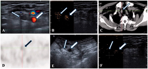
Figure 1. A 55-year-old female patient with a recurrent and ectopic SHPT nodule in the suprasternal fossa 3 years after parathyroidectomy that was treated by microwave ablation (MWA). (A) A hypoechoic nodule (thin arrow) without a blood signal beside the carotid artery (thick arrow) was disclosed by ultrasound. (B) A uniform hyper-enhancement of the nodule (thin arrow) beside the carotid artery (thick arrow) was displayed in CEUS pre-ablation. (C) The CT scan showed that the nodule is in the suprasternal fossa (thin arrow). (D) The nodule has radioactivity concentration (black arrow) in the late phase on MIBI scan. (E) The hyperechoic signal emerging inside the nodule (thin arrow) beside the carotid artery (thick arrow) during ablation. (F) A non-enhancement area covered the nodule (thin arrow) beside the carotid artery (thick arrow) after MWA, suggesting complete ablation was achieved by MWA.
Procedure
MWA was performed in an inpatient regimen by a well-trained, experienced radiologist. Before ablation, intravenous access was obtained via an antecubital vein. Patients were placed in the supine position with the neck extended. After the neck was sterilised, local anaesthesia with 2% lidocaine hydrochloride was performed. Next, a lidocaine and normal saline mixture (1:3, 20 mL) was injected into the area around the hyperplasia nodule for heat insulation, nerve isolation, and local anaesthesia [Citation14], even in the case of adhesion and uneven dispersion. We used a microwave generator and a 17-gauge internally cooled antenna with a 0.4-cm tip (Intelligent Basic Microwave Tumour Ablation System; Nanjing ECO Microwave System, Nanjing, China). The antenna was freehandedly inserted into the parathyroid nodule under ultrasound guidance using a 10-MHz linear probe. Before each ablation the needle tip was displayed by ultrasound to ensure that it was located inside the nodule. Because nodules are usually small with a maximum diameter of about 1 cm, the needle tip was held in a quiescent state for 15–25 s for every microwave radiation session with a power of 25 W, and the repeat radiation was performed two to four times with 5-s intervals to prevent heat injury to surrounding critical structures [Citation24]. After ablation in one unit, the antenna was inserted into another unit to continue ablation. The power was suspended if patients could not tolerate the pain during ablation. Ablation was terminated when transient hyperechoic zones could be identified inside the whole nodule. CEUS was applied 30 min later to evaluate the complete ablation of the parathyroid gland [Citation25]. If the non-enhanced zone covered the ablated nodule, complete ablation was achieved; if there was nodular enhancement inside the nodule, a follow-up ablation would be performed immediately.
During the procedure, an anaesthetist assisted to manage agonising pain, vasovagal reaction, or tracheal stenosis caused by uncontrollable haematoma. The total ablation time was recorded.
At the end of the procedure, mild compression with bagged normal saline at 4 °C was applied to the site of the needle path for 20 min. All patients remained under observation for 2 h to monitor the occurrence of complications.
Follow-up
Ultrasound was performed at 2 and 24 h after ablation for possible haematoma or other complications, and then at 3-month intervals for 1 year. Major and minor complications were those defined by the Society of Interventional Radiology [Citation26]. The iPTH, serum calcium, phosphorus and alkaline phosphatase (ALP) levels were measured at 1 and 7 days, as well as 1, 3, 6, 9, and 12 months after ablation. SHPT-related symptoms were recorded and analysed 1–2 days before and 1 month after MWA using a questionnaire.
Statistical analysis
The data were analysed by SPSS version 17.0. Baseline data were expressed as mean ± SD. The distribution of the data was tested by the Kolmogorov-Smirnov test. One-way repeated measures analysis of variance (ANOVA) was used to determine differences among values of iPTH, calcium, phosphate and ALP before, 1 day after MWA, and at the end of follow-up. The level of statistical significance was set at p < 0.05. The assumption of sphericity was assessed by the test of sphericity; any violations were adjusted for by using the Greenhouse–Geisser correction.
Results
Microwave ablation
Sixteen hyperparathyroidism nodules (eight patients with one nodule each, one patient with two nodules, and two patients with three nodules each) in 11 patients matching the inclusion criteria were treated by MWA in 11 sessions (). All 16 nodules had radioactivity concentration in the early and late phases on MIBI scans () and hyper-enhancement in the artery phase on pre-ablation CEUS (). Detailed information regarding nodule parameters, individual energy doses, the number of ablation units, and side effects in the 11 patients is reported in . The ablation time was 147–484 s (mean 305 ± 131 s) in a single nodule, and the ablation units were 4–8 (mean 4.8 ± 1.3) (). After ablation all of the nodules showed non-enhancement in CEUS, representing complete ablation (). One haematoma occurred during ablation, and 2 U haemocoagulase atrox (Penglai Nuokang Pharmaceutical, Shandong, China) was intravenously injected immediately. The hematoma had not increased in size 5 min later, so the ablation was continued. The haematoma was absorbed 7 days after MWA according to ultrasound examination. Transient mild to moderate pain was encountered in nine patients during ablation, but spontaneous remission occurred after ablation was suspended. No other side effect was encountered during ablation.
Change in laboratory test results after MWA
Detailed data regarding serum iPTH, serum calcium, phosphate and ALP levels before MWA, 1 day after MWA and at the end of follow-up are reported in . Before MWA, the mean value of iPTH in 11 patients was 1570 pg/mL (range 651–6800 pg/mL), which markedly decreased to 287 pg/mL (range 17.5–665 pg/mL) 1 day after MWA (p < 0.05), and 419 pg/mL (range:21–1500 pg/mL) at the end of follow-up (p < 0.05). It is worth noting that nine patients (81.8%) demonstrated iPTH values in the range recommended by the recent Kidney Disease-Improving Global Outcomes Chronic Kidney Disease – Mineral and Bone Disorder (KDIGO CKD-MBD) Work Group guidelines (600 pg/mL) at the end of follow-up [Citation4], and six patients (54.5%) had iPTH values less than 300 pg/mL, which was within the standard for successful treatment according to a prior report [Citation11]. Similar results were achieved for serum calcium levels, which were 2.51 ± 0.23 mmol/L before MWA, 2.06 ± 0.27 mmol/L 1 day after MWA and 2.32 ± 0.15 mmol/L at the end of follow-up; the decreased levels post-ablation were statistically significant (p < 0.01 and p < 0.05, respectively). On further analysis four patients had hypercalcaemia before MWA (4/11, 36.4%), six patients had hypocalcaemia 1 day after MWA (6/11, 54.5%), and only one patient had hypercalcaemia at the end of follow-up; most patients (90.9%) had calcium values in the normal range at the end of follow-up. A continuing trend downwards after MWA could be observed in the phosphorus levels, which were 1.80 ± 0.43 mmol/L before MWA, 1.48 ± 0.32 mmol/L 1 day after MWA, and 1.32 ± 0.37 mmol/L at the end of follow-up (p < 0.05 and p < 0.05 for decreased levels post-ablation). In addition, hyperphosphataemia occurred in nine patients (81.8%) before MWA, four patients (36.4%) 1 day after MWA, and two patients (18.2%) at the end of follow-up. There were no significant differences in ALP values before and after MWA (p > 0.05).
Drug therapy against SHPT before and after MWA
Before ablation nine of eleven patients had taken medication for high iPTH levels or hypercalcaemia (). After MWA eight patients still required drugs and the rest of the patients had normal blood calcium levels without medical intervention. The doses of the medicines taken by patients before and after MWA were adjusted based on the iPTH, calcium and phosphate levels. For hypocalcaemia just after MWA, calcitriol at 0.5 μg BID combined with calcium carbonate at 2.25 g TID were given for a blood calcium concentration of >1.8 mmol/L, while a high-calcium dialysate with 1.75 mmol/L was added for a blood calcium concentration of ≤ 1.8 mmol/L. The rate of hypercalcaemia decreased from 36.4% to 9.1% at the end of follow-up. Before MWA, three patients took medication to correct hyperphosphataemia, but one still had hyperphosphataemia. After MWA two patients (66.7%) had phosphate values in the normal range without taking medication, and one other (33.3%) still had hyperphosphatemia. Fortunately, five of six patients (83.3%) who could not afford the drug for hyperphosphataemia before ablation had a normal range of phosphataemia without drug intervention at the end of follow-up.
Table 4. Medication details for treatment of SHPT patients prior to microwave ablation (MWA) and afterwards.
Improvement in SHPT-related symptoms after MWA
There is a clear improvement in symptoms related to SHPT after MWA according to follow-up (). Ostalgia, the most common symptom, occurred in 9 of 11 patients (81.8%) before MWA, but the symptom has disappeared in four patients (44.4%), obviously remitted in four cases (44.4%) and non-improved in one case (11.1%) after MWA. Seven patients (63.6%) had symptoms of pruritus before MWA, which went into remission in two patients (28.6%) and disappeared in five patients (71.4%). Eight patients had a myasthaenia before MWA, three patients (37.5%) had distinct remission, and five patients (62.5%) showed complete recovery after MWA. Restless legs occurred in four patients (36.4%) before MWA, and all had disappearance of symptoms after MWA (100%). Coexistent hypertension related to SHPT occurred in seven patients (63.6) before MWA; however, two patients (28.6%) fully recovered without medical intervention, and the other five patients (71.4%) showed no change.
Table 5. The improvement of hyperparathyroidism-related symptoms in patients with recurrent and persistent SHPT before and after MWA.
MWA-related complications
One haematoma occurred during ablation and was successfully managed with haemostatics (9.1%). Transient hoarseness was encountered in two patients (18.2%), and hypocalcaemia occurred in six patients after MWA (54.5%); all were rapidly corrected with calcium supplements. No other complication was reported during the follow up of 4–12 months (6.8 ± 2.4 months).
Discussion
Subtotal and total parathyroidectomy with forearm autograft offers a higher cure rate for SHPT than all other medical and surgical treatments. However, recurrent hyperparathyroidism can be observed in 10–70% of patients with SHPT and in 5–10% of those with primary hyperparathyroidism (PHPT) depending on the follow-up time [Citation27–29]. The most common reasons for failed parathyroid surgery are lack of experience in parathyroidectomy, unrecognised multiglandular disease, ectopic localisation of parathyroid adenoma, inadequate extent of parathyroid tissue resection, parathyroid capsule rupture causing parathyromatosis, or (rarely) parathyroid cancer [Citation30]. Nawrot et al. have reported that the missed hyperfunctioning parathyroid gland was found on reoperation in the eutopic position in 55.5% of patients, and in the ectopic position in 44.3% of patients [Citation30], compared with 62.5% and 37.5%, respectively, in the present study. Reoperations for persistent or recurrent hyperparathyroidism compared with the initial operations are associated with higher complication rates [Citation6,Citation31]. Moreover, co-morbidities and the high surgical anaesthetic risk might contraindicate parathyroidectomy in some elderly patients. Finally, many patients with few or no symptoms refuse surgery. This explains the considerable interest in identifying therapeutic alternatives to surgery [Citation16].
The present pilot study enrolled 11 cases, representing the first experience with the MWA technique in patients with recurrent and persistent SHPT. Eleven sessions were applied to successfully manage 16 hyperplastic parathyroid nodules which did not need further sessions for the same nodule – usually one nodule requires more than one session for PEI, HIFU or laser ablation of hyperparathyroidism [Citation11,Citation16,Citation17].
Our report illustrates the potential benefits of this method in the setting of recurrent and persistent SHPT. A marked decrease in serum iPTH was achieved in all 11 patients 1 day after MWA. At the end of follow-up, most patients (81.8%) had iPTH reduced to the iPTH range recommended by the recent KDIGO CKD–MBD guideline [Citation4]. However, 50% of patients had an iPTH value less than 300 pg/mL at the end of follow-up, this percentage is lower than that of 91.8% reported by a prior study in which PEI therapy (PEIT) was applied to manage recurrent SHPT. But the reported PEIT procedure requires three to five injections at 7-day intervals for each nodule [Citation11], which is more complicated than that of MWA. Concomitantly, the control of serum calcium was also improved, as demonstrated by the fact that 36.4% of patients had hypercalcaemia pre-ablation, 54.5% had hypocalcaemia 1 day after ablation and 90.9% were in the normal range at the end of follow-up. By contrast, among the 50–80% of patients with a normal range of calcium values after PEIT, laser or HIFU was used to treat those with PHPT or SHPT in multiple sessions [Citation16,Citation17,Citation32]. The results of phosphorus were also encouraging, as evidenced by the decreasing tendency after MWA. In general, the success rate in the present study was inferior to the 90–100% of thoracoscopy of the mediastinal parathyroid glands [Citation33–35] and 76–95% of open surgery for persistent and recurrent SHPT [Citation30,Citation36]. However, MWA has a shorter operation time, fewer complications, and a wider indication than surgery. By contrast regarding drug therapy, the most promising drug, cinacalcet, was tentatively used for management of patients with SHPT or recurrent SHPT. Only 20.3% (26/128) of patients who had undergone a prior parathyroidectomy and 18.2% (253/1388) of those who did not, achieved serum PTH and Ca × P values within the recommended KDOQI(TM) target ranges [Citation28]; these rates were lower than those in the present study. At the end of follow-up, various SHPT-related symptoms showed marked improvement or disappeared in all cases, most likely due to the optimisation of calcium–phosphorus metabolism following MWA. These changes in symptoms are beneficial for the quality of life of patients.
The minor complications and side effects encountered in the present study include haematoma (9%), transient hoarseness (18.2%), and hypocalcaemia (54.5%). By contrast, 5–30.6% transient hoarseness and 40.8% hypocalcaemia were encountered in PEIT of recurrent or persistent SHPT [Citation11,Citation32], and 16.7% transient hoarseness after laser ablation of PHPT [Citation16]. The minor complications or side effects following HIFU treatment of SHPT include mild subcutaneous oedema (60%), prolonged vocal cord mobility impairment (40%), prolonged bitonal voice (20%) and difficulty in swallowing water (20%) [Citation17]. However, the rates of major complications and mortality in surgery are 6–21.6% and 1.7% respectively; the major complications include permanent unilateral or bilateral recurrent laryngeal nerve injury, permanent hypoparathyroidism, wound infection, post-operative bleeding, and pneumonia [Citation30,Citation34,Citation36,Citation37].
Limitations of our study are the relatively short follow-up duration and the low number of patients. A definitive conclusion regarding MWA of recurrent and persistent SHPT could be reached in a further study involving a greater number of cases and a longer follow-up duration
In conclusion, MWA as a minimally invasive technique may be safe and effective for the management of recurrent and persistent SHPT. A further prospective study enrolling a large population will be carried out to reach a definitive conclusion.
Declaration of interest
This work was supported by the Science Foundation of China-Japan Friendship (2014-2-QN-16) and China-Japan Friendship Hospital Youth Science and Technology Excellence Project (2015-QNYC-B-12).
The authors have declared there are no financial conflicts of interest or commercial involvement in regard to this work. The authors alone are responsible for the content and writing of the paper.
References
- Cannata-Andía JB, Fernández-Martín JL, Zoccali C, London GM, Locatelli F, Ketteler M, et al. Current management of secondary hyperparathyroidism: A multicenter observational study (COSMOS). J Nephrol 2008;21:290–8
- He Q, Zhuang D, Zheng L, Fan Z, Zhou P, Zhu J, et al. Total parathyroidectomy with trace amounts of parathyroid tissue autotransplantation as the treatment of choice for secondary hyperparathyroidism: A single-center experience. BMC Surg 2014;14:26
- Sarfati E, Drueke TB. Surgical management of secondary hyperparathyroidism. In: Olgaard K, Silver J, Salusky IB, eds. The Spectrum of Mineral and Bone Disorders in Chronic Kidney Disease, 2nd ed. Oxford: Oxford University Press, 2010, pp. 543–59
- Kidney Disease-Improving Global Outcomes (KDIGO) CKD-MBD Work Group. KDIGO clinical practice guideline for the diagnosis, evaluation, prevention, and treatment of chronic kidney disease – mineral and bone disorder (CKD-MBD). Kidney Int Suppl 2009;113:S1–130
- Richards ML, Wormuth J, Bingener J, Sirinek K. Parathyroidectomy in secondary hyperparathyroidism: Is there an optimal operative management? Surgery. 2005;139:174–80
- Simental A, Ferris RL. Reoperative parathyroidectomy. Otolaryngol Clin N Am 2008;41:1269–74
- Yen TW, Wang TS, Doffek KM, Krzywda EA, Wilson SD. Reoperative parathyroidectomy: An algorithm for imaging and monitoring of intraoperative parathyroid hormone levels that results in a successful focused approach. Surgery 2008;144:611–21
- Fletcher S, Kanagasundaram NS, Rayner HC, Irving HC, Fowler RC, Brownjohn AM. Assessment of ultrasound guided percutaneous ethanol injection and parathyroidectomy in patients with tertiary hyperparathyroidism. Nephrol Dial Transplant 1998;13:1311–17
- Giangrande A, Castiglioni A, Solbiati L, Allaria P. Ultrasound-guided percutaneous fine-needle ethanol injection into parathyroid glands in secondary hyperparathyroidism. Nephrol Dial Transplant 1992;7:412–21
- Kakuta T, Fukagawa M, Fujisaki T, Hida M, Suzuki H, Sakai H. Prognosis of parathyroid function after successful percutaneous ethanol injection therapy guided by color Doppler flow mapping in chronic dialysis patients. Am J Kidney Dis 1999;33:109–19
- Chen HH, Lin CJ, Wu CJ, Lai CT, Lin J, Cheng SP, et al. Chemical ablation of recurrent and persistent secondary hyperparathyroidism after subtotal parathyroidectomy. Ann Surg 2011;253:786–90
- Nakamura M, Fuchinoue S, Teraoka S. Clinical experience with percutaneous ethanol injection therapy in hemodialysis patients with renal hyperparathyroidism. Am J Kidney Dis 2003;42:739–45
- Douthat WG, Orozco SE, de Arteaga J, Massari PU. Treatment of refractory secondary hyperparathyroidism with ethanol injection: The importance of glandular volume. Kidney Int 2003;63(Suppl 85):S101–4
- Wang R, Jiang T, Chen Z, Chen J. Regression of calcinosis following treatment with radiofrequency thermoablation for severe secondary hyperparathyroidism in a hemodialysis patient. Intern Med 2013;52:583–7
- Carrafiello G, Laganà D, Mangini M, Dionigi G, Rovera F, Carcano G, et al. Treatment of secondary hyperparathyroidism with ultrasonographically guided percutaneous radiofrequency thermoablation. Surg Laparosc Endosc Percutan Tech 2006;16:112–16
- Andrioli M, Riganti F, Pacella CM, Valcavi R. Long-term effectiveness of ultrasound-guided laser ablation of hyperfunctioning parathyroid adenomas: Present and future perspectives. Am J Roentgenol 2012;199:1164–8
- Kovatcheva RD, Vlahov JD, Stoinov JI, Kirilov GG, Krivoshiev SG, Arnaud F, et al. High-intensity focussed ultrasound (HIFU) treatment in uraemic secondary hyperparathyroidism. Nephrol Dial Transplant 2012;27:76–80
- Yang YL, Chen CZ, Zhang XH. Microwave ablation of benign thyroid nodules. Future Oncol 2014;10:1007–14
- Feng B, Liang P, Cheng Z, Yu X, Yu J, Han Z, et al. Ultrasound-guided percutaneous microwave ablation of benign thyroid nodules: Experimental and clinical studies. Eur J Endocrinol 2012;166:1031–7
- Noussios G, Anagnostis P, Natsis K. Ectopic parathyroid glands and their anatomical, clinical and surgical implications. Exp Clin Endocrinol Diabetes 2012;120:604–10
- Roy M, Mazeh H, Chen H, Sippel RS. Incidence and localization of ectopic parathyroid adenomas in previously unexplored patients. World J Surg 2013;37:102–6
- Kim HS, Choi BH, Park JR, Hahm JR, Jung JH, Kim SK, et al. Delayed surgery for parathyroid adenoma misdiagnosed as a thyroid nodule and treated with radiofrequency ablation. Endocrinol Metab (Seoul) 2013;28:231–5
- Yang J, Hao R, Yuan L, Li C, Yan J, Zhen L. Value of dual-phase (99m)Tc-sestamibi scintigraphy with neck and thoracic SPECT/CT in secondary hyperparathyroidism. Am J Roentgenol 2014;202:180–4
- Monchik JM, Donatini G, Iannuccilli J, Dupuy DE. Radiofrequency ablation and percutaneous ethanol injection treatment for recurrent local and distant well-differentiated thyroid carcinoma. Ann Surg 2006;244:296–304
- Machi J. Radiofrequency ablation for hyperparathyroidism: Can it be a new treatment? Surg Laparosc Endosc Percutan Tech 2006;16:116
- Sacks D, McClenny TE, Cardella JF, Lewis CA. Society of Interventional Radiology clinical practice guidelines. J Vasc Interv Radiol 2003;14(9/2):S199–202
- Henry JF. Reoperation for primary hyperparathyroidism: Tips and tricks. Langenbecks Arch Surg 2010;395:103–9
- Zitt E, Rix M, Ureña Torres P, Fouque D, Jacobson SH, Pétavy F, et al. Effectiveness of cinacalcet in patients with recurrent/persistent secondary hyperparathyroidism following parathyroidectomy: Results of the ECHO study. Nephrol Dial Transplant 2011;26:1956–61
- Stracke S, Keller F, Steinbach G, Henne-Bruns D, Wuerl P. Long-term outcome after total parathyroidectomy for the management of secondary hyperparathyroidism. Nephron Clin Pract 2009;111:c102–9
- Nawrot I, Chudziński W, Ciąćka T, Barczyński M, Szmidt J. Reoperations for persistent or recurrent primary hyperparathyroidism: Results of a retrospective cohort study at a tertiary referral center. Med Sci Monit 2014;20:1604–12
- Wells SA Jr, Debenedetti MK, Doherty GM. Recurrent or persistent hyperparathyroidism. J Bone Miner Res 2002;17:N158–62
- Singh Ospina N, Thompson GB, Lee RA, Reading CC, Young WF Jr. Safety and efficacy of percutaneous parathyroid ethanol ablation in patients with recurrent primary hyperparathyroidism and multiple endocrine neoplasia type 1. J Clin Endocrinol Metab 2015;100:E87–90
- Lu HI, Chou FF, Chi SY, Huang SC. Thoracoscopic removal of hypertrophic mediastinal parathyroid glands in recurrent secondary hyperparathyroidism. World J Surg 2015;39:400–9
- Alesina PF, Moka D, Mahlstedt J, Walz MK. Thoracoscopic removal of mediastinal hyperfunctioning parathyroid glands: Personal experience and review of the literature. World J Surg 2008;32:224–31
- Ravipati NB, McLemore EC, Schlinkert RT, Argueta R. Anterior mediastinotomy for parathroidecotmy. Am J Surg 2008; 195:799–802
- Karakas E, Müller HH, Schlosshauer T, Rothmund M, Bartsch DK. Reoperations for primary hyperparathyroidism – improvement of outcome over two decades. Langenbecks Arch Surg 2013;398: 99–106
- Yue W, Chen L, Wang S, Yu S. Locoregional control of recurrent papillary thyroid carcinoma by ultrasound-guided percutaneous microwave ablation: A prospective study. Int J Hyperthermia 2015;31:403–8