Dear Editor,
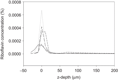
We thank Spoerl and co-workers for their interest in our recent paper on riboflavin distribution in the corneal stroma.Citation1 As correctly pointed out by Spoerl et al., we published relatively low peak riboflavin concentrations of 0.012 mg/ml. Unfortunately, in Figures 3 and 4 an error occurred in the scaling of the y-axes, where the correct unit should be % and not mg/ml, as stated in our erratum below. Thus, the maximum achieved peak concentration is 0.12 mg/ ml, corresponding to 0.012%. In measurements performed by Spoerl et al. (in the preceding letter) and Kampik et al.,Citation2 peak concentrations of up to 0.06% are reported and it is suggested that incomplete epithelial removal in our study could contribute to the observed differences in riboflavin concentration. Our experiments were conducted in porcine and human corneas ex vivo with complete epithelial removal. However, we also measured riboflavin concentrations using the same confocal fluorescence microscopy method in five porcine corneas with intact epithelium (). These scans demonstrate a significantly diminished riboflavin uptake compared to the de-epithelialized specimen. Thus, the slightly lower peak concentrations observed in our study relative to the studies by Kampik et al. and Spoerl et al. appear not to be due to incomplete epithelial removal.
FIGURE 1 Riboflavin distribution in five porcine eyes with intact epithelium, following the preparation phase of 30 min 0.1% riboflavin application, one drop every 5 min.
Spoerl et al. demonstrated, in the preceding letter, the presence of some riboflavin in deeper corneal layers, although the exact depth may be subject to considerable variation in cut thickness. Further, the absorption values represent averages from the overall absorption of a stromal flap of varying thickness. Kampik et al.Citation2 used two-photon fluorescence microscopy to measure the distribution of riboflavin, and further normalized the measured fluorescence intensities at varying corneal depths, using the intensity loss of the second harmonic generation by collagen fibers. With this method, Kampik et al. found very high concentrations of riboflavin in the posterior corneal stroma. This normalization for depth-associated signal loss might play an increasingly significant role when measuring throughout the stromal thickness. However, Kampik et al. do not state how the normalization procedure was verified. Verification could be performed by scanning the corneal buttons from the endothelial side, comparing the obtained fluorescence intensities with the normalized data. In our series, we performed scans from both sides of the corneal button to verify the distribution of riboflavin within the stroma. When the tissue was inverted and placed with the endothelium closest to the objective, the scans showed the same distribution of riboflavin as scans from the epithelial side. Furthermore, the peak concentration was reduced only approximately 20%. Thus, scanning through the cornea caused only minor loss of signal and confirmed that in our series riboflavin only passed into the anterior approximately 200 µm tissue.
REFERENCES
- Søndergaard AP, Hjortdal J, Breitenbach T, et al. Corneal distribution of riboflavin prior to collagen cross-linking. Curr Eye Res. 2010;35:116–121.
- Kampik D, Ralla B, Keller S, et al. Influence of corneal collagen crosslinking with riboflavin and ultraviolet-A irradiation on excimer laser surgery. Invest Ophthalmol Vis Sci. 2010;51(8): 3929–3934. [Epub ahead of print]
- Spoerl E, Mrochen M, Sliney D, et al. Safety of UVA-riboflavin cross-linking of the cornea. Cornea. 2007;26:385–389.
- Wollensak G, Aurich H, Wirbelauer C, et al. Significance of the riboflavin film in corneal collagen crosslinking. J Cataract Refract Surg. 2010;36:114–120.