
Abstract
Objective. Scandinavian guidelines recommend controlling middle-ear effusion (MEE) after acute otitis media. The study aim was to determine whether nurses without otoscopic experience can reliably exclude MEE with tympanometry or spectral gradient acoustic reflectometry (SG-AR) at asymptomatic visits. Design. Three nurses were taught to perform examinations with tympanometry and SG-AR. Pneumatic otoscopy by the study physician served as the diagnostic standard. Setting. Study clinic at primary health care level. Patients. A total of 156 children aged 6–35 months. Main outcome measures. Predictive values (with 95% confidence interval) for tympanometry and SG-AR, and the clinical usefulness, i.e. the proportion of visits where nurses obtained the exclusive test result from both ears of the child. Results. At 196 visits, the negative predictive value of type A and C1 tympanograms (tympanometric peak pressure > −200 daPa) was 95% (91–97%). Based on type A and C1 tympanograms, the nurse could exclude MEE at 81/196 (41%) of visits. The negative predictive value of SG-AR level 1 result was 86% (79–91%). Based on SG-AR level 1 results, the nurse could exclude MEE at 29/196 (15%) of visits. Conclusion. Tympanograms with tympanometric peak pressure > −200 daPa (types A and C1) obtained by nurses are reliable test results in excluding MEE. However, these test results were obtained at less than half of the asymptomatic visits and, thus, the usefulness of excluding MEE by nurses depends on the clinical setting.
Scandinavian guidelines recommend controlling middle-ear effusion (MEE) after acute otitis media. Nurses’ role in this practice is unknown.
Type A and C1 tympanograms (tympanometric peak pressure > −200 daPa) obtained by nurses are reliable test results in excluding MEE.
With type A and C1 tympanograms, nurses can exclude MEE at less than half of the visits.
In primary care, the usefulness of excluding MEE by nurses depends on the clinical setting.
Introduction
Middle-ear effusion (MEE) may affect hearing and the development of speech [Citation1]. Therefore, controlling the resolution of MEE after acute otitis media (AOM) is recommended in Finland and Norway [Citation2,Citation3]. In Sweden, routine ear control is recommended in selected cases [Citation4]. Since AOM is prevalent, these control visits take up a lot of general practitioners’ time. General practitioners’ time for other duties in primary health care might be enhanced if nurses could reliably perform routine ear controls after the diagnosis of AOM.
To detect the presence or absence of MEE, tympanometry and spectral gradient acoustic reflectometry (SG-AR) are adjunctive diagnostic tools for pneumatic otoscopy [Citation5–11]. The tympanometer is a useful tool for general practitioners [Citation12,Citation13], and it has been found to be slightly more accurate than SG-AR [Citation9]. However, the disadvantages of tympanometry include the requirement of an airtight seal to the ear canal and, thus, the cooperation of the young child. On the other hand, SG-AR is easy to perform without these requirements [Citation9]. Even though previous studies have investigated the predictive values, none of the studies has evaluated the clinical usefulness of tympanometry and SG-AR in primary health care, i.e. the proportion of visits where reliable results are obtained from both ears of the child [Citation5,Citation8–11,Citation14–17].
Our aim was to determine whether nurses without otoscopic experience can use tympanometry or SG-AR as a diagnostic tool to exclude MEE in young asymptomatic children. Our second aim was to investigate the proportion of visits where nurses could exclude MEE in asymptomatic children on the basis of tympanometry or SG-AR.
Material and methods
Study population and visits
This study was part of a project examining the optimal diagnosis and treatment of AOM in children aged 6–35 months at primary health care level (http://www.clinicaltrials.gov, identifier NCT00299455) in Turku, Finland [Citation18]. The selection criteria of visits for this study were: between the years 2006 and 2009, control visits where the children had no symptoms (i.e. were asymptomatic); visits at least three days apart; maximum six visits per one child; and visits with successful SG-AR, tympanometry, and pneumatic otoscopy by a physician. Since most of the children were at preverbal age and could not describe their symptoms, parental evaluation was used to assess the symptoms of the child. According to the parents, these children did not have any symptoms or signs during the two days preceding the visit.
Written informed consent was obtained from the parents of each child. The study protocol was approved by the Ethical Committee of the Hospital District of Southwest Finland.
Diagnostic procedures
We trained three nurses to perform tympanometry (MicroTymp2™, Welch Allyn, Skaneateles Falls, NY) and SG-AR (EarCheck PRO Otitis Media Detector™, Innovia Medical LLC, Omaha, NE). The nurses had no experience with pneumatic otoscopy, tympanometry, or SG-AR. In one training session lasting approximately two hours, we taught the nurses the principles of tympanometry and SG-AR and how to perform examinations with these devices. After teaching, we checked how they used the devices. During the study visits, one of the three nurses performed examinations. The examinations were performed independently without any guidance from the study physician.
Children were examined in an upright position. The nurse first performed SG-AR and then tympanometry. After that, the study physician performed SG-AR, tympanometry, pneumatic otoscopy (Macroview otoscope Model 23810™, Welch Allyn, Skaneateles Falls, NY), and video otoscopy (Jedmed, St. Louis, MO). Cerumen was carefully removed before pneumatic otoscopy. The order of diagnostic procedures was chosen to optimize the cooperation of the children during SG-AR and tympanometry.
Pneumatic otoscopy performed by the study physician was used as the diagnostic standard. The diagnosis of MEE was based on the presence of effusion in the middle ear shown by reduced mobility of the tympanic membrane or by visible air-fluid interface; retracted or normal (i.e. slightly concave) position of the tympanic membrane; and the absence of acute inflammatory signs on the tympanic membrane (i.e. distinct erythematous patches or streaks). Of the five study physicians, three made over 90% of the otoscopic examinations and had excellent inter-observer agreement (kappa values ranging from 0.80 to 0.92) [Citation11].
Classification of diagnostic test results
All the tympanograms were evaluated by two study physicians (ML and AR) who were blinded to the results of the otoscopic examination. When there was disagreement, AR made the final decision. We classified the tympanograms according to the classifications of Jerger and Fiellau-Nikolajsen and Lous [Citation5,Citation19]. We separated markedly wide (> 300 daPa) or low peaked (static acoustic admittance < 0.2 mmho) tympanograms into the class Cs because these tympanograms have been shown to associate with increased likelihood of MEE [Citation8]. The five tympanogram types were: Type A (tympanometric peak pressure greater than −100 daPa); type C1 (the pressure between −100 and −199 daPa); type C2 (the pressure −200 daPa or less); type Cs (width > 300 daPa or static acoustic admittance < 0.2 mmho); and type B (flat). Flat tympanograms were repeated three times whenever possible.
We divided the results of SG-AR into five manufacturer determined levels: < 49˚ (level 5); 49–59˚ (level 4); 60–69˚ (level 3); 70–95˚ (level 2); and > 95˚ (level 1). We recorded SG-AR examination as failed if the device repeatedly showed an error symbol or the angle value was seen only for a moment.
Statistical analyses
Test characteristics for the diagnostic test results of the nurses were calculated by comparing the pneumatic otoscopic diagnosis of MEE by the study physician (the positive reference standard) to the healthy middle ear (the negative reference standard). Sensitivity, specificity, positive predictive value, and negative predictive value were calculated with their respective 95% confidence intervals (CI).
The tympanometric diagnostic test result for MEE was the grouped result of type C2, Cs, and B tympanograms (the positive test result) which was contrasted with type A and C1 tympanograms (the negative test result). Correspondingly, test characteristics for SG-AR were calculated for levels 2–5 (≤ 95˚; the positive test result) vs. level 1 (> 95˚; the negative test result). For the reliable exclusion of MEE, we considered that the negative predictive value for the diagnostic test result should be at least 95%.
To estimate the clinical usefulness, we calculated the proportion of visits where the nurses could exclude MEE from both ears of the child with tympanometry or SG-AR. The statistical analyses were generated using SAS™ software (version 9.3 for Windows, SAS Institute, Cary, NC).
Results
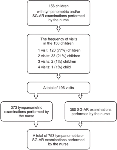
This study included 156 children who had 196 asymptomatic visits when a nurse performed tympanometry and/or SG-AR on one or both ears (). The prevalence of MEE with pneumatic otoscopy by the study physician was 206/753 (27%) of all ears examined by the nurse.
Figure 1. Flow chart of the included children, visits, and tympanometric and spectral gradient acoustic reflectometry (SG-AR) examinations.
The median age of the children was 13 months (range 6–35 months); 58% of the children were male; the median number of previous AOM episodes was 1 (range 0–10); and the median age at first AOM episode was 9 months (range 0–27).
Tympanometric examinations
The nurses performed a total of 373 tympanometric examinations, 272 (73%) of which were successful, i.e. the children were cooperative during the examination. The three nurses succeeded in 35/58 (60%), 149/206 (72%), and 88/109 (81%) of performed tympanometric examinations, respectively. The proportions of MEE with different tympanogram types are presented in . The negative predictive value of type A and C1 tympanograms was 95% (95% CI 91–97%) ().
Table I. Successful tympanometric examinations (n = 272) performed by the nurses.
Table II. Exclusion of middle-ear effusion (MEE) by nurses.a
SG-AR examinations
The nurses performed 332/380 (87%) successful SG-AR examinations. The three nurses succeeded in 46/60 (77%), 183/208 (88%), and 103/112 (92%) of performed SG-AR examinations, respectively. The proportions of MEE with different SG-AR levels are presented in . The negative predictive value of the SG-AR level 1 result was 86% (95% CI 79–91%) (see ).
Table III. Successful spectral gradient acoustic reflectometry (SG-AR) examinations (n = 332) performed by the nurses.
Clinical usefulness
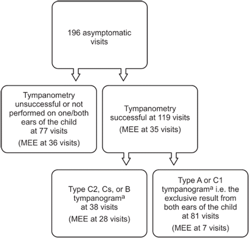
Of the 196 visits, tympanometry was successfully performed on both ears of the child at 119 (61%) visits. The nurses obtained type A and C1 tympanogram (peak pressure > −200 daPa) from both ears of the children at 81 visits. Thus, the exclusive result was obtained at 41% (81/196) of all visits (). Of these 81 visits, MEE was diagnosed with pneumatic otoscopy at seven (9%) visits.
Figure 2. Flow chart of the clinical usefulness of excluding middle-ear effusion (MEE) based on tympanometry performed by the nurses at asymptomatic visits (n = 196). Type A and C1 tympanograms from both ears of the child were regarded as the exclusive test result for MEE. Notes: aTympanogram types: Type A (tympanometric peak pressure greater than −100 daPa); type C1 (the pressure between −100 and −199 daPa); type C2 (the pressure −200 daPa or less); type Cs (width > 300 daPa or static acoustic admittance < 0.2 mmho); and type B (flat).
SG-AR was successfully performed on both ears of the child at 158/196 (81%) visits. The nurses obtained the exclusive result i.e. level 1 from both ears of the child at 29/196 (15%) visits. Of these 29 visits, MEE was diagnosed with pneumatic otoscopy at five (17%) visits.
Discussion
Statement of principal findings
We found that tympanograms with tympanometric peak pressure > −200 daPa (types A and C1) obtained by nurses were reliable test results in excluding MEE. However, the clinical usefulness is reduced by the fact that these test results were obtained only at less than half of the asymptomatic visits due to young uncooperative children, inexperienced nurses, and the relative rarity of exclusive test results.
Strengths and weaknesses of the study
The strengths of our study include the quality of reference diagnostics by trained otoscopists. Furthermore, participating children at the primary health care level represented the age group where most AOM episodes are diagnosed. Thus, this study setting reflects the reality in which routine ear controls are actually performed. The use of pneumatic otoscopy as diagnostic standard instead of myringotomy can be considered a limitation of this study. However, pneumatic otoscopy is the only diagnostic standard that can be used in uncomplicated AOM episodes in primary health care. Furthermore, the relatively high prevalence of MEE may underestimate the usefulness of tympanometry and SG-AR. In addition, only a few nurses performed examinations. On the other hand, the variation in the success rates of the three nurses with tympanometry and SG-AR improves the generalizability of our results and correlates with primary health care centres where nurses have different levels of experience with these diagnostic tools.
Findings in relation to other studies
In this study, tympanometric and SG-AR success rates were affected by the children's age, the nurses’ experience, the tympanometer used, and whether examinations (ears) or visits were analysed. The tympanometric success rate was lower than in most of the previous studies [Citation5,Citation14,Citation16,Citation20]. On the other hand, the tympanometric success rate corresponds to some of the studies with children aged 6–35 months [Citation9,Citation10]. These children aged less than three years are most challenging to examine, and better success rates can be expected when examining older children [Citation20]. Notably, in our current study, the variation of success rates among the three nurses was wide, and the nurse who performed least examinations had the lowest success rate. The nurses were inexperienced when starting to perform examinations, and they gradually became more experienced during the study. Thus, in clinical practice, nurses would perform better if they were already experienced with the devices when starting to perform routine ear controls. Furthermore, the tympanometer (MicroTymp2) we used in this study has been found slightly difficult to handle, and better success rates could be obtained with a more easily handled tympanometer (e.g. GSI 37, Grason-Stadler, Eden Prairie, MN, USA) [Citation21].
Analysing the clinical usefulness, i.e. success with tympanometry and SG-AR in both ears of the child at a visit, was a new practical perspective which none of the previous studies has investigated. Type A and C1 tympanograms obtained by the nurses excluded MEE at less than half of asymptomatic visits, which limits the clinical usefulness. Major causes seem to be young uncooperative children, inexperienced nurses, and the relative rarity of exclusive test results. Even though adding type C2 and Cs tympanograms as exclusive test results for MEE would seem to be tempting, we and others have found that wide tympanograms and tympanograms with marked negative peak pressure (≤ −200 daPa) are unreliable in excluding MEE [Citation8,Citation10]. Furthermore, in our study, SG-AR was not reliable in the exclusion of MEE. In study settings with a low prevalence of MEE and/or an older age group of children, higher negative predictive values for SG-AR have been reported [Citation15,Citation17]. However, because level 1 is a rare result, excluding MEE with SG-AR cannot be considered useful in routine ear controls.
Meaning of the study
This study has a practical meaning for both Scandinavian guideline makers and primary health care centres. The Scandinavian guideline makers could consider recommending that nurses are involved in excluding MEE because our results show that nurses can use tympanometry reliably. On the other hand, when individual primary health care centres consider applying such a practice they should survey their prevalence of MEE, choose an appropriate tympanometer and exclusive test result for their use, and evaluate the success rates of their nurses.
Acknowledgements
This work was supported by the Fellowship Award of the European Society for Paediatric Infectious Diseases to Dr Aino Ruohola and by grants from the Research Funds from Specified Government Transfers; the Foundation for Paediatric Research; Jenny and Antti Wihuri Foundation; the Maud Kuistila Memorial Foundation; University of Turku; the Turku University Foundation; the Finnish Medical Foundation; the Finnish Cultural Foundation, Varsinais-Suomi Regional Fund; the Turku University Hospital Research Foundation; the Paulo Foundation; the Outpatient Care Research Foundation; and the Finnish-Norwegian Medical Foundation.
The study protocol (http://www.clinicaltrials.gov, identifier NCT00299455) was approved by the Ethical Committee of the Hospital District of Southwest Finland.
Declaration of interest
All authors declare to have no competing interests. The authors alone are responsible for the content and writing of the paper.
References
- Rosenfeld RM, Schwartz SR, Pynnonen MA, Tunkel DE, Hussey HM, Fichera JS, et al. Clinical practice guideline: Tympanostomy tubes in children. Otolaryngol Head Neck Surg 2013;149:S1–35.
- Heikkinen T, Huovinen P, Jero J, Pitkäranta A, Renko M, Sumanen M, et al. Update on Current Care Guidelines: Acute otitis media. January 15, 2010. Available at: http://www.kaypahoito.fi/web/english/kaypa-hoito (accessed October 10, 2014).
- Norsk Forening for Otorhinolaryngologi, Hode- og Halskirurgi. Veileder for øre-nese-halsfaget/Otologi/Akutt otitt [Recommendations for ear-nose-throat diseases/ Otology/Acute otitis media]. March 8, 2012. Available at: http://legeforeningen.no/fagmed/norsk-forening-for-otorhinolaryngologi-hode-og-halskirurgi/veileder-for-ore-nese-halsfaget/otologi/akutt-otitt/ (accessed October 10, 2014).
- Läkemedelsverket. Diagnostik, behandling och uppföljning av akut mediaotit (AOM) – ny rekommendation [Diagnosis, management, and follow-up of acute otitis media – new recommendation]. Information från Läkemedelsverket 2010;21:13–59.
- Jerger J. Clinical experience with impedance audiometry. Arch Otolaryngol 1970;92:311–24.
- Teele DW, Teele J. Detection of middle ear effusion by acoustic reflectometry. J Pediatr 1984;104:832–8.
- Kimball S. Acoustic reflectometry: Spectral gradient analysis for improved detection of middle ear effusion in children. Pediatr Infect Dis J 1998;17:552–5; discussion 580.
- Smith CG, Paradise JL, Sabo DL, Rockette HE, Kurs-Lasky M, Bernard BS, et al. Tympanometric findings and the probability of middle-ear effusion in 3686 infants and young children. Pediatrics 2006;118:1–13.
- Chianese J, Hoberman A, Paradise JL, Colborn DK, Kearney D, Rockette HE, et al. Spectral gradient acoustic reflectometry compared with tympanometry in diagnosing middle ear effusion in children aged 6 to 24 months. Arch Pediatr Adolesc Med 2007;161:884–8.
- Helenius KK, Laine MK, Tähtinen PA, Lahti E, Ruohola A. Tympanometry in discrimination of otoscopic diagnoses in young ambulatory children. Pediatr Infect Dis J 2012;31:1003–6.
- Laine MK, Tähtinen PA, Helenius KK, Luoto R, Ruohola A. Acoustic reflectometry in discrimination of otoscopic diagnoses in young ambulatory children. Pediatr Infect Dis J 2012;31:1007–11.
- Lous J, Ryborg CT, Damsgaard JJ, Munck AP. Tympanometry in general practice: Use, problems and solutions. Fam Pract 2012;29:726–32.
- Lous J. Use of tympanometry in general practice in Denmark. Int J Pediatr Otorhinolaryngol 2014;78:124–7.
- Palmu A, Puhakka H, Rahko T, Takala AK. Diagnostic value of tympanometry in infants in clinical practice. Int J Pediatr Otorhinolaryngol 1999;49:207–13.
- Teppo H, Revonta M, Linden H, Palmu A. Detection of middle-ear fluid in children with spectral gradient acoustic reflectometry: A screening tool for nurses? Scand J Prim Health Care 2006;24:88–92.
- Blomgren K, Haapkyla J, Pitkaranta A. Tympanometry by nurses: Can allocation of tasks be optimised? Int J Pediatr Otorhinolaryngol 2007;71:7–10.
- Puhakka T, Pulkkinen J, Silvennoinen H, Heikkinen T. Comparison of spectral gradient acoustic reflectometry and tympanometry for detection of middle ear effusion in children. Pediatr Infect Dis J 2014;33:e183–6.
- Tähtinen PA, Laine MK, Huovinen P, Jalava J, Ruuskanen O, Ruohola A. A placebo-controlled trial of antimicrobial treatment for acute otitis media. N Engl J Med 2011;364:116–26.
- Fiellau-Nikolajsen M, Lous J. Prospective tympanometry in 3-year-old children: A study of the spontaneous course of tympanometry types in a nonselected population. Arch Otolaryngol 1979;105:461–6.
- Koivunen P, Alho OP, Uhari M, Niemela M, Luotonen J. Minitympanometry in detecting middle ear fluid. J Pediatr 1997;131:419–22.
- Patricoski C, Ferguson S. Which tympanometer is optimal for an outpatient primary care setting? J Fam Pract 2006; 55:946–52.