Bevacizumab (Avastin®) in association with chemotherapeutic agents has become a standard treatment for patients with metastatic colorectal cancer reaching a median overall survival of 22.7 months [Citation1]. However, bevacizumab-therapy can be associated with some adverse events, including bleeding (3%), gastrointestinal perforation (2%), arterial thromboembolism (1%), hypertension (5.3%), proteinuria (1%) and wound-healing complications (1%) [Citation2–5]. Bleeding complications have been observed as pulmonary hemorrhage and/or hemoptysis, mainly in patients with lung cancer; gingival and vaginal bleeding have also been reported, but are less common [Citation2].
Herein, the occurrence of a (pseudo-)aneurysm within an asymptomatic renal angiomyolipoma in a patient with colorectal liver metastases treated with FOLFIRI and bevacizumab is reported.
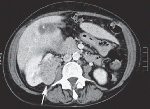
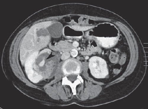
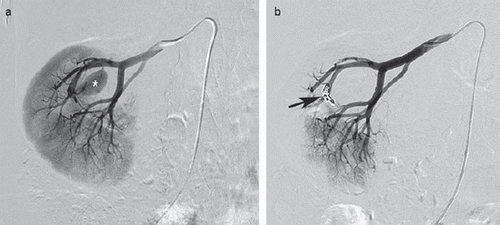
A 57-year-old woman underwent a laparoscopic low anterior resection for obstructing rectosigmoid carcinoma with concommitant diffuse liver metastases. At pre-operative screening CT, a fat-containing mass lesion was detected suggestive of an angiomyolipoma (). A true-cut biopsy of the renal lesion was performed. Pathological analysis confirmed the diagnosis of an angiomyolipoma. One month postoperatively, chemotherapeutic treatment with FOLFIRI and buvacizumab was started to manage the liver metastases. Control CT-scan three months after start of the chemotherapeutic treatment showed a marked involution of the liver metastases, but also a (pseudo-) aneurysm within the angiomyolipoma. The (pseudo-) aneurysm had a diameter of 4.5 cm with peripheral thrombus-formation (). It was agreed to exclude the (pseudo-)aneurysm by transcatheter embolization. Under local anesthesia, the feeding vessel was selectively catheterized () and occluded by 250 μ microparticles (Embozène, CeloNova, Newnan, GA, USA) and microcoils (Microtornado, Cook Medical, Bjaeverskov, Denmark) (). FU-CT two months later depicted a further shrinkage of the liver metastases and a full exclusion of the (pseudo-) aneurysm.
Figure 1. CT-scan of the abdomen, performed one month after surgical resection of the primary tumor, shows liver metastases (asterisk) and an incidentally found angiomyolipoma (white arrow) in the right kidney.

Figure 2. CT-scan of the abdomen three months after chemotherapy associated with bevacizumab reveals the appearance of a (pseudo-)aneurysm within the angiomyolipoma (black arrowhead).

Figure 3. a. Selective angiography of the right kidney confirms a large (pseudo-)aneurysm (asterisk) in the hilum of the kidney. b. Selective angiography of the right kidney after coil-embolization (arrow): complete exclusion of the (pseudo-)aneurysm.

Bleeding complications associated with bevacizumab-therapy potentially can be explained by the anti-angiogenic effect, thus inhibiting the growth of the endothelium which might result in vessel wall contiguity and pseudoaneurysm formation. In the present case, the exact pathophysiological mechanism for the development of the (pseudo-)aneurysm within the angiomyolipoma is not very clear. A vessel wall lesion might be induced initially by the true-cut biopsy. However, FU-CT one month after the biopsy showed no radiological sign of intralesional aneurysm formation. Bevacizumab therapy might have jeopardized endothelialization at a biopsy-injured vessel, a phenomenon which is similar to late bevacizumab-related surgical site complications [Citation6]. Also, bevacizumab therapy might have a direct effect on the physiology of the endothelium of the vascular components of the angiomyolipoma. Angiomyolipoma may harbor subtle microaneurysms not visible on imaging and a pseudoaneurysm may have developed spontaneously at the site of such a microaneurysm with a susceptible and fragile endothelial lining [Citation7].
In summary, the exact pathophysiological mechanism for the development of the (pseudo-) aneurysm within the angiomyolipoma is not very clear. However, it is not excluded that bevacizumab might be a potential trigger.
Based on this observation it is suggested that patients presenting with a renal angiomyolipoma and selected for anti-angiogenic treatment, should be monitored closely with imaging techniques and whenever a (pseudo-)aneurysm occurs or increases in size, selective embolization must be performed. Selective transcatheter embolization of angiomyolipoma is safe, effective and durable in experienced hands [Citation8,Citation9].
In conclusion, a yet unreported case of (pseudo-)aneurysm formation within a renal angiomyolipoma in a patient under bevacizumab therapy is described, stressing the need for close radiological follow-up during anti-angiogenic therapy, although there is no proof of a direct relation between bevacizumab therapy and (pseudo-)aneurysm formation in an angiomyolipoma. Transcatheter embolotherapy is the preferred treatment whenever a (pseudo-) aneurysm becomes apparent.
Declaration of interest: The authors report no conflicts of interest. The authors alone are responsible for the content and writing of the paper.
References
- Van Cutsem E, Rivera F, Berry S, Kretzschmar A, Michael M, Dibartolomeo M, . Safety and efficacy of first line bevacizumab with FOLFOX, XELOX, FOLFIRI and fluoropyrimidines in metastatic colorectal cancer: The BEAT study. Ann Oncol 2009;20:1842–7.
- Gordon MS, Cunningham D. Managing patients treated with bevacizumab combination therapy. Oncology 2005; 69:25–33.
- Heinzeling JH, Huerta S. Bowel perforation from bevacizumab for the treatment of metastatic colon cancer: Incidence, etiology, and management. Curr Surg 2006;63: 334–7.
- Kesmodel SB, Ellis LM, Chang GJ, Abdalla EK, Kopetz S, Vauthey JN, . Preoperative bevacizumab does not significantly increase postoperative complication rates in patients undergoing hepatic surgery for colorectal cancer liver metastases. J Clin Oncol 2008;26:5254–60.
- Collins D, Ridgway PF, Winter DC, Fennelly D, Evroy D. Gastrointestinal perforation in metastatic carcinoma: A complication of bevacizumab therapy. Eur J Surg Oncol 2009; 35:444–6.
- Bège T, Lelong B, Viret F, Turrini O, Guiramand J, Topart D, . Bevacizumab-related surgical site complications despite primary resection in colorectal cancer patients. Ann Surg Oncol 2009;16:856–60.
- Bissler JJ, Kingswood JC. Renal angiomyolipomata. Kidney Int 2004;66:924–34.
- Ramon J, Rimon U, Garniek A, Golan G, Bensaid P, Kitrey ND, . Renal angiomyolipomata: Long-term results following selective arterial embolization. Eur Urol 2009;55: 155–62.
- Kothary N, Soulen MC, Clark TW, Wein AJ, Shlansky-Goldberg RD, Crino PB, . Renal angiomyolipomata: Long-term results after arterial embolization. J Vasc Interv Radiol 2005;16:45–50.
