Abstract
In preparation for studies of dose volume of ionizing radiation and long-term side effects, we assessed both variation in position and volume of organs at risk in the small pelvis. Material and methods. On 10 men and seven women we delineated the sigmoid, rectum, anal sphincter, bladder, penile bulb, and cavernous bodies in two CT scans taken between five to 69 days apart. Results. The measured overlap of the two delineated volumes divided by the maximum possible overlap, was below 50% for the sigmoid in six of 17 patients, for the distal 4 cm of the sigmoid in five of 17 patients, for the rectum in none of 17 patients, for the anal sphincter in three of 17 patients and for the urinary bladder in none of 17 patients. The smaller volume divided by the larger volume was below 50% in three of 17 patients for the sigmoid, in six of 17 patients for the 4 distal cm of the sigmoid, in two of 17 patients for the rectum, in two of 17 patients for the anal sphincter and in seven of 17 patients for the urinary bladder. For the urinary bladder the largest deviation was found cranially, 4.0 cm (SD 2.0 cm), the caudal part being relatively fixed. For the rectum the largest deviation was found in the anterior wall, 1.8 cm (SD 0.7 cm), with maximum documented variation in cranial direction of 3.2 cm (SD 1.8 cm). Conclusions. The sigmoid varies considerably in documented position with the largest deviation anteriorly, the urinary bladder change in volume with the extension mainly located cranially and for the rectum the anterior wall is the most mobile with the distension becoming more pronounced cranially. In modeling dose-volume effects one may consider our results.
Being subjected to radiotherapy for malignant disease in the small pelvis unfortunately entails receiving unwanted ionizing radiation to the organs surrounding the tumor site, the organs at risk [Citation1]. Using high-quality equipment and delineating the organs at risk we try to avoid administering unwanted ionizing radiation without tampering with tumor control [Citation2]. Still we have limited information on how well the calculated dose and volume to an organ at risk correlate with what really happens during the course of radiotherapy in the real-life situation.
Organs, such as the sigmoid, rectum, and bladder change in size, shape, and position during the treatment [Citation3–9]. Movements occur due to peristalsis, bladder and rectal filling, affecting the relation to surrounding organs [Citation10–13]. Tumor shrinkage may affect the anatomy and radiotherapy can cause hyperirritability of the bowels.
The aim of this study is to obtain data about the movements of the pelvic organs. We will use this information in large ongoing studies in women treated for gynecological cancers and men treated for prostate cancer, to better understand the relation between dose volume and unwanted long-term side-effects that impair quality of life for long-term cancer survivors treated with pelvic radiotherapy. In order to try to evaluate the uncertainties of the organ movements, we analyzed the maximum variation of an organ at risk. We delineated anatomically the organs at risk in the small pelvis, and assessed their position and volume in 17 patients radically treated with radiotherapy. Two computed tomography (CT) investigations, performed immediately before or during radiotherapy, were compared.
Material and methods
Study population
We identified 17 patients who received pelvic radiotherapy as primary treatment and who, in addition to the dose planning CT scan, CT 1, were exposed to a second CT scan. Ten of the patients were men treated for prostate cancer, and seven were women treated for gynecological malignancies (). The second CT scan, CT 2, was made due to dose-escalation to a smaller target volume in the male patients and for the women the reasons were; adding a para-aortic field (no. 11, 12); the first CT not covering the planned volume (13, 15, 16); or continuation to a higher dose for a smaller field (14, 17). All patients were scanned in the supine position and a knee-cushion was used for immobilization of the pelvis. No standardized instruction for bladder filling was given over the time period, nor was there any specific routine regarding emptying of the rectum. The radiotherapy was given between 1997 to 2005 at the Sahlgrenska University Hospital in Göteborg, Sweden.
Table I. Demography.
Imaging
Low density bladder contrast was administered in all patients. Computed tomography was performed by obtaining contiguous transverse sections with a Hi Speed FX/i scanner (General Electric Medical Systems). A scanning technique using 120 kV, 200 mAs, and a section thickness of 5 mm (in two female patients, 7 mm) was employed. The CT sections had a 512 × 512 pixel resolution. The scanned area included the whole pelvis from the iliac crest to the ischial tuberosity. The two CT scans for each patient were matched according to pelvic bony structures.
Definition of organs at risk
The organs at risk investigated were (1) the sigmoid (cranial limit: the transition to the descending colon); (2) the distal 4 cm of the sigmoid; (3) the rectum (cranial limit located at the sigmoid junction where the rectum turns horizontally into the sigmoid; (4) the anal sphincter; (5) the bladder; (6) the penile bulb; and (7) the cavernous bodies (intracorporeal parts). When delineating the organs we followed the outer border of the structure where this was possible. Where the CT scan was suboptimal for proper visualization of the structures, interpolation between adjacent slices was made by the eye. To keep variations of delineation due to interpersonal bias to a minimum, the same two oncologists (A.C.W. and D.A.) together delineated the organs at risk, reaching consensus on the outlining in all cases (17 + 17). The two CT scans of the same patient were not blinded. In the CT slices where it was difficult to differ between the sigmoid colon and the small intestine, we were helped by the second scan to a certain extent. The organ definitions were confirmed by a senior radiologist.
Analysis of position
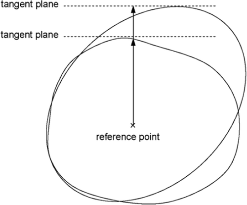
To assess the absolute magnitude of variation in position and volume of the organs at risk between the two CT examinations, and to locate the direction in which this variation had taken place, we first identified the extreme points in six anatomical directions, the cranial, caudal, dexter, sinister, anterior, and posterior directions. As extreme point, we considered the point through which a perpendicular tangent intersected the organ surface in each of the six directions (). This resulted in a definition of the “smallest box” volume which contained the organ at risk being assessed. To estimate the variation in position, we measured the absolute difference (in cm) of the extreme points compared with a given reference point of each organ at risk in all six directions in every patient. For the rectum and the anal sphincter, we conducted a more thorough assessment, where we measured the variation in the dexter, sinister, anterior, and posterior direction in each CT slice.
Figure 1. The variation in position measured as the absolute difference (cm) of extreme points compared to a given reference point of each organ at risk in two consecutive CT-scans.
Analysis of volume
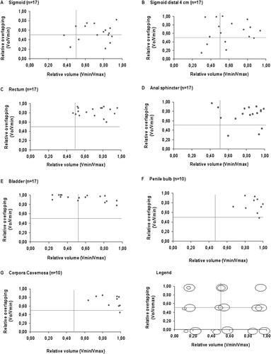
The volume of the delineated structures in the two CT scans of the 17 patients was calculated by the radiotherapy planning system. Using the Varian Eclipse Treatment Planning System (TPS), we calculated the relative volume of each organ at risk comparing the two CT scans for the same patient. The smaller volume of the two was put in the numerator in order to obtain a value between 0.0 and 1.0. A value of 1.0 shows that the two documented volumes are equal, and any value between 0.0 and 1.0 shows a volume difference. The value of 0.0 cannot be reached, unless the organ at risk is absent in one of the CT scans (, Legend).
Figure 2. Relative volume (Vmin/Vmax) of the smaller volume compared to the larger volume and relative overlapping (Vo/Vmin) for the organs at risk. The legend shows how the diagrams display relative volume and overlapping of the organs at risk. A value of 1 in relative volume means that the two volumes are equally large. Values approaching 0 means that there is a large difference in volume. A value of 1 in relative overlapping means that the smaller volume lies within the larger volume. A value of 0 means that the two volumes are separated.
Analysis of overlap
The maximum possible overlap (VOmax) occurs when the smaller volume is completely included in the larger volume. In this case VOmax is equal to the smaller volume (Vmin). To calculate the relative overlap, we compared the overlap volume (VO) of each organ at risk with the maximum possible overlap (VOmax) in each patient for all organs at risk.
A value of 1.0 indicates a perfect overlap, and a value of 0.0 indicates that the volumes are completely separated (). The overlap volumes were obtained using Boolean functions in the TPS.
Results
Among the 17 patients, the average age of the men was 68 years (range 55–76 years). The average age of the women was 65 years (range 32–78 years). The time between the two CT investigations varied from 40 to 57 days (mean 68 days) for men and from five to 69 days (mean 27 days) for women. The delivered target dose at CT 2 varied from 32 to 58 Gy, with an average of 47 Gy for men, and from 0 to 46 Gy, with an average of 11 Gy, for women (). The largest volume variations were seen for the sigmoid and for the urinary bladder (, ). Among the seven studied organs at risk, the urinary bladder had the largest mean volume (179 cm3). The penile bulb had the smallest mean volume (5.5 cm3) ().
Table II. Absolute volume (cm3), relative volume† and relative overlapping* in two consecutive CT-scans for seven organs at risk in the small pelvis.
Table III. Mean, median and range of baseline characteristics given in .
Variation in position in six anatomical directions
The absolute variation in the six anatomical directions for each organ is shown in . Large variations in position of the organs were found in one or more directions for the sigmoid, the distal 4 cm of the sigmoid, and the bladder. For the sigmoid, the absolute variation in position was generally large, except for the posterior direction. The largest deviation of the sigmoid, 4.9 cm (standard deviation, SD, 1.5 cm), was found in the extreme anterior points; the deviation of the extreme cranial points was 3.5 cm (SD 1.4 cm). For the distal 4 cm of the sigmoid, the largest deviation, 4.8 cm (SD 1.8 cm), was found in the extreme anterior points. For the urinary bladder, the largest deviation, 4.0 cm (SD 2.0 cm), was found in the extreme cranial points.
Figure 3. Deviation of the extreme points in the organs at risk, CT 2 compared to CT 1. The extreme points in the six directions together define the smallest “box-volume” that encloses the organ at risk. A positive value represents movement in the direction indicated on the x-axis. The diagrams show the mean±1SD and the maximum deviation measured.
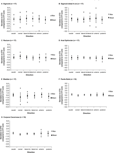
Smaller variations were found for the other organs. For the whole rectum, the largest deviation, 1.8 cm (SD 0.7 cm), was found in the extreme anterior points, located in the superior part of the rectum. For the cavernous bodies, the largest deviation, 1.6 cm (SD 0.7 cm), was found in the extreme anterior points.
Variation in overlap and volume
The agreement in relative overlap was >0.5 in 13 of 17 patients () for the rectum, the anal sphincter, and the bladder. It was >0.5 in eight of 10 patients for the penile bulb and the cavernous bodies. The agreement was also >0.5 in relative volume for the sigmoid, the rectum, the anal sphincter, the penile bulb, and the cavernous bodies. Looking at the specific organs at risk, this means that for the sigmoid, the agreement in relative volume was >0.5 in 14 of 17 patients (), while the agreement in relative overlap was >0.5 only in 11 of 17 patients. For the rectum, the agreement in relative overlap was >0.6 in all 17 patients and the agreement in relative volume was >0.5 in 16 of 17 patients. For the anal sphincter, the agreement in relative overlap was >0.5 in 14 of 17 patients. With regard to the relative volume, the agreement was >0.5 in 15 of 17 patients. For the bladder, all 17 patients reached an agreement in relative overlap, >0.8, with perfect overlap in four patients, while the relative volume varied in agreement from 0.2 to 0.9. This shows that the cranial part of the bladder moves while the caudal part is more fixed. The penile bulb and the cavernous bodies showed little variation in relative overlap and relative volume.
Table IV. Proportion of delineated organs at risk that reaches >0.5 in agreement.
Variation in position of the anal sphincter and rectum
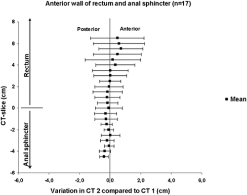
Analysis of the variation in each CT slice for the rectum and the anal sphincter showed that the overall variation was largest in the anterior wall (). Cranially, we only included CT slices where we had data from 10 or more patients. The variation was larger in the more cranial CT slices. In this material, the CT slice with the largest variation was found 4.5 cm cranially of the anorectal transition, where the anterior wall varied by up to 3.2 cm (SD 1.8 cm).
Figure 4. Variation of the anterior wall extreme points of rectum and anal sphincter measured in each CT-slice (0.5 cm thickness) in two consecutive CT-scans in 17 patients (mean and one standard deviation).
Discussion
We found that the documented relative overlap was good for the rectum, the anal sphincter, the urinary bladder, the penile bulb, and the cavernous bodies. The agreement on documented relative volume was good for the sigmoid, the rectum, the anal sphincter, the penile bulb, and the cavernous bodies. Our results show that the variation in relative overlap and relative volume for the sigmoid mostly occurs in the anterior and cranial directions. For the distal 4 cm of the sigmoid, the variation in relative overlap and relative volume mostly occurs in the anterior direction. The variation in relative overlap and relative volume for the rectum is primarily found in the anterior rectal wall, for the urinary bladder in the cranial part and for the cavernous bodies anteriorly. The variation in relative overlap of the anterior rectal wall increases in the cranial direction.
For the sigmoid the agreement in relative volume was good, but the variation in relative overlap was large. Given that the delineation was accurate, the sigmoid was very mobile. For the distal 4 cm of the sigmoid, the agreement was neither good in relative volume nor was it good for relative overlap, indicating difficulties in determining this arbitrary volume anteriorly, or, which is more probable, considering the result for the whole sigmoid, indicating a true variation in both volume and position. Our results show that the sigmoid is a mobile organ and that one CT scan gives little information about the location of the sigmoid during the course of radiotherapy.
For the rectum as a whole, we found good agreement in relative overlap together with good agreement in relative volume for 14 of 17 patients. However, when we analyzed each CT slice, the anterior rectal wall varied in relative overlap. The average volume was 77 cm3 (range 21–170 cm3). Roeske et al. [Citation14] found an average rectal volume of 79 cm3 (range 35–182 cm3) in 10 patients with localized carcinoma of the prostate followed with four to seven weekly CT scans before and during radiotherapy. Their average rectal volume was only 2 cm3 larger than what we found. Lebesque et al. [Citation9] studied 11 prostate cancer patients with four CT scans before and during radiotherapy and found rectal volumes ranging from 58 to 157 cm3, with an average of 94 cm3, 17 cm3 larger than our results.
Measuring the position of the rectal wall in each CT slice for all our patients, we found a large variation of the anterior wall, the largest variation being 3.2 cm. The variations in the other three directions analyzed were small, if not negligible. Also, the distension of the anterior wall becomes more pronounced cranially. Fiorino et al. [Citation11] followed nine patients previously operated with radical prostatectomy, with weekly CT-scans, during treatment with adjuvant radiotherapy. Six of nine patients showed a systematic anterior shift of both the anterior and the posterior rectal walls in the cranial half of the rectum.
For the anal sphincter, we found good agreement in relative overlap and relative volume between the two CT investigations. The anal canal lying inside the sphincter is empty under normal conditions, and both are surrounded by the levator ani muscle in the cranial part. Fecal leakage has been associated with absorbed dose to the anal sphincter region [Citation15]. It is unclear which anatomical structure is most important for anal continence.
The good agreement in relative overlap, but poor agreement in relative volume for the urinary bladder reflects the bladder extending cranially with increasing bladder filling, the caudal part being relatively fixed. We found bladder volumes between 54 cm3 and 424 cm3, with an average of 171 cm3. In a study comparing empty bladder and full bladder in 50 men with prostate cancer, Pinkawa et al. [Citation4] found significant bladder wall displacement only at the superior and anterior borders. Fiorino et al. detected very large variations in bladder filling, with large variability in the cranial and anterior direction, while the caudal border was not affected by different filling. Lebesque et al. found bladder volumes ranging from 94 to 317 cm3, with an average of 220 cm3. In the study by Roeske et al. the observed bladder volume varied from 70 to 509 cm3, with an average of 187 cm3. Consequently, available data indicate that the bladder varies considerably, the largest variation being in the cranial direction.
The penile bulb and the cavernous bodies showed little variation in relative overlap and relative volume in spite of some uncertainty when dealing with volumes of small cranial and caudal extension in comparison with slice thickness. The variation in the cavernous bodies, seen in the anterior direction, probably reflects the difficulty in determining where the intraabdominal part ends when delineating the structure.
The method chosen is more sensitive for the extreme position of the organs which we found to be relevant for our future study of dose-volume and late effects of radiotherapy. The study is limited due to the small number of patients, different diagnoses and limited CT scans available, and also by bladder and rectal volumes not being standardized. However, with standardized bladder and rectal filling, the variation not only in size but in position becomes less and our aim was to study the maximum possible variation. In the clinical situation interactions between the pelvic organs including the prostate/uterus also need further investigation. Partial volume effect due to the transverse orientation of the CT scans may have influenced the measurements of the cranial and caudal organ position and volume and may in part explain the larger variations in this direction. It is uncertain to what extent the variation in cranial and caudal directions can be compared with the variation in the anterior, posterior, and lateral directions. The effect on smaller structures, such as the bulb, is more obvious than that of larger organs like the bladder. The matching procedure according to skeletal structures, may make a difference of up to 0.5 cm in some parts of the pelvis, depending on different patient set-ups at the scanner and differing thickness of the CT sections.
Implications of the study
When the sigmoid, rectum, and bladder are present in an area with a steep dose gradient, their changes in position and volume may have an important effect on the radiation dose to these organs. The variation in documented position and volume presented here can be considered when looking at the relation between dose volume histograms for organs at risk and self-assessed long-term treatment related symptoms and quality of life.
Declaration of interest statement
Any actual or potential conflicts of interest do not exist.
References
- Pinkawa M, Fischedick K, Asadpour B, Gagel B, Piroth MD, Eble MJ. Low-grade toxicity after conformal radiation therapy for prostate cancer—impact of bladder volume. Int J Radiat Oncol Biol Phys 2006;64:835–41.
- Fokdal L, Honore H, Hoyer M, Meldgaard P, Fode K, von der Maase H. Impact of changes in bladder and rectal filling volume on organ motion and dose distribution of the bladder in radiotherapy for urinary bladder cancer. Int J Radiat Oncol Biol Phys 2004;59:436–44.
- Stasi M, Munoz F, Fiorino C, Pasquino M, Baiotto B, Marini P, . Emptying the rectum before treatment delivery limits the variations of rectal dose–volume parameters during 3DCRT of prostate cancer. Radiother Oncol 2006;80:363–70.
- Pinkawa M, Asadpour B, Siluschek J, Gagel B, Piroth MD, Demirel C, . Bladder extension variability during pelvic external beam radiotherapy with full or empty bladder. Radiother Oncol 2007;83:163–7.
- Muren LP, Smaaland R, Dahl O. Organ motion, set-up variation and treatment margins in radical radiotherapy of urinary bladder cancer. Radiother Oncol 2003;69:291–304.
- Miralbell R, Taussky D, Rinaldi O, Lomax A, Canales S, Escude L, . Influence of rectal volume changes during radiotherapy for prostate cancer: A predictive model for mild-to-moderate late rectal toxicity. Int J Radiat Oncol Biol Phys 2003;57:1280–4.
- Nuyttens JJ, Robertson JM, Yan D, Martinez A. The variability of the clinical target volume for rectal cancer due to internal organ motion during adjuvant treatment. Int J Radiat Oncol Biol Phys 2002;53:497–503.
- Turner SL, Swindell R, Bowl N, Marrs J, Brookes B, Read G, . Bladder movement during radiation therapy for bladder cancer: Implications for treatment planning. Int J Radiat Oncol Biol Phys 1997;39:355–60.
- Lebesque JV, Bruce AM, Kroes AP, Touw A, Shouman RT, van Herk M. Variation in volumes, dose-volume histograms, and estimated normal tissue complication probabilities of rectum and bladder during conformal radiotherapy of T3 prostate cancer. Int J Radiat Oncol Biol Phys 1995;33:1109–19.
- Kirisits C, Lang S, Dimopoulos J, Oechs K, Georg D, Potter R. Uncertainties when using only one MRI-based treatment plan for subsequent high-dose-rate tandem and ring applications in brachytherapy of cervix cancer. Radiother Oncol 2006;81:269–75.
- Fiorino C, Foppiano F, Franzone P, Broggi S, Castellone P, Marcenaro M, . Rectal and bladder motion during conformal radiotherapy after radical prostatectomy. Radiother Oncol 2005;74:187–95.
- Hellebust TP, Dale E, Skjonsberg A, Olsen DR. Inter fraction variations in rectum and bladder volumes and dose distributions during high dose rate brachytherapy treatment of the uterine cervix investigated by repetitive CT-examinations. Radiother Oncol 2001;60:273–80.
- van Herk M, Bruce A, Kroes AP, Shouman T, Touw A, Lebesque JV. Quantification of organ motion during conformal radiotherapy of the prostate by three dimensional image registration. Int J Radiat Oncol Biol Phys 1995;33:1311–20.
- Roeske JC, Forman JD, Mesina CF, He T, Pelizzari CA, Fontenla E, . Evaluation of changes in the size and location of the prostate, seminal vesicles, bladder, and rectum during a course of external beam radiation therapy. Int J Radiat Oncol Biol Phys 1995;33:1321–9.
- al-Abany M, Helgason ÁR, Cronqvist AK, Lind B, Mavroidis P, Wersäll P, . Dose to the anal sphincter region and risk of fecal leakage. Acta Oncol 2004;43:117–8.