Phyllodes tumors are rare neoplasm of the breast that account 2 to 3% of fibroepithelial breast tumors [Citation1,Citation2]. Their prognosis and treatment are still subject of discussion. They occur most frequently between the ages of 30 and 50.
The majority tend to be less than 5 cm in size. However, in some cases the tumor growth increases rapidly. They are associated with skin ulceration in some cases.
In this case report, we discuss a patient who developed a rapidly expanding malignant phyllodes tumor.
Case report
A 32-year-old young woman arrived at the emergency room with a huge tumor of the left breast. No family history of breast cancer was found. She told us that it began one year earlier, when she discovered a solid mass in the left breast that she neglected. She presented herself, when she observed that the tumor was growing rapidly, and ulceration and bleeding occurred.
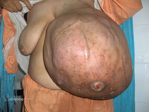
We were surprised by the size and the smell of the tumor. It measured 60 × 38 × 18 cm. It had an inflammatory aspect with ulcerations (). Palpation of the axillary lymph nodes revealed an adenopathy of 6 cm fix to the deep plan.
Figure 1. Preoperative view.
The examination of the right breast was normal.
Neither a mammography nor an ultrasound study was able to be realized because of the tumor size. Due to the physical nature of the mass and suspicion of malignancy remained, a wide local resection of the mass was recommended. The resection was large extending, from the clavicle superiorly, the sternum medially, laterally to the anterior axillary line and to 3 cm lower than the inframammary fold. The deep limit was pre-costal infiltrating the minor and major pectoralis. The axillary's lymph nodes resection was then completed ( and ).
Figure 2. Postoperative result.
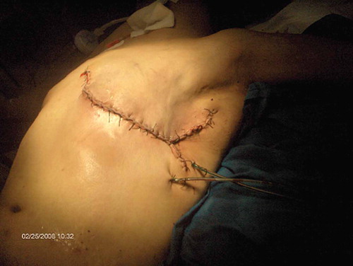
Figure 3. Operative piece.
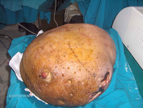
The total body scintigraphy, the thoracic x-ray and the abdominal sonography were normal. The histology revealed a phyllodes tumor of high grade. One of the 22 dissected axillary nodes indicated malignant involvement. Because of the high grade of the tumor, the oncologists decided to continue the treatment by radiotherapy. The one-year check up with a mammography did not show any recurrence.
Discussion
Phyllodes tumors of the breast are unusual; they count for 0.4% of all breast tumors [Citation1,Citation2]. It is well established that there is a wide range in the age at diagnosis, with an average of 45 years [Citation2,Citation3] at presentation.
Phyllodes tumors are fibroepithelial breast tumors that are characterized by proliferation of both stromal and epithelial cells [Citation4,Citation5]. The small phyllodes tumors are very similar to fibroadenomas in terms of clinical and histological aspects. But the distinction is important to make preoperatively, as simple enucleation is relevant for adenomas.
Patients typically present with a firm, seldom painful breast mass that is smooth, mobile and well circumscribed [Citation4,Citation6]. The tumor exhibits continuous growth, but it may also increase rapidly in size. This may result in shiny stretched skin that becomes translucent to display underlying veins [Citation6]. As a result of stretching and increased pressure; the attenuated skin ulcerates secondary to ischemia [Citation7].
In the literature, the tumor size varies between 5 mm and 450 mm [Citation1–4]. The mean volume of the phyllodes tumors was significantly higher in the high-grade group compared with the low-grade group. This is probably a reflection of the greater proliferative activity of the high-grade phyllodes tumors [Citation3].
The treatment of phyllodes tumors is surgical. A security resection margin of 1 to 2 cm is necessary [Citation2,Citation5]. When the tumor is larger than 5 cm or malignant, a mastectomy is recommended [Citation2–4].
The resection of the axillary's lymph nodes does not need to be systematic [Citation8]. This is in accordance with the fact that lymph node involvement is rarely described in phyllodes tumors [Citation8] and metastatic spread occurs often haematogenously.
Radiotherapy does not routinely play a role in conventional treatment modalities for phyllodes tumors. Belkacémi et al. study [Citation9] has reported a benefit from adjuvant radiotherapy in patients exhibiting poor prognostic features. He observed, after a median follow-up of 106 months, in the malignant group and borderline group (n = 159), that radiotherapy decrease local recidive. The local control with radiotherapy was 86% versus 59% without (p = 0.02). Moreover, negative margins and non-residual disease were observed after initial treatment and radiotherapy. However, it had no impact in survival. The chemotherapy does not improve the survival rate but it is useful for palliative treatment [Citation10]. This was included in Ben Hassouna et al.'s study [Citation1] of metastasis cases, but the evolution was always fatal.
Karim et al.'s study [Citation3] summarized the clinical outcome data of 19 series of phyllodes tumors. The recurrence rate reported by the 19 studies varied widely (benign 1.5–12%, borderline 0–38.5% and malignant 3–50%) but has an upward trend with increasing grade.
Ben Hassouna et al.'s study [Citation1] showed that the risk of distant spread is correlated with histotype, tumor size, cytonuclear atypia, presence of necrosis and hemorrhage, tumor margins, stromal overgrowth, and number of mitosis. No relationship between age, type of surgery, recurrences, and risk of metastasis was found in study [Citation1].
The overall five year disease-free survival rate ranged from 78–91% [Citation1,Citation3,Citation4,Citation8]. The total time to recurrence, observed in Karim et al.'s study [Citation3] was significantly lower in the high-grade group compared with the low-grade group. This may reflect the greater proliferative potential and tendency for local recurrences in high grade phyllodes tumors.
Postmastectomy breast reconstruction is being offered to more and more patients with early breast cancer, for whom breast conservation is not possible or not desired by the patient. There are just a few reports of breast reconstruction after treatment for phyllodes tumors [Citation11]. The interest in reconstruction after excision for phyllodes tumors arose because of a young unmarried female who came with a very large tumor, and who refused treatment elsewhere, because she did not want to lose her breast [Citation11].
Conclusion
Phyllodes tumors of the breast are rare. This case of malignant phyllode tumor is particular by its wide size. It shows women's carelessness, related to their shyness and fear of breast cancer.
The mainstay of treatment is surgical resection with 1–2 cm margins. Total mastectomy for tumors larger than 5 cm or malignant phyllodes tumors is suggested. Radiotherapy can be beneficial and chemotherapy can be useful in palliation.
Declaration of interest: The authors report no conflicts of interest. The authors alone are responsible for the content and writing of the paper.
NOTICE OF CORRECTION
The Early Online version of this article published online ahead of print on 15 09 2010 contained errors on page 1. The authors name have been reversed. This have been corrected for the current version.
References
- Ben Hassouna J, Damak T, Gamoudi A, Chargui R, Khomsi F, Mahjoub S, . Phyllodes tumors of the breast: A case series of 106 patients. Am J Surg 2006;192:141–7.
- Walravens C, De Greef C. Giant phyllodes tumor of the breast. J Plastic Reconstruct Aesthet Surg 2008;61: e9–e11.
- Karim RZ, Gerega SK, Yang YH, Spillane A, Carmalt H, Scolyer RA, . Phyllodes tumors of the breast: A clinicopathological analysis of 65 cases from a single institution. Breast 2009;18:165–70.
- Guerrero MA, Ballard BR, Grau AM. Malignant phyllodes tumor of the breast: Review of the literature and case report of stromal overgrowth. Surg Oncol 2003;12:27–37.
- Sabban F, Collinet P, Lucot JP, Boman F, Leroy JL, Vinatier D. Tumeurs phyllodes du sein. À propos de 8 patientes. J Gynecol Obstet Biol Reprod 2005;34(cahier 1):252–6.
- Konstantakos AK, Raaf JH. Cystosarcoma phyllodes. Emedicine. Available from: www.emedicine.com/med/topic500.htm. 2001;2.
- Petrek JA. Phyllodes tumors. Diseases of the breast. 2nd. Philadelphia: Lippincott Williams & Wilkins; 2000. 669–74.
- Lenhard MS, Kahlert S, Himsl I, Ditsch N, Untch M, Bauerfeind I. Phyllodes tumor of the breast: Clinical follow-up of 33 cases of this rare disease. Eur J Obstet Gynecol Reproduct Biol 138 (2008) 217–221.
- Belkacémi Y, Bousquet G, Marsiglia H, Ray-Coquart I, Magne N, . Phyllodes tumor of the breast. Int J Radiat Oncol Biol Phys 2008;70:492–500.
- Chen WH, Cheng SP, Tzen CY, . Surgical treatment of phyllodes tumors of the breast: Retrospective review of 172 cases. J Surg Oncol 2005;91:185e94.
- Singh G, Sharma RK. Immediate breast reconstruction for phyllodes tumors. Breast 2008;17:296–301.