To the Editor,
For decades, CT scans have been considered sufficient and reliable for staging lymphoma. CT offers high specificity and sensitivity in pretreatment staging [Citation1,Citation2], while specificity appears to be lower in response assessment [Citation3].
However, FDG-PET scan (positron emission tomography) has proven to be highly sensitive in detecting nodal and extranodal involvement by most histologic subtypes of lymphoma prior to and following treatment [Citation4–6]. Most common subtypes of lymphoma including Hodgkin's lymphoma (HL) are FDG-sensitive, demonstrating sensitivity of over 80% and specificity of approximately 90%, which is superior to CT [Citation1,Citation2]. Moreover, the combined modality PET/CT has demonstrated even more sensitive and specific imaging than either modality alone [Citation7].
We report a case of newly diagnosed HL, with an unusual discrepancy concerning the initial staging performed by clinical and CT evaluation and the FDG-PET/CT scan which followed, which ultimately influenced therapy.
Case report
A 42-year-old previously healthy man was admitted with cervical lymphadenopathy, which had presented two months earlier, along with low grade evening fever, night sweats and 6 kg weight loss. Examination revealed left supraclavicular lymphadenopathy, without other peripheral lymph nodes or hepatosplenomegaly. Laboratory tests demonstrated serum albumin 3.8 g/dL, Hb 12.2 g/dL, WBC 12 900/mm3, without lymphocytopenia. ESR was 59 mm/h. Viral serology and tumour markers were negative. Lymph node biopsy revealed classical HL, nodular sclerosis type, grade II, according to the WHO classification.
CT scan (multislice 5 mm thickness with IV contrast) revealed multiple abnormally enlarged left cervical and supraclavicular lymph nodes. Lymph nodes detected in the chest, abdomen and pelvis were not pathological according to CT criteria. Bone marrow biopsy was negative for disease, with an increase in eosinofils, not uncommon in HL. According to these findings disease stage was determined as IB (one site of lymphadenopathy on one side of the diaphragm, with systemic B symptoms). Intended therapy involved four cycles of ABVD (adriamycin, bleomycin, vinblastine, dacarbazine) followed by involved field radiotherapy (IFRT).
Routine PET/CT scan was performed before chemotherapy, with 10 mCi of 18FDG, initiated after 60 min (field of view from the base of the cranium to the middle of the femora). CT was performed with 140 kV at 80 mAs (low dose four-slice, slice thickness 3.5mm). Images of both PET and CT were reconstructed on three planes and combined with fusion ():
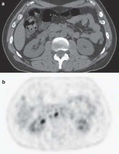
Figure 1. Comparison between normal abdominal CT (a) and PET/CT which demonstrates abnormal uptake in paraaortic lymph nodes (b).

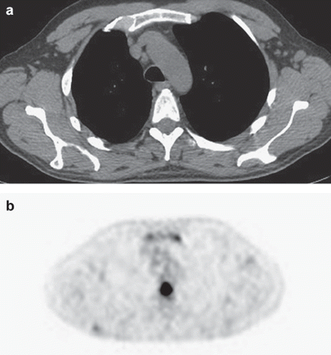
Figure 2. Comparison between normal CT image (a) and PET/CT which demonstrates abnormal uptake in the sternum and in thoracic vertebrae (b).

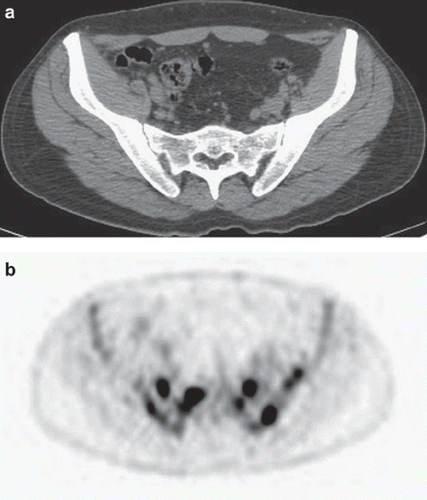
Figure 3. Unremarkable CT scan of lower abdomen and pelvis (a) compared to intense uptake in the sacrum and pelvic bones on PET/CT (b).

Avid FDG uptake in multiple lymph nodes: A block of left cervical lymph nodes, left supraclavicular nodes, aortopulmonary window nodes, hilar nodes bilaterally, left axillary, left paraesophageal, aortocaval node at the level L1–L2 ( and ), right external iliac nodes, inguinal bilaterally, and hepatic nodes. With the exception of the left cervical nodes originally described in the diagnostic CT as well, all nodes described with PET/CT were <1 cm transverse diameter.
Avid FDG uptake in a mass of soft tissue density between the left trapezoid and the supraspinal muscle.
Avid FDG uptake in multiple bone sites: left scapula, sternum ( and ), T6–T8, T10, L1, L3, L5, sacrum, pelvis ( and ), upper third of both humeri, upper third of the right femur. The CT performed with PET demonstrated no lesions on bone algorithm in the areas of increased FDG uptake.
The stage of disease was re-evaluated as IVB (extensive disease), with an International Prognostic Score (IPS) of 3 (IPS 2 before re-evaluation). Treatment began with aggressive chemotherapy (escalated BEACOPP) [Citation8]. After two cycles of therapy, PET/CT was repeated, demonstrating very weak uptake of FDG (SUV max: 2.3) in a small right inferior tracheobronchial node. The overall image was that of complete response. The patient received two more cycles of BEACOPP then four cycles of ABVD. Follow-up has demonstrated complete remission.
Discussion
Agreement of PET and CT in staging of HL is only about 60–80%. Disagreement in findings has similar frequency (10–20%) in both directions [Citation9,Citation10]. However it has been reported that while PET identified more lymph nodes than physical examination or CT in 15 of 60 patients with non-HL (NHL) and HL, in only two was there a change in stage (IA to IIA in one HL patient, II to III in one NHL patient) with no change in treatment [Citation5]. In other studies PET altered stage and therapy in only 8% of patients [Citation11].
Although most studies show that PET-negative/CT positive findings are less common than the opposite, it seems that PET alone cannot replace CT for pretreatment staging of HL [Citation9,Citation10]. However, despite the fact that PET/CT scanning has not been incorporated in pretreatment staging of lymphoma, it is encouraged in patients with HL as a reference point for posttherapy PET/CT [Citation12]. The unusual contradiction between CT and PET/CT in this case demonstrates the potential for therapeutic error, considering that without the PET/CT findings, the original protocol would have been unsatisfactory for advanced disease [Citation8].
PET can detect focal or multifocal bone and marrow involvement in lymphoma patients with negative bone marrow biopsy, subsequently confirmed by histopathology or magnetic resonance imaging (MRI) [Citation13]. However, PET alone is considered unreliable in detecting bone marrow involvement, particularly of limited extent (10–20% of marrow space), while some estimates of PET sensitivity for detecting marrow infiltration in HL are 76% [Citation13]. What is unusual in this case is that bone marrow biopsy was initially negative, while PET/CT uptake was observed in pelvic and multiple bone sites. No bone lesions were detected on diagnostic CT nor on PET/CT in areas of increased FDG uptake. There were no factors that would explain these sites as false positives (e.g. G-CSF), moreover, these sites disappeared after chemotherapy.
This patient presented an unusual site of FDG uptake in soft tissue mass between thoracic muscles, which on CT appears as slightly increased muscle dimensions in the affected area compared to contralateral muscles. This was not described in the diagnostic CT but retrospectively was vaguely apparent. PET has been found to detect more occult lymphomatous sites than contrast-enhanced CT and bone marrow biopsy [Citation6,Citation10,Citation14].
Taken together, this case indicates that more research is needed concerning the role of PET/CT in the initial staging of HL, potentialy upgrading first-line therapy if necessary and preventing treatment failure.
Declaration of interest: The authors report no conflicts of interest. The authors alone are responsible for the content and writing of the paper.
References
- Newman JS, Francis JR, Kaminski MS, Wahl RL. Imaging of lymphoma with PET with 2-[F-18]-fluoro-2-deoxy-D-glucose: Correlation with CT. Radiology 1994;190:111–6.
- Thill R, Neuerburg J, Fabry U, Cremerius U, Wagenknecht G, Hellwig D, . Comparison of findings with 18-FDG PET and CT in pretherapeutic staging of malignant lymphoma. Nuklearmedizin 1997;36:234–9.
- Stumpe KD, Urbinelli M, Steinert HC, Glanzmann C, Buck A, von Schulthess GK. Whole-body positron emission tomography using fluorodeoxyglucose for staging of lymphoma: Effectiveness and comparison with computed tomography. Eur J Nuclear Med 1998;25:721–8.
- Schaefer NG, Hany TF, Taverna C, Seifert B, Stumpe KD, von Schulthess GK, . Non-Hodgkin lymphoma and Hodgkin disease: Coregistered FDG PET and CT at staging and restaging: Do we need contrast-enhanced CT? Radiology 2004;232:823–9.
- Jerusalem G, Warland V, Najjar F, Paulus P, Fassotte MF, Fillet G, . Wholebody 18F-FDG PET for the evaluation of patients with Hodgkin's disease and non-Hodgkin's lymphoma. Nucl Med Commun 1999;20:13–20.
- Moog F, Bangerter M, Diederichs CG, Guhlmann A, Merkle E, Frickhofen N, . Extranodal malignant lymphoma: Detection with FDG PET versus CT. Radiology 1998;206: 475–81.
- Raanani P, Shasha Y, Perry C, Metser U, Naparstek E, Apter S, . Is CT scan still necessary for staging in Hodgkin and non-Hodgkin lymphoma patients in the PET/CT era? Ann Oncol 2006;17:117–22.
- Franklin J, Diehl V. Current clinical trials for the treatment of advanced-stage Hodgkin's disease: BEACOPP. Ann Oncol 2002;13(Suppl 1):98–101.
- Bangerter M, Moog F, Buchmann I, Kotzerke J, Griesshammer M, Hafner M, . Wholebody 2-[18F]-fluoro-2-deoxy-D-glucose positron emission tomography (FDG-PET) for accurate staging of Hodgkin's disease. Ann Oncol 1998; 9:1117–22.
- Jerusalem G, Beguin Y, Fassotte MF, Najjar F, Paulus P, Rigo P, . Whole-body positron emission tomography using 18F-fluorodeoxyglucose compared to standard procedures for staging patients with Hodgkin's disease. Haematologica 2001;86:266–73.
- Rodríguez-Vigil B, Gómez-León N, Pinilla I, Hernández-Maraver D, Coya J, Martín-Curto L, . PET/CT in lymphoma: Prospective study of enhanced full-dose PET/CT versus unenhanced low-dose PET/CT. J Nucl Med 2006; 47:1643–8.
- Juweid ME, Stroobants S, Hoekstra OS, Mottaghy FM, Dietlein M, Guermazi A, . Use of positron emission tomography for response assessment of lymphoma: Consensus recommendations of the Imaging Subcommittee of the International Harmonization Project in Lymphoma. J Clin Oncol 2007;25:571–8.
- Pakos EE, Fotopoulos AD, Ioannidis JP. 18FFDG PET for evaluation of bone marrow infiltration in staging of lymphoma: A meta-analysis. J Nucl Med 2005;46:958–63.
- Naumann R, Beuthien-Baumann B, Reiss A, Schulze J, Hänel A, Bredow J, . Substantial impact of FDG PET imaging on the therapy decision in patients with early-stage Hodgkin's lymphoma. Br J Cancer 2004;90:620–5.
