Abstract
Objective. To examine temporal trends in the incidence of hydatidiform mole (HM) in relation to maternal age, the occurrence of choriocarcinoma in women with a history of HM and the extent of underreporting of HM to the Swedish Cancer Register (SCR). Methods. Women registered with a diagnosis of HM were identified in the Swedish Cancer Register and the Swedish Inpatient Register (IPR). Record linkage to the Medical Birth Register provided information to assess the incidence of HM. Results. We identified 3 844 unique cases of HM in the SCR and in the IPR combined between 1973 and 2004, yielding an incidence of 1.2 per 1 000 deliveries. The incidence of HM increased during the period under study. The highest incidence was observed in women below 20 and above 39 years of age. Of all registered cases of choriocarcinoma, 37% occurred in women with a previous history of HM. The risk of choriocarcinoma following HM was 1.3%, compared to 0.005% in women without a previous molar diagnosis. The records of the Cancer Register included 83.2% of all identified cases of HM. Conclusion. The incidence of HM in Sweden has increased over time, and is characterized by a bimodal pattern with distinctive peaks in the youngest and oldest women of reproductive age. More than one third of all women registered with choriocarcinoma had a previous diagnosis of HM. Despite mandatory reporting, there was evidence of underreporting of HM to the SCR that remained virtually unchanged over calendar time.
Hydatidiform mole (HM) is a genetically abnormal pregnancy and can be subdivided into a complete and partial form. The etiology behind hydatidiform mole is still not fully known. All molar pregnancies have a possible malignant potential depending on local proliferation, invasion of the myometrium and metastasis, i.e. invasive mole or choriocarcinoma. The majority of women with a previous history of hydatidiform mole will have perfectly normal subsequent pregnancies and have no increased risk of other obstetric complications. However, the risk of a new molar pregnancy and choriocarcinoma is substantially higher than in women without previous HM [Citation1,Citation2]. In Sweden reporting of HM to the Swedish Cancer Register (SCR) is mandated by law. Cases can also be identified in the nationwide Inpatient Register (IPR) covering all in-hospital care. Results from one earlier Swedish report based on regional data indicate that there is an underreporting of HM to the Cancer Register [Citation3].
Using information from two separate sources: the National Swedish Cancer Register and the Inpatient Register, the purpose of the present study was threefold. Firstly, to assess the reporting of HM to the Swedish Cancer Register over time, secondly to examine incidence trends of HM by maternal age and thirdly to examine the occurrence of choriocarcinoma in women with a history of HM.
Material and methods
Data sources and ascertainment of outcome
Cases of HM [International classification of diseases, ICD-7: 173 combined with pathoanatomical diagnosis, [Citation4] (PAD: 801) and choriocarcinoma (PAD: 806, 803) were identified in the SCR which was established in 1958. Notification of newly diagnosed tumors to the SCR is mandated by Swedish law and more than 95% of all incident cases of cancer are reported separately by both a clinician and a cytologist or pathologist [Citation5]. The SCR does not differentiate between complete and partial hydatidiform mole. A repeat molar pregnancy is considered as recurrent disease and is not registered separately.
Information on hospital admissions and discharges has been recorded in the IPR from 1964 and onwards and was used to identify additional cases of HM that had not been reported to the SCR. This database had partial national coverage between 1964 and 1986, achieving complete coverage from 1987 [Citation6]. For each hospital admission the patient receives up to nine discharge diagnoses classified according to the International Classification of Disease system. Any first primary or secondary discharge diagnosis of HM (ICD-8: 634.2, ICD9: 630x, ICD10: O010, O011, O019) was considered in the present study. To assess the incidence of HM in relation to number of deliveries, information on reproductive history was obtained from the Swedish Medical Birth Register. This database started in 1973 and contains information on both live births and stillbirths (from gestational week 28 or later) and encompasses 98–99% of all births in Sweden [Citation7]. Record linkage between registers was facilitated by the individually unique national registration number, assigned to all Swedish residents at time of birth or first permanent residency.
The study protocol was approved by the Institutional Review Board at Karolinska Institutet, Stockholm.
Study population
For our cohort we selected HM cases registered from 1973 through 2004 in both the SCR and the IPR. Amongst women with multiple reported events in the IPR, we only selected the first recorded event. This way, for all years under study, we were able to identify 3 844 unique cases of HM; 3 198 (83.2%) identified in the SCR while 646 (16.8%) additional unique cases were reported exclusively to the Inpatient Register. In the IPR, the distribution between primary and secondary diagnoses of HM was 82.7% and 17.3%, respectively. For our incidence estimates we used nearly 3.2 million births (n = 3 199 695) recorded in the Medical Birth register between 1973 and 2004. One woman with HM was excluded in the assessment of incidence due to missing information on age.
Statistical methods
To assess possible underreporting of HM to the SCR we investigated what proportion of patients had been reported to the SCR and the IPR, respectively, and also what proportion that had been recorded in both registers. These calculations were performed by period of diagnosis (1973–1976, 1977–1980, 1981–1984, 1985–1988, 1989–1992, 1993–1996, 1997–2000 and 2001–2004) and in total over the entire study period. Incidence of HM was defined as the number of HM divided by the sum of HM and deliveries. The incidence is reported per 1 000 deliveries (including moles), and was calculated by age at diagnosis < 20, 20–24, 30–34, 35–39, > 39 years and calendar period of diagnosis 1973–1980, 1981–1988, 1989–1996, 1997–2004.
Standard errors for single sample proportions were used to obtain 95% confidence intervals for the incidence. Relative risks and 95% confidence intervals for the period effect were estimated. The standard error for the log of the relative risk was used in the calculations of the confidence interval.
The occurrence of choriocarcinoma among women with a molar pregnancy was compared to that of women with no history of HM and a crude relative risk was estimated.
Results
During the whole study period, the SCR included 83.2% of all identified cases of HM. With the exception of the earliest periods under study, the underreporting remained relatively unchanged over time (, ). The overall incidence of HM during the complete period under study was 1.2 per 1 000 deliveries (including moles), and increased from 1.01 between 1973 and 1980 to 1.43 between 1997 and 2004 (). In all time periods, the highest incidence was found among women below the age of 20 and over the age of 39. Even though the incidence in women over 39 years of age was high it remained fairly stable during the study period. The most pronounced increase in incidence was observed in the youngest age group ().
Figure 1. Graphical illustration of distribution of moles (numbers) by source of registration, SCR: Swedish Cancer Register, IPR: Swedish Inpatient Register.
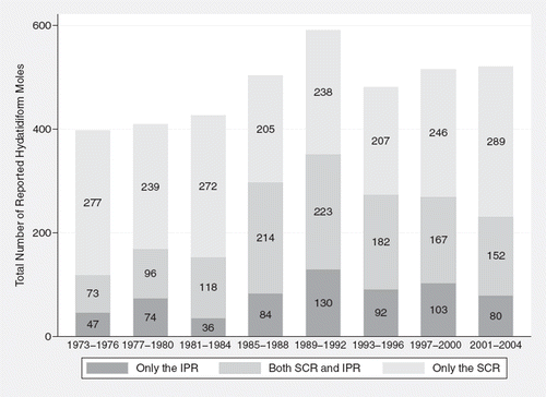
Figure 2. Graphical illustration of distribution of moles (percent) by source of registration, SCR: Swedish Cancer Register, IPR: Swedish Inpatient Register.
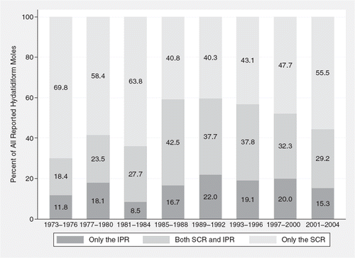
Figure 3. Age specific incidence of Hydatidiform mole per 1000 deliveries (including moles) by calendar period.

Table I. Incidence of Hydatidiform mole per 1000 deliveries (including moles) 1973-2004.
One hundred and thirty one women with a diagnosis of choriocarcinoma were identified in the Cancer Register of which 48 (37%) had a previous recorded diagnosis of hydatidiform mole in the SCR or in the Inpatient Register. The risk of choriocarcinoma following HM was 1.3%, compared to 0.005% in women without a previous molar diagnosis, yielding a risk ratio of 247 (95% CI: 173–352) (data not shown).
Discussion
There are broad international variations in the incidence of hydatidiform mole with the highest incidence rates observed in some regions of Asia. In Europe, 100 cases of HM per 100 000 pregnancies have been reported, while the corresponding estimates from Asia range from 100 to 1000 per 100 000 pregnancies [Citation8]. It remains unclear if these differences are due to regional reporting routines, genetic factors or an influence of socioeconomic conditions that may affect dietary and reproductive patterns. At present there is little definite knowledge regarding the etiology of HM, partly because of the rarity of the condition. The risk factor most consistently associated with HM is maternal age, and possibly also paternal age [Citation9], which may reflect that both ovum and spermatozoa quality depends on age [Citation10]. Other suggested risk factors include smoking and body mass index. Also, there is some evidence that twinning [Citation11,Citation12] and high offspring birth weight increase the risk of developing HM, possibly because of exposure to elevated levels of pregnancy hormones [Citation13].
We utilized information from both the SCR and the National Inpatient Register to assess the reporting of HM to the Cancer Register. Of all cases identified during the period under study, about 20% were not found in the SCR. Thus, clinicians fail to conform to the mandatory reporting of HM, with little improvement over time.
Taken together, our results show that the incidence of hydatidiform mole in Sweden has increased during the three decades under study. The incidence estimates observed in our study are comparable with findings in other Western countries [Citation8], but were higher than what has previously been reported in an earlier Swedish study [Citation14]. The incidence had a bimodal pattern with distinctive peaks in young and older women, possibly reflecting an increased risk of fertilization of an abnormal oocyte early and late during childbearing years. The highest incidence was found in women 39 years or older and the highest incidence increase was observed in the youngest age group. Medical rather than surgical treatment of spontaneous and legal abortions is available today which makes it more difficult to obtain pathological confirmation. Thus, some HM cases may go unrecognized, contributing to spuriously low incidence rates. On the other hand, it cannot be excluded that the observed temporal change in incidence have been influenced by a more frequent use of karyotyping during the later part of the period under study.
A South Korean study found that the hospital-based incidence of HM declined from about 40 per 1 000 deliveries 1971–1975 to around two per 1 000 deliveries 1991–1995. The authors speculated that this trend could be correlated to economic developments during the same time period [Citation15]. A Finnish study showed no marked changes in incidence during the same time period [Citation16]. Previous Swedish studies in this area have been restricted to regional databases or the National Swedish Cancer Register only. One earlier study based on information obtained from the National Swedish Cancer Register covering the period 1958–1965 estimated the incidence of hydatidiform mole to 0.7 per 1 000 deliveries [Citation14] compared to 1.2 per 1 000 deliveries in our study. A Swedish study based on Regional Cancer Register and hospital data from Stockholm County between 1975 and 1988 estimated the incidence of HM to be about two times higher (1.5 per 1 000 deliveries and moles), roughly equivalent to the incidence estimates in the in the most recent period covered by the present study [Citation3]. In the same study there was evidence of considerable underreporting of HM, with no record in the Cancer Register of 25% of all identified cases in the Stockholm region.
In the previous Swedish study based on the National Cancer Register data only, 40% of all cases of choriocarcinoma were preceded by a molar pregnancy [Citation14], compared to 37% in the present study. Our estimate corresponds to a 247-fold increased risk of choriocarcinoma. While high, this risk estimate is lower than what has been reported from some other studies [Citation12], and may have been influenced by initiation of treatment of persistent trophoblastic disease without confirmation whether it represented a case of invasive mole or choriocarcinoma.
Strengths of the present study include the size of the material, the population-based design, the possibility to retrieve information regarding HM diagnosis from two separate databases and a virtually complete follow-up. Limitations included the incomplete coverage of the Inpatient Register during the early part of the study, and the inability to differentiate between the distinct clinical entities of partial and complete hydatidiform mole precluding systematic comparisons between the two entities or detailed studies of possible risk factors. It cannot be excluded that our study design using the incidence per delivery including moles rather than per pregnancy, has contributed to the observed bimodal pattern incidence, since young women more often discontinue a pregnancy and the age of primiparas has increased dramatically between the first and last period under study.
Ideally, the denominator used for calculating the incidence rate should encompass all possible pregnancy outcomes. However data on the number of miscarriages and legal abortions on an individual level are not available in Sweden and may have contributed to an overestimation of the incidence rates observed in the present study.
In conclusion, we found clear evidence of an underreporting of HM to the SCR that has remained virtually unchanged over calendar time, indicating that official cancer statistics continue to underestimate the true incidence of HM. Furthermore, our results indicate that the Inpatient Register represent the less complete data source with regard to registration of HM. Thus, the detected level of underreporting in the Swedish Cancer Register represents a minimum level.
In the present material based on information retrieved from two separate sources our results indicate that the incidence of HM has increased over time together with a bimodal age specific incidence pattern with the highest rates observed among younger and older women. More than one third of all recorded cases of choriocarcinomas were preceded by molar pregnancy, corresponding to a more than 200 times increased risk of choriocarcinoma in women with a history of HM.
Acknowledgements
This study was supported by the Swedish Cancer Society, Grant no 07-0526.
Declaration of interest: The authors report no conflicts of interest. The authors alone are responsible for the content and writing of the paper.
References
- Sebire NJ, Fisher RA, Foskett M, Rees H, Seckl MJ, Newlands ES. Risk of recurrent hydatidiform mole and subsequent pregnancy outcome following complete or partial hydatidiform molar pregnancy. BJOG 2003;110:22–6.
- Garrett LA, Garner EI, Feltmate CM, Goldstein DP, Berkowitz RS. Subsequent pregnancy outcomes in patients with molar pregnancy and persistent gestational trophoblastic neoplasia. J Reprod Med 2008;53:481–6.
- Flam F, Rutqvist LE. Under-registration of gestational trophoblastic disease in the Swedish Cancer Registry. Eur J Epidemiol 1992;8:683–6.
- PAD. WHO World Health Statistical Code for Human Tumours, WHO/US/CANC, 24.1 and 24.2, August 15. Geneva 1956.
- Swedish Cancer Registry. Cancer Incidence in Sweden 2005. Stockholm: Centre for Epidemiology. National Board of Health and Welfare. Official Statistics of Sweden. Health and Diseases 2007:3. Available from: http://www.socialstyrelsen.se/Publicerat/2007/9514/2007-42-3.htm.
- Patientregistret. Utskrivningar från sluten vård 1964–2005. Kvalitet och innehåll [In English: The Hospital Discharge Register. Discharges from in-patient care 1964–2005. Quality and Contents]. Centre for Epidemiology, National Board of Health and Welfare. December 2006. Available from: http://www.socialstyrelsen.se/NR/rdonlyres/550EBD44-91B7-47C0-81B0-0087D24F50C7/0/Kvalitetochinnehåll19642005.pdf.
- The Swedish Medical Birth Register. A summary of content and quality. Centre for Epidemiology, National Board of Health and Welfare, Stockholm, Sweden 2004.
- http://www.sos.se/fulltext/112/2003-112-3/2003-112-3.pdf.
- Altieri A, Franceschi S, Ferlay J, Smith J, La Vecchia C. Epidemiology and aetiology of gestational trophoblastic diseases. Lancet Oncol 2003;4:670–8.
- La Vecchia C, Parazzini F, Decarli A, Franceschi S, Fasoli M, Favalli G, . Age of parents and risk of gestational trophoblastic disease. J Natl Cancer Inst 1984;73:639–42.
- Levitas E, Lunenfeld E, Weisz N, Friger M, Potashnik G. Relationship between age and semen parameters in men with normal sperm concentration: Analysis of 6022 semen samples. Andrologia 2007;39:45–50.
- De George FV. Hydatidiform moles in other pregnancies of mothers of twins. Am J Obstet Gynecol 1970;108:369–71.
- Buckley JD, Henderson BE, Morrow CP, Hammond CB, Kohorn EI, Austin DF. Case-control study of gestational choriocarcinoma. Cancer Res 1988;48:1004–10.
- Kaijser M, Granath F, Jacobsen G, Cnattingius S, Ekbom A. Maternal pregnancy estriol levels in relation to anamnestic and fetal anthropometric data. Epidemiology 2000; 11:315–9.
- Ringertz N. Hydatidiform mole, invasive mole and choriocarcinoma in Sweden 1958–1965. Acta Obstet Gynecol Scand 1970;49:195–203.
- Kim SJ, Bae SN, Kim JH, Kim CJ, Han KT, Chung JK, . Epidemiology and time trends of gestational trophoblastic disease in Korea. Int J Gynaecol Obstet 1998;60(Suppl 1):S33–8.
- Loukovaara M, Pukkala E, Lehtovirta P, Leminen A. Epidemiology of hydatidiform mole in Finland, 1975 to 2001. Eur J Gynaecol Oncol 2005;26:207–8.