Abstract
Background. To investigate the patterns of relapse following intensity modulated radiotherapy (IMRT) given after radical surgery for oral and oropharyngeal squamous cell cancer. Patients and methods. One hundred and two patients with oral or oropharyngeal cancer were treated with radical surgery followed by IMRT up to a mean total dose of 60 Gy between years 2001 and 2007. Thirty-nine of the patients (%) also received concomitant weekly cisplatin. Forty of the patients had oral and 62 had oropharyngeal cancer. Data on the tumour, patient and treatment factors were collected. Following therapy the patients were followed by clinical examination, endoscopy and MRI/CT at 2- to 3-months interval up to 2 years and thereafter at 6-month intervals. Results. The mean follow-up time of the patients was 55 months (range, 26–106 months). The rate for local tumour control for the whole cohort was 92.2%: 87.5% for oral cancer patients and 96.7% for oropharyngeal cancer patients. The 5-year disease specific survival was 90.2% and 5-year overall survival 84.3%. During the follow-up eight locoregional recurrences were observed, three at the primary tumour site and one at regional nodal site and four at both sites. The mean time to primary tumour recurrence was seven months (range, 2–10 months) and to nodal recurrence seven months (range, 2–12 months). Distant metastasis occurred in six (6%) patients. The factors associated with poor prognosis were the primary tumour size and tumour site with oral cancers having worse outcome. The treatment was well tolerated with no unexpected toxicities. The most frequent late toxicity was dysphagia necessitating permanent PEG in five patients. This was correlated with the advanced primary tumour size and resulting in wide tumour excision and reconstruction. Conclusions. Surgery combined with postoperative radiotherapy given as IMRT results in low level of tumour recurrence.
In the treatment of head and neck squamous cell cancer (HNSCC) the main treatment modalities have been surgery and radiotherapy and often a combination of these modalities is needed. In early stage cancers surgery alone as a single modality treatment is often considered adequate. There are no randomised trials comparing directly surgery and postoperative RT versus surgery alone in HNSCC treatment. In retrospective studies, the local control, and also survival, has been better especially in patients with a high risk of local recurrence, when surgery is combined with postoperative radiotherapy.
Intensity modulated radiotherapy (IMRT) is one of the most important advances in the treatment of HNSCC during the last decade. It provides an effective tool to reduce the dose to the surrounding sensitive normal structures and, in addition allows dose escalation at a given level of normal tissue damage. In postoperative RT for head and neck cancer IMRT enables better coverage of the operation area and elective lymph node areas together with better sparing of the surrounding normal structures.
Concomitant chemoRT is the established standard therapy in the treatment of advanced head and neck cancer [Citation1]. Also in the postoperative setting chemoRT has proved to be more effective than RT alone in the treatment of patients with high risk of recurrence [Citation2,Citation3]. The best investigated single agent both in definitive and postoperative treatment has been cisplatin. In our institute the standard postoperative RT consists of IMRT, which is combined with weekly cisplatin at the dose level of 40 mg/m2 in patients with high risk of locoregional recurrence. The aim of this study was to evaluate the patterns of relapse following surgery and postoperative IMRT in the treatment of oral and oropharyngeal squamous cell cancer.
Patients and methods
Patients
This study is based on a cohort of 102 patients with oral (n = 40, 39%) or oropharyngeal (n = 62, 61%) squamous cell cancer treated with surgery followed by postoperative IMRT between the years 2001 and 2007. The mean age of the patients at the time of diagnosis was 57 years (range, 30–79 years). Thirty-four patients (33%) were female and 68 (67%) male. The mean follow-up time was 55 months (range, 26–106 months). The main patient and tumour characteristics are presented in .
Table I. Patient and tumour characteristics.
Pretreatment evaluation of all patients was done by clinical examination, imaging (MRI/CT) and endoscopy, and the diagnosis was confirmed by histopathological tissue biopsies and surgical speimens. The tumours were staged according to the International Union Against Cancer (UICC) Tumour-Node-Metastasis (TNM) classification. The preoperative stage was down- or up-staged according to resection specimens of the primary tumour and neck dissection specimens. The stage announced here is based on the final histopathological examination of each tumour. A prophylactic percutaneus endoscopic gastrostomy (PEG) was applied to 45 (43%) of the patients to ensure nutrition after surgery.
Treatment related toxicity was scored according to the Common Terminology Criteria for Adverse Events (CTCAE) version 3.0.
The study is a part of a IMRT quality assurance study approved by the Research Ethics Board at the Helsinki University Central Hospital.
Operative therapy
Oral cancer patients typically had either partial resection of the tongue alone or in combination with resection the floor of mouth. Eighty-five percent of these surgical defects were reconstructed (). Majority of the oropharyngeal cancer patients underwent lateral wall resection and some of them resection of tongue base or posterior wall. Fifty-eight percent of these underwent reconstruction (). The current established technique for the reconstruction of oral and oropharyngeal oncological defects is the use of a microvascular transfer. Anterolateral thigh free flap and radial forearm free flap were most frequently in the present series ().
Table II. Surgical treatment of the primary tumour.
An ipsilateral modified radical neck dissection or a selective neck dissection was performed in 65% and 66.5% of the oral and oropharyngeal cancer cases, respectively ().
Table III. Surgical treatment of the neck.
Radiotherapy
All patients were treated by IMRT. In patients treated before the year 2001 a conventional thermoplastic mask (Posicast®, Sinmed BV, EM Reenwijk, the Netherlands) was used for immobilisation and a stereotactic head and neck immobilisation device (BrainLab, Heimstetten, Germany) thereafter. The treatment isocentre was localised following computation of the stereotactic coordinates with the BrainScan® stereotactic treatment planning system (BrainLab). A treatment planning CT was performed using a thickness of 5.0 mm until the year 2005 and of 2.5 mm thereafter. Irradiation was performed with a 6 MV linear accelerator using a dynamic multileaf collimator (dMLC) with the sliding window principle.
The treatment planning CT images and preoperative CT or MRI images were used to delineate the target volumes. The preoperative images were used to determine the exact site of the primary tumour and nodal metastases. The clinical target volume (CTV1) typically first included the resection area of the primary tumour with a 10 mm margin and the locoregional nodal areas. All patients in this series were treated with bilateral irradiation of nodal areas. A 3–5 mm margin was added to the CTV1 to obtain the planning target volume (PTV). This target volume was irradiated to a cumulative dose of 50 Gy in 2 Gy daily fractions in five weeks. Thereafter, the primary tumour resection area and nodal areas with high risk of recurrence (PTV2-3) were boosted up to a mean total dose of 58 Gy (range, 56–70 Gy). In 97 patients one volume reduction was done after completion of PTV1 and in five patients another reduction after completion of PTV2 was done and the final boost was given to a further reduced volume (PTV3). All dose prescriptions were based on ICRU 50 recommendations. The organs at risk (OAR) were defined in all CT slices. In all patients the spinal cord was defined as OAR and the maximum dose to spinal cord allowed during the IMRT course was 40 Gy. The contralateral parotid gland was defined as OAR in all patients. In patients with oropharyngeal cancer the opposite submandibular gland was also spared when it was considered to be safe. In patients with oral cancer no attempt to spare the submandibular glands was made because of the high risk of level I nodal metastasis. Unnecessary irradiation of normal mucosa was also avoided. The principles of our salivary gland sparing radiotherapy have been published earlier [Citation4,Citation5].
Chemotherapy
Weekly cisplatin 40 mg/m2 was scheduled to 39 (38%) patients concomitantly with RT. The indications for concomitant chemotherapy were marginal resection, more than one metastatic lymph node, and extracapsular spread of nodal metastasis. The number of planned cycles was six in all patients. The cisplatin was given as a 30 min infusion in 500 ml 0.9% NaCl and preceded by a 1000 ml 0.9% NaCl infusion. Laboratory tests including complete blood count, C-reactive protein and renal and liver function tests were checked before the onset of treatment and weekly during the treatment. Prophylactic antiemetics were prescribed for all patients before chemotherapy.
Statistical analyses
The SPSS statistical program (Chicago, IL, USA) version 17.0 was used for statistical calculations. Logistic regression analysis was used to calculate the significance of patient and tumour characteristics. The Kaplan-Meier product-limit method was used to estimate the local control and survival rates. Durations were calculated from the end of RT. Local control was defined as absence of primary tumour and regional nodal metastasis on physical examination, endoscopy and MRI/CT scans.
Results
Local control and patterns of relapse
After the radiation therapy the follow up was arranged at the Department of Otorhinolaryngology – Head and Neck Surgery, Helsinki University Central Hospital. The mean follow-up time was 55 months (range, 26–106 months).
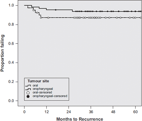
During the follow-up period eight locoregional failures (8%) were observed: five in patients with oral cancer (12.5%) and three in patients with oropharyngeal cancer (4.8%). Three of recurrences occurred at the primary tumour site, one at nodal site regional only and four locoregional at both primary tumour and nodal sites. Six of the seven recurrences at the primary tumour bed occurred within the high dose irradiation area, five within CTV1 and CTV2 with a mean dose of 58 Gy at the area of recurrence. One of the primary recurrences occurred within CTV1 inside the 50 Gy dose volume. Only one primary recurrence was observed outside CTV1 and CTV in patient with T3N1 cancer at the anterior part of the floor of mouth. All five regional recurrences were within CTV1 and two also within CTV2 with a mean dose of 53 Gy (range, 50–60 Gy) at the level of recurrence. All but one nodal recurrence occurred at ipsilateral site. In one patient with a T4N2c oral cancer bilateral nodal recurrence was observed together with failure at the operation bed of the primary tumour. The mean time from the end of RT to local recurrence at primary tumour site was seven months (range, 2–10 months) and to nodal recurrence also seven months (range, 2–12 months). Locoregional tumour control following surgery and postoperative IMRT was achieved in 35 of the 40 oral cancer patients (87.5%) and in 59 of the 62 oropharyngeal cancer patients (95.2%). In one patient with T4 tonsillar cancer local control at primary tumour site was achieved following salvage surgery and thus the ultimate local control for patients with oropharyngeal cancer was 96.7%. The local control figures are presented in .
Distant metastasis was observed in six patients during the follow-up period. In three patients distant metastasis occurred together with locoregional failure and in three patients without local tumour relapse. The location of metastasis was pulmonary in three patients, hepatic in two and multiple sites in one patient.
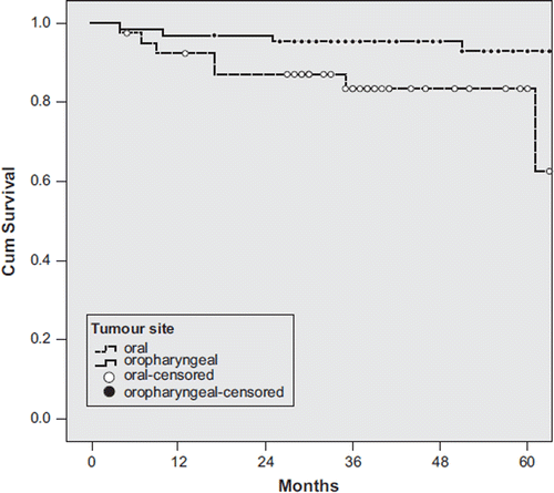
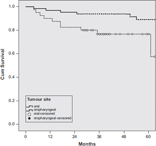
The 5-year overall survival (OS) for the whole cohort was 84.3%; 90.3% for oropharyngeal cancer patients and 75% for oral cancer patients. The corresponding numbers for 5-year disease specific survival (DSS) were 90.2%, 95.2% and 82.5%, respectively. The Kaplan-Meier curves for DSS and OS of the patients are presented in and . The causes of death were local recurrence in four patients (4%), local recurrence together with distant metastasis in three patients (3%) and distant metastasis without local recurrence in three patients (3%). Six patients (6%) died of causes not related with cancer (lung cancer in one patient, rectal cancer in one, pneumonia in one, accident in one and coronary heart disease in two patients). The fatal pneumonia occurred 17 months after the end of RT and was not treatment-related.
Thirty-nine patients with high risk of local recurrence were scheduled to receive weekly cisplatin during the RT course. The amount of stage IV tumours in patients treated by chemoRT was 79% (31/39) and in those treated by RT alone 52% (33/63), p = 0.007 (Fisher's exact test). The mean total dose in patients treated by chemoRT was 60 Gy (range 56–70 Gy) and in patients treated by RT alone 57 Gy (range 56–66 Gy). The amount of local failures in the chemoRT group was 3/39 (8%) and in the RT group 5/63 (8%). In the chemoRT patients four patients (10%) died during the follow-up period and in all of these patients the cause of death was recurrent cancer. Twelve (19%) of the patients treated by RT died, six of them of recurrent cancer (9.5%) and six of causes not related with cancer (9.5%). The differences in LC (p = 0.95), OS (p = 0.42) or DSS (p = 0.76) between the patients treated by chemoRT or RT alone were not statistically significant. The differences remained non-significant also when analysed separately for oral and oropharyngeal tumours.
In univariate analysis factors that predicted poor prognosis were the primary tumour site and the T stage. Tumours with oral origin had worse outcome than oropharyngeal tumours (p = 0.01 for LC, p = 0.02 for DSS and p = 0.01 for OS). The corresponding values for T4 versus T1-3 tumours were p = 0.08 for LC, p = 0.01 for DSS and p = 0.09 for OS. The same factors remained significant in multivariate analysis testing the significance of tumour site, T-stage, N-stage and TNM stage. The patient age and gender were found to be insignificant in both univariate and multivariate analysis.
Treatment related acute toxicity
The most common radiation-related side effects were local skin and mucosal reactions. Dermatological toxicity was mild in all patients, Grade 1 in 91 (90%) and Grade 2 in 11 (10%) of the patients. Mucositis was Grade 1 in six (6%), Grade 2 in 70 (69%) and Grade 3 in 26 (25%) of the patients. Strong opioids for mucosal pain were needed in 26 (25%) and non-opioid analgesics in 16 (15%) of the patients.
Six patients (6%) needed hospitalisation during the treatment period. The mean duration of hospitalisation was five days (range, 3–7 days). The reasons were infection in four patients, nausea and dehydration in one and mucositis in one patient. Peroral antibiotics were prescribed to four (4%) patients and three patients (3%) required intravenous antibiotics. Peroral antimycotics were prescribed to 11 (11%) of the patients.
Thirty-nine patients were scheduled to receive six cycles of weekly cisplatin during the RT. All six cycles could be given in 16 (41%) of the patients, four to five cycles in 10 (26%) and less than four in the remaining 13 (33%) of the patients. The most important causes for altered administration schedule were renal insufficiency manifested as a rise in serum creatinine in four (10%) and chemotherapy induced cytopenia in four (10%) patients. None of the patients developed permanent renal insufficiency. One patient needed treatment for neutropenic infection during the chemoRT.
Treatment related late morbidity
At the end of the follow-up period, five patients (5%) had difficulties in swallowing necessitating a permanent PEG. Three of these patients were totally dependent on PEG with no peroral food intake and the remaining two patients could take part of the nutrition as peroral liquids and softened food. The strongest factor correlating with permanent PEG dependence was the pretreatment tumour T-stage (p = 0.03, Fisher's exact test) necessitating wide resection and reconstruction. In the PEG dependent patients the T-stage was T2 in one, T3 in one and T4 in three patients. The symptoms of xerostomy following radiotherapy were mild, Grade 0-1 in 70% and Grade 2 in 30%. None of the patients was reported to have higher than Grade 2 xerostomy. No cases of osteoradionecrosis were reported. The problems reported in phonation (n = 5) were associated with operative treatment (partial glossectomy or hemiglossectomy and reconstruction), and no cases associated with RT doses to laryngeal structures were identified.
Discussion
We investigated the patterns of failure following surgery and postoperative IMRT in the treatment of oral and oropharyngeal cancer. Locoregional tumour control was achieved in 87.5% patients with oral cancer and in 96.7% patients with oropharyngeal cancer. Only one recurrence occurred outside the high dose CTV. The corresponding overall survival figures were 75% and 90.3%. Local recurrence was the main cause of death in four patients (4%) and together with distant metastasis in additional three patients (3%). Three patient died of distant metastasis without local cancer recurrence.
Thirty-nine high-risk patients were treated with radiotherapy combined with weekly cisplatin. The LC, DSS and OS figures of these patients were similar to the 63 patients with lower risk of recurrence treated without concomitant chemotherapy. It is possible that without weekly cisplatin the outcome in high-risk patients would have been worse. This can also be partly due to the somewhat higher RT dose to the areas of high risk of recurrence in the chemoRT patients (mean total doses 60 Gy vs. 57 Gy). The total amount of failures in this series is, however, too low to make any firm calculations between subgroups. As only one of the recurrences occurred outside the defined target volumes, it can be concluded that the target volumes were correctly defined. It can be speculated if escalation of RT doses to areas of high risk of recurrence would have improved the achieved local control rates even further. The LC rates with the used dose levels were, however, good in both chemoRT and RT patients.
The failure patterns in this study are in line with earlier studies on head and neck IMRT. Marginal recurrences outside the high dose treatment area are rare and the local control within the primary tumour area and also within the nodal sites is good as compared to historical series. The observation of oral tumours having worse outcome is also in line with earlier results. Human papillomavirus (HPV) is a well recognised factor in the pathogenesis in the head and neck cancer, particularly in those arising from the lingual and palatine tonsils. The HPV-positive tumours tend to have better prognosis than HPV-negative tumours [Citation6]. In our series the HPV-status was not determined and therefore no conclusions on the possible significance of the HPV status on the prognosis of these patients can be made.
In HNSCC the most important cause of death has been local tumour recurrence. In patients treated by definitive RT the recurrence occurs almost always within the site of original primary tumour or macroscopic nodal metastases [Citation7]. Following surgery the recurrences are most frequent at the margins of the operation area. Therefore it is important in the postoperative RT for HNSCC to use radiotherapy techniques enabling adequate coverage of the whole operation area and elective nodal sites. In our institution the postoperative RT was traditionally given in most instances first by lateral wedged photon portals to the primary tumour bed and the upper cervical lymph nodes, and the lower neck was treated by a separate anterior field up to a total dose of 50 Gy. After the total dose of 38–40 Gy to spinal cord the RT was continued to the posterior cervival lymph nodes by 9–12 MeV electrons to avoid over-dosage in spinal cord. After a total dose of 50 Gy a boost dose of 6–16 Gy was given to the tumour bed and nodal areas of high risk of recurrence. The problems associated with this kind of field arrangements are high doses to the major salivary glands, mandible and mucosal membranes and areas of dose uncertainty near the fields junctions. A much more conformal dose distribution to the tumour bed and neck lymphatics with avoidance of high doses to radiosensitive normal structures can be achieved by IMRT. Junctional recurrences have been reported also following RT given by IMRT to primary tumour site and upper neck lymphatics and by an separate anterior field to lower neck [Citation8]. By the use of comprehensive intensity modulated photon fields to irradiate the whole treatment volume, tumour recurrence at field junctions may be avoided. This can be expected to lead in better local control figures. This was observed in the present series with only few local recurrences together with acceptable acute and late toxicity. Acute mucositis accompanying high-dose RT has been the most important cause of treatment interruptions in the RT. Treatment gaps during definitive or postoperative RT of HNSCC have been considered a major cause of treatment failures. By using IMRT, the areas of normal mucosa irradiated to a high dose can be reduced, which can have an impact on treatment tolerability.
The most prominent late effect of RT of HNSCC has been permanent xerostomy resulting from damage to parotid and submandibular glands. The D50 for salivary glands has been estimated to be in the range of 26–39 Gy by conventional fractionation, and lowering the total dose below these values has in several trials been observed to result in better salivary gland function measured by total saliva secretion or by scintigraphy, and the subsequent symptoms of xerostomy [Citation4,Citation5,Citation9,Citation10]. In our series no higher than Grade 2 xerostomy was observed. Another well recognised problem following RT of this area has been mandibular osteoradionecrosis. In a study by Glanzmann and Gratz on 189 patients treated for oral or oropharyngeal cancer, a 11% incidence of osteoradionecrosis was observed [Citation11]. The factors associated with this complication are total dose higher than 66 Gy, and the volume of irradiated mandible, field size, and tumour tonsillar or retromolar location [Citation11,Citation12]. By IMRT the volume of the mandible irradiated to a high dose can be reduced [Citation13]. No cases of mandibular osteoradionecrosis were observed in our series of 102 patients. The theoretical benefit resulting from the use of an separate anterior field is a lower RT dose to laryngeal structures. By IMRT dose optimisation programs the laryngeal dose can, however, be kept low enough to avoid postirradiation problems in phonation, which was also observed in the present study. Difficulties in swallowing following surgery and postoperative RT may also have an impact on the patient's quality of life following treatment. In our series five patients needed a permanent PEG. This is a problem that seems not to be totally avoidable after radical surgery and reconstruction followed by high dose RT. Posttreatment dysphagia is affected by location and stage of tumour, type of surgery and radiotherapy doses and also by the treatment technique used. In a study by Borggreven et al. the factors predicting posttreatment dysphagia associated with surgery for oral cavity and oropharynx cancer were comorbid conditions, large tumours and the resections of the base of tongue and soft palate combined [Citation14]. Radiotherapy associated factors are RT schedule, field size, and the use of concomitant chemotherapy [Citation15,Citation16]. Eisbruch et al. have shown that IMRT can reduce the dose in structures important in swallowing resulting in better swallowing function after RT [Citation17]. Another important factor contributing to better swallowing function after IMRT is sparing of the major salivary glands with less posttreatment xerostomy.
Conclusions
Surgery combined with postoperative IMRT results in excellent local control with low posttreatment morbidity.
Declaration of interest: The authors report no conflicts of interest. The authors alone are responsible for the content and writing of the paper.
References
- Pignon JP, le Maitre A, Bourhis J. Meta-Analyses of Chemotherapy in Head and Neck Cancer (MACH-NC): An update. Int J Radiat Oncol Biol Phys 2007;69(2 Suppl): S12–S114.
- Bernier J, Domenge C, Ozsahin M, Matuszewska K, Lefebvre JL, Greiner RH, . Postoperative irradiation with or without concomitant chemotherapy for locally advanced head and neck cancer. N Engl J Med 2004;350:1945–52.
- Cooper JS, Pajak TF, Forastiere AA, Jacobs J, Campbell BH, Saxman SB, . Postoperative concurrent radiotherapy and chemotherapy for high-risk squamous-cell carcinoma of the head and neck. N Engl J Med 2004;350:1937–44.
- Saarilahti K, Kouri M, Collan J, Hamalainen T, Atula T, Joensuu H, . Intensity modulated radiotherapy for head and neck cancer: Evidence for preserved salivary gland function. Radiother Oncol 2005;74:251–8.
- Saarilahti K, Kouri M, Collan J, Kangasmaki A, Atula T, Joensuu H, . Sparing of the submandibular glands by intensity modulated radiotherapy in the treatment of head and neck cancer. Radiother Oncol 2006;78:270–5.
- Fakhry C, Gillison M. clinical implications of human papillomavirus in head and neck cancer. J Clin Oncol 2006; 24:2606–11.
- Pigott K, Dische S, Saunders MI. Where exactly does failure occur after radiation in head and neck cancer? Radiother Oncol 1995;37:17–9.
- Daly ME, Le QT, Maxim PG, Loo BW, Jr., KaplanMJ, Fischbein NJ, . Intensity-modulated radiotherapy in the treatment of oropharyngeal cancer: Clinical outcomes and patterns of failure. Int J Radiat Oncol Biol Phys 2010;76:1339–46.
- Roesink JM, Moerland MA, Battermann JJ, Hordijk GJ, Terhaard CH. Quantitative dose-volume response analysis of changes in parotid gland function after radiotherapy in the head-and-neck region. Int J Radiat Oncol Biol Phys 2001; 51:938–46.
- Eisbruch A, Ten Haken RK, Kim HM, Marsh LH, Ship JA. Dose, volume, and function relationships in parotid salivary glands following conformal and intensity-modulated irradiation of head and neck cancer. Int J Radiat Oncol Biol Phys 1999;45:577–87.
- Glanzmann C, Gratz KW. Radionecrosis of the mandibula: A retrospective analysis of the incidence and risk factors. Radiother Oncol 1995;36:94–100.
- Murray CG, Herson J, Daly TE, Zimmerman S. Radiation necrosis of the mandible: A 10 year study. Part I. Factors influencing the onset of necrosis. Int J Radiat Oncol Biol Phys 1980;6:543–8.
- Studer G, Studer SP, Zwahlen RA, Huguenin P, Gratz KW, Lutolf UM, . Osteoradionecrosis of the mandible: Minimized risk profile following intensity-modulated radiation therapy (IMRT). Strahlenther Onkol 2006;182:283–8.
- Borggreven PA, Verdonck-de Leeuw I, Rinkel RN, Langendijk JA, Roos JC, . Swallowing after major surgery of the oral cavity or oropharynx: A prospective and longitudinal assessment of patients treated by microvascular soft tissue reconstruction. Head Neck 2007;29:638–47.
- Caudell JJ, Schaner PE, Meredith RF, Locher JL, Nabell LM, Carroll WR, . Factors associated with long-term dysphagia after definitive radiotherapy for locally advanced head-and-neck cancer. Int J Radiat Oncol Biol Phys 2009;73: 410–5.
- Poulsen MG, Riddle B, Keller J, Porceddu SV, Tripcony L. Predictors of acute grade 4 swallowing toxicity in patients with stages III and IV squamous carcinoma of the head and neck treated with radiotherapy alone. Radiother Oncol 2008; 87:253–9.
- Eisbruch A, Schwartz M, Rasch C, Vineberg K, Damen E, Van As CJ, . Dysphagia and aspiration after chemoradiotherapy for head-and-neck cancer: Which anatomic structures are affected and can they be spared by IMRT? Int J Radiat Oncol Biol Phys 2004;60:1425–39.


