To the Editor,
Pure Red Cell Aplasia (PRCA) is a disorder characterized by anemia with almost complete absence of red-cell precursors but normal platelet and white blood cell precursors. It is a recognized complication of malignancies, particularly Large Granulocyte Lymphomas, CLL, Hodgkins Lymphoma and thymomas [Citation1]. PRCA has also been associated with other solid organ malignancies, including breast cancer although the occurrence is very rare [Citation2,Citation3].
Treatment of PRCA is usually based on oral corticosteroids and cyclosporine, although other immunosuppressive agents have been used successfully [Citation4]. Relapse, however, is common and often prolonged courses of immunosuppression are required [Citation5]. There have been a number of cases where treatment of an underlying malignancy has resulted in sustained remission of the PRCA [Citation3,Citation6].
We will describe a case of breast cancer associated with PRCA, in which treatment of the underlying breast cancer in conjunction with brief course of oral prednisolone has caused lasting remission.
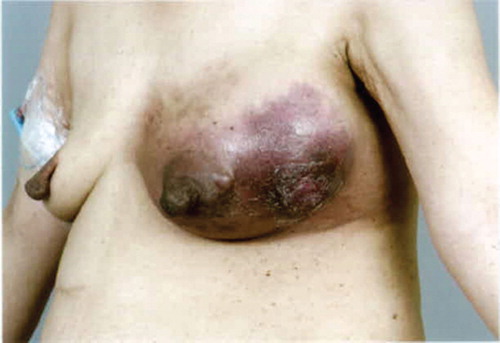
A 54-year-old lady presented with a 10 cm left fungating inflammatory breast mass and fevers (). Biopsy confirmed a basal-like invasive ductal carcinoma HER-2 negative ER/PR negative. CT scanning on presentation revealed mild left-sided lymphadenopathy suspicious for locally advance disease. Whole body bone scan did not reveal any evidence of metastatic disease. The initial hemoglobin (Hb) was 72 g/l. The white cell count was 21.7 × 10⁹ (neutrophils 18.6 × 10⁹) and platelets were 532 × 109. The C reactive protein was elevated at 179. A thorough septic screen did not reveal any source of sepsis. Iron studies, B12 and folate studies were normal. Bone marrow aspirate and trephine markedly reduced erythropoiesis (absent in some sections) with granulocytic hyperplasia with left shift and normal megakaryocyte populations.
Figure 1. Initial presentation.
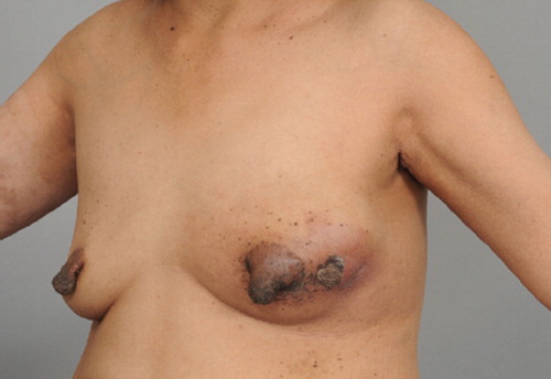
There was no evidence of metastatic disease. There were no clonal B cells or abnormal T-lymphocyte populations seen with flow cytometry. The bone marrow aspirate and trephine was consistent with PRCA. Investigations for other potential causes of PRCA were normal, including negative parvovirus serology. A total of 8 units of packed red blood cells (PRBC) (2 units on four separate occasions) were required in the first month after presentation to maintain the hemoglobin above 85 g/l. Neoadjuvant chemotherapy was administered, consisting of five cycles of three weekly doxorubicin and cyclophosphamide with a 25% dose reduction after an episode of neutropenia following the first cycle. Prednisolone 40 mg per day was also commenced concurrently with the first cycle of chemotherapy, with the intention of controlling the PRCA. This was continued for eight weeks. There was an excellent clinical response with an impressive reduction in size of the breast cancer, a resolution of fever and normalization of the inflammatory markers (see ). The patient's Hb remained stable at around 95–100 g/dl.
Figure 2. Following initial chemotherapy.
The chemotherapy regimen was switched to single agent weekly paclitaxel as per institutional practice (as per previous published series.) Unfortunately there was a progression of the breast cancer, with and increase in size of the breast mass. paclitaxel was changed to nanoparticle-albumin bound paclitaxel (nab paclitaxel) to which gemcitabine was added after a further two cycles due to further growth of the breast mass. There was also an increase in inflammatory markers and a recurrence of low grade fevers. During this time of progression of the cancer a further three transfusions of 2 units PRBC were required. Due to ongoing local progression of the breast lesion, mastectomy was undertaken with axillary clearance. Pathology confirmed a grade III 115mm ductal breast carcinoma which was ER/PR/HER-2 negative, All of the resected lymph nodes were clear of carcinoma. Following the mastectomy, the patient received chest wall radiotherapy. As of the most recent review, ten months following mastectomy no further blood transfusions have been required. Restaging investigations have not revealed any evidence of metastatic disease.
The association of PRCA and breast cancer has been described in three previous cases. Two cases were briefly described in 1971 as part of a larger review of seven cases of PRCA and malignancy [Citation2]. In these cases, breast cancer had been preceded by PRCA by three months and one year. Neither case was reported to have responded to treatment and both were also associated with a benign thymoma. A more detailed report from 1976 described a case in which PRCA preceded the discovery of the breast cancer by over six months [Citation3]. Treatment was with oral prednisolone, which was ineffective. This was followed by CMF chemotherapy for six months, once the cancer was detected. This produced a lasting remission of the PRCA despite eventual progression of the breast cancer.
Our case differed in that there appeared to be a direct link between cancer activity and the PRCA. There was an initial reduction in the requirements for PRBC transfusions after the initiation of chemotherapy and prednisolone, however when the disease began to progress, requirements for transfusions increased. Resection however, resulted in a remission of the PRCA, with no further requirement for transfusions.
Declaration of interest: The authors report no conflicts of interest. The authors alone are responsible for the content and writing of this paper
References
- Fisch P, Handgretinger R, Schaefer HE. Pure red cell aplasia. Br J Haematol 2000;111:1010–22.
- Mitchell AB, Pinn G, Pegrum GD. Pure red cell aplasia and carcinoma. Blood 1971;37:594–7.
- Slater LM, Schlutz MJ, Armentrout SA. Remission of pure red cell aplasia associated with nonthymic malignancy. Cancer 1979;44:1879–81.
- Sawada K, Fujishima N, Hirokawa M. Acquired pure red cell aplasia: Updated review of treatment. Br J Haematol 2008; 142:505–14.
- Sawada K, Hirokawa M, Fujishima N, Teramura M, Bessho M, Dan K, . Long-term outcome of patients with acquired primary idiopathic pure red cell aplasia receiving cyclosporine A. A nationwide cohort study in Japan for the PRCA Collaborative Study Group. Haematologica 2007;92:1021–8.
- Hirokawa M, Sawada K, Fujishima N, Kawano F, Kimura A, Watanabe T, . Acquired pure red cell aplasia associated with malignant lymphomas: A nationwide cohort study in Japan for the PRCA Collaborative Study Group. Am J Hematol 2009;84:144–8.