Abstract
Background. Securin is an oncogene with functions in cell proliferation, tumour initiation and progression. Its prognostic value in rectal cancer is somewhat unknown. Accordingly, we studied securin expression together with Ki-67 in rectal cancer in relation to preoperative (chemo)radiotherapy (RT) and disease outcome. Material and methods. Biopsies (n = 65 for securin; n = 57 for Ki-67) and operative specimens (n = 207) from 211 patients treated with short-course RT (n = 87), long-course RT (n = 54) or surgery only (n = 70) were studied with immunohistochemistry (IHC) for securin and Ki-67 expression. In the long-course RT group, 45 patients received chemotherapy (5-fluorouracil or capecitabine) concomitantly with RT. The results of IHC were related to clinicopathological variables, disease outcome and tumour regression grade (TRG) after long-course RT. Results. Both markers showed significant reduction after RT (p < 0.001). No differences in expression was seen in the long-course RT group between the patients with or without concomitant chemotherapy (p = 0.23 for securin; p = 0.31 for Ki-67). Low Ki-67 expression, but not that of securin, in operative specimens was significantly related to excellent TRG (p = 0.02 for Ki-67; p = 0.21 for securin). In univariate survival analysis, excellent TRG predicted longer disease-specific survival (DSS; p = 0.03). In multivariate Cox analysis, high securin expression after long-course (chemo)RT was an independent predictor of shorter DSS (p = 0.036) together with patient age (p = 0.043) and disease recurrence (local or distant; p = 0.009), whereas no similar appearance was seen in other treatment groups. Conclusion. Securin expression in rectal cancer is significantly reduced after RT. High securin expression and poor TRG after long-course (chemo)RT are indicators of unfavourable disease outcome.
In rectal cancer, preoperative short-course radiotherapy (RT) has been shown to decrease local recurrence rate when compared to surgery alone [Citation1]. If the aim of RT is downstaging to facilitate complete surgical removal of the tumour, long-course RT regimens are preferable [Citation2]. Despite the improved local control rates, patients with the same disease stage may manifest with fairly differing outcomes. Accordingly, tumour molecular profile has been studied increasingly in relation to tumour regression grade (TRG) and patient outcome after RT [Citation3].
Cancer cell proliferation has been intensely studied attempting to find predictive and prognostic markers for colorectal cancer (CRC). The results using conventional proliferation marker Ki-67 have, however, remained inconclusive [Citation4]. Human securin (pituitary tumour transforming gene 1, PTTG1) is a protein essential for sister chromatid separation, thereby regulating cell proliferation and chromosome stability [Citation5,Citation6]. In contrast to Ki-67, which is detectable in all active phases of the cell cycle [Citation7], securin expression peaks essentially in G2/M phases [Citation5,Citation6]. The securin gene is an oncogene shown to induce anchorage-independent cell transformation [Citation8] and relate to tumour angiogenesis and invasiveness [Citation9,Citation10]. Securin expression is up-regulated in many neoplasms [Citation11] including CRC [Citation12]. In CRC, high securin expression has been demonstrated to induce genetic instability and correlate with higher tumour stage [Citation12,Citation13], invasiveness, lymph node involvement, and microvascular density [Citation12]. Furthermore, securin expression has been demonstrated to be regulated by β-catenin/T-cell factor-signalling pathway important in adenoma-to-carcinoma sequence [Citation14].
To our knowledge, no data is available on the relation of securin expression and disease outcome in rectal cancer, and the only studies examining the effects of RT on securin expression have been made with cell lines [Citation15,Citation16]. In the present study, we examined securin expression at protein level together with Ki-67 in 211 rectal carcinomas treated with or without preoperative (chemo)radiotherapy. The expression of these markers was related to clinicopathological characteristics, disease outcome and TRG.
Material and methods
Patients and study material
The study material consisted of formalin-fixed, paraffin-embedded tissue specimens from 211 patients operated for rectal cancer at Turku University Hospital between 2000 and 2009. Specimens were retrieved from the archives of the Department of Pathology, Turku University Hospital. Altogether 65 preoperative biopsies and 211 operative samples were available for this study. The biopsies were taken during colonoscopy for diagnostic purposes before any oncological or surgical treatments. To ensure a biologically and therapeutically homogenous study population, only tumours of the middle and lower rectum were included. Patients with superficial tumours treated with excision only, as well as patients with distant metastases were excluded. The use of archival tissue material was approved by the National Supervisory Authority for Welfare and Health (permission # Dnro 1709/32/300/02, May 13 2002).
Selection of treatment was based on tumour preoperative staging which included computerised tomography (CT) or magnetic resonance imaging (MRI) of the rectum, CT of the abdomen, and x-ray or CT of the chest. According to the common guidelines [Citation17], patients were treated either with short-course preoperative RT (n = 87), long-course preoperative (chemo)RT (n = 54) or received no treatment before surgery (n = 70). Short-course RT consisted of five 5 Gy fractions within one week with surgery on the following week. Long-course RT was given in 1.8 Gy fractions to a total dose of 50.4 Gy over a six week period with (n = 45) or without (n = 9) concomitant chemotherapy, and operation was performed in about five to seven weeks after RT. Chemotherapy regimens were either bolus 5-fluorouracil (n = 5) or capecitabine (n = 40). Anterior resection was performed in 117 cases (56%), and abdominoperineal resection in 91 cases (43%). In three cases (1%), some other technique, such as low Hartmann's procedure, was used. The presence of vascular invasion was assessed in 154 cases. Of them, cancer cells could be detected in 44 cases (29%) either in the extramural lymphatic or blood vessels. Postoperative adjuvant chemotherapy was given to patients with lymph node-positive or high risk lymph node-negative tumours according to the standard clinical practice [Citation17]. The key demographic and clinical characteristics of the patients are summarised in .
Table I. The clinical characteristics of the patients in the three groups.
Detection of securin and Ki-67 expression with immunohistochemistry
Four tumours with complete response after long-course RT were not immunostained. Securin expression was analysed in 65 preoperative biopsies and 207 operative specimens, and Ki-67 expression was analysed in 57 preoperative biopsies and 207 operative specimens. The most representative blocks were selected and cut into 5 μm sections. Antigen retrieval was performed by heating in microwave oven in 10 mM sodium citrate, pH 6, two times for seven minutes. Endogenous peroxidase activity was blocked with incubating the slides in 0.3% hydrogen peroxide in Tris-buffered saline (TBS). The sections were subjected to immunohistochemical (IHC) staining with monoclonal antibody for securin (clone DCS-280, ab3305, Abcam, Cambridge, UK) and Ki-67 (clone MIB-1, M7240, DakoCytomation, Glostrup, Denmark). Securin was stained manually with a concentration of 1:100, and the detection was performed using biotin-avidin reaction (Vectastain ABC reagent, Vector Laboratories, Burlingame, CA, USA) with diaminobenzidine chromogen (Sigma, St Louis, MO, USA). Ki-67 was stained with a concentration of 1:100 using LabVision automatic stainer 480 (Thermo Fisher Scientific, Fremont, CA, USA), and the detection was performed with Envision + Dual Link System-HRP kit (Dako, Glostrup, Denmark).
Analysis of nuclear securin and Ki-67 expression
Securin showed both nuclear and cytoplasmic staining, while Ki-67 stained only nuclei. As the nuclear localisation of the protein is thought to represent the biological activity of securin [Citation11], only nuclear expression was assessed. The IHC stainings were individually evaluated by two observers (TA and JS for securin; EK and JS for Ki-67) blinded to all clinical data. First, using 10 × objective, three areas with the highest proportion of positive cancer cells were chosen. Thereafter, using 40 × objective and a 10 × 10 craticle (0.0484 mm2 for TA, 0.0625 mm2 for JS and 0.0324 mm2 for EK), the proportion of positive cancer cell nuclei was calculated in relation to the number of all cancer cell nuclei (%) within a craticle in these three areas. In normal rectal epithelium, cells showing securin and Ki-67 expression were confined predominantly to the base of crypts. The proportion of them was not, however, analysed in detail.
The staining results of securin and Ki-67 in biopsy and operative specimens in whole study population were graded using three approaches: 1) the mean nuclear expression (%) from three areas showing the most immunopositive cells, 2) grading the mean expression into three categories: for securin < 25%, 25–50% and > 50%, and for Ki-67 < 60%, 60–80% and > 80% and 3) the staining intensity of securin using three categories: 1 = weak (a faint positive staining in the nucleus), 2 = moderate (intermediate between weak and strong) and 3 =strong staining (corresponding to the nuclear staining intensity of Ki-67). The staining intensity of Ki-67 was almost uniform and was not separately assessed.
For statistical purposes, securin and Ki-67 expression in each treatment group was also studied as dichotomous variables with median as a cut-off. For securin, the cut-off was 30% (short-course RT group), 22% (long-course (chemo)RT group) and 34% (control group), and for Ki-67, 51%, 58% and 70%, respectively.
Evaluation of TRG after long-course (chemo)RT
Tumour response to long-course RT was defined in all 54 tumours of the long-course (chemo)RT group. TRG was defined as poor, moderate or excellent according to the modified Dworak and Rödel scales, as described recently [Citation18]. Briefly, poor TRG was defined as minimal or no tumour regression after (chemo)RT. In this case, a considerable tumour mass was left after treatment. In tumours with moderate response, there were only a few tumour cells or tumour cell groups left in the primary tumour, lymph nodes or perirectal fat. In tumours with excellent response, very few or no tumour cells could be detected.
Statistical analysis
Statistical analyses were run using PASW Statistics® 18.01 for Windows (SPSS, Inc., Chicago, USA) software package. Frequency tables were analysed using the χ2-test, with the likelihood ratio (LR) or Fisher’s exact test for categorical variables. Expression of securin was normally distributed, and ANOVA was used to compare the mean expression between the treatment groups. For Ki-67, non-parametric tests (Mann-Whitney, Kruskal-Wallis) were used as the expression of Ki-67 was not normally distributed. Pairwise concordance of securin and Ki-67 expression between preoperative biopsies and operative specimens was analysed using a non-parametric paired-samples test (Wilcoxon signed-ranks test). For non-paired correlations, bivariate correlation test (Spearman's rho) was used. Inter-observer reproducibility of the securin and Ki-67 assessments was tested using weighted kappa. It was calculated using the intra-class correlation coefficient (ICC) test, in parallel mode with a two-way random model, using consistency assumption and the average-measures option to interpret the ICC (95% CI). The ICC of securin and Ki-67 assessments was very good with weighted kappa ranging from 0.83 to 0.91.
Univariate survival analysis for disease-free survival (DFS) and disease-specific survival (DSS) was based on the Kaplan-Meier method where stratum-specific outcomes were compared using log-rank (Mantel-Cox) statistics. To adjust for the covariates, a Cox proportional hazards regression model was used, covariates (as listed separately) being entered in a stepwise backwards manner. Validity of the proportional hazards assumption was verified by log-minus-log plot. Power analyses for Cox regression was done using STATA/SE 11.1 Software (StataCorp, 4905 LakeWay Drive, College Station, TX, USA). All statistical tests were two-sided and declared significant at a p-value of < 0.05.
Results
General aspects of securin and Ki-67 expression
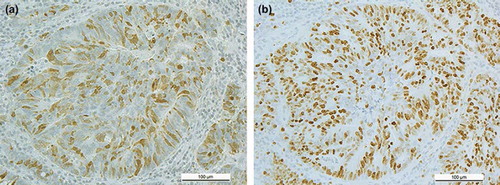
Examples of positive IHC staining for securin and Ki-67 in rectal cancer are illustrated in . Altogether 98% of the biopsies (n = 64/65) and operative specimens (n = 202/207) showed securin expression (≥1%) in cancer cell nuclei with a mean percentage of 47% (95% CI 42.1–52.2) and 31% (95% CI 28.7–32.6; p < 0.001), respectively. All the biopsies (n = 57/57) and 99% of the operative specimens (n = 204/207) showed Ki-67 expression with mean percentages of 83% (95% CI 79.8–86.4) and 56% (95% CI 53.2–64.7; p < 0.001), respectively.
Figure 1. Immunohistochemical staining of securin (a) and Ki-67 (b) in rectal cancer. Staining pattern of Ki-67 was almost exclusively nuclear, while securin had a combination of nuclear and cytoplasmic staining with a clear prominence of nuclear one.
Correlation between securin and Ki-67 expression
Securin and Ki-67 expression were closely correlated in biopsy and operative specimens (R = 0.38; p = 0.004; R = 0.47, p < 0.001, respectively. Supplementary to be found online at http://www.informahealth care.com/doi/abs/10.3109/0284186X.2011.584327). In both specimens, Ki-67 expression was higher than securin expression (p < 0.001). The level of securin expression and the staining intensity were closely correlated both in biopsies (R = 0.33, p = 0.01) and in operative specimens (R = 0.29; p < 0.001).
Securin and Ki-67 related to clinicopathological variables and disease outcome
Securin or Ki-67 expression in biopsies, nor securin staining intensity, did not correlate to clinicopathological variables. The expression of securin and Ki-67 in operative specimens related to key clinicopathological variables is shown in . Patients over 70 years showed higher expression of both markers than patients under 70 years (p = 0.04 for securin; p = 0.02 for Ki-67), but the difference was preserved only in the control group for securin (p = 0.036 for securin; p = 0.08 for Ki-67 in the same group) when the treatment groups were analysed separately.
Table II. The expression of securin and Ki-67 in the operative specimens related to key clinicopathological variables.
In operative specimens, T1-2 tumours showed the highest expression of securin (34%), followed by a lower expression in T3-tumours (29%) and the lowest expression in T4-tumours (24%; p = 0.01). These differences were not, however, preserved in any of the subgroups when analysed separately. No association was seen with Ki-67 expression and depth of tumour invasion ().
In the whole series, securin or Ki-67 did not significantly predict DFS or DSS in univariate (Kaplan-Meier) analysis, and thus were not included in the multivariate Cox regression model, where the following variables were entered: treatment group, sex, age, disease recurrence (only for DSS), pre- and postoperative T, lymph node status, tumour grade, circumferential margin and vascular invasion. For DFS, independent predictors were: tumour circumferential margin (HR = 2.4; 95% CI 1.1–5.1; p = 0.025) and vascular invasion (HR = 3.5; 95% CI 1.6–7.5; p = 0.002). For DSS, independent predictors were: patient age (HR = 1.1; 95% CI 1.0–1.2; p = 0.007), postoperative T (HR = 7.4; 95% CI 1.6–35.5; p = 0.01) and disease recurrence (HR = 19.8; 95% CI 4.3–91.8; p < 0.001).
Securin and Ki-67 in response to (chemo)RT and expression in the treatment groups
In the pairwise-comparison of pre-treatment biopsies and post-treatment operative specimens, securin expression was decreased in 51 cases (78%), and the pairs were discordant both in the short- (p < 0.001) and long-course RT group (p = 0.001) as shown in . Ki-67 expression was decreased in 55 cases (96%), the pairs being discordant both in the short- and long-course RT groups (p < 0.001, ).
Table III. The pairwise-comparison of biopsy and operative specimens according to securin and Ki-67 expression.
The comparison of securin and Ki-67 expression in operative specimens between the three treatment groups is shown in . In subgroup comparison, patients in the long-course (chemo)RT group had significantly less expression of securin than patients in the short-course RT or control groups (p < 0.001), whereas no difference existed between the short-course RT and the control groups (p = 0.65). For Ki-67, patients both in the short- and long-course RT-groups had significantly less expression than patients in the control group (p < 0.001; p = 0.003) while no difference was seen between the two RT groups (p = 0.95). No differences existed in either marker expression in the long-course RT group between the patients with and without concomitant chemotherapy (p = 0.23; p = 0.31).
TRG in relation to securin and Ki-67
TRG was poor in 50% (n = 27), moderate in 28% (n = 15) and excellent in 22% (n = 12) of the tumours. Of the 12 tumours with excellent TRG, four showed complete response to RT. In biopsies, the pre-RT securin or Ki-67 expression did not predict TRG. TRG according to securin and Ki-67 expression in operative specimens is shown in . Only Ki-67 expression was significantly related to TRG (p = 0.02 for Ki-67; p = 0.21 for securin).
Disease outcome in the long-course (chemo)RT group
In the short-course RT and control group, securin or Ki-67 expression did not predict either DFS or DSS (data not shown). As shown in and , no significant difference was seen in DFS between long-course (chemo)RT patients with high (over median) and low (under median) securin expression (p = 0.33), whereas DSS was significantly (p = 0.019) shorter in patients with high securin expression. For Ki-67, although showing a similar trend, no statistical significant differences existed in DFS or DSS (p = 0.72; p = 0.07; data not shown). Patients with excellent TRG tended to have longer DFS compared to patients with moderate or poor TRG (p = 0.06), and had the longest DSS (p = 0.03; and ).
Figure 2. Securin expression in operative specimens of the long-course (chemo)RT patients related to DFS and DSS (a–b). Tumour regression grade (TRG) after long-course (chemo)RT related to DFS and DSS (c–d).
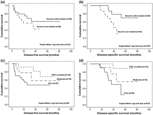
The multivariate Cox-model included following variables: sex, age, postoperative T and lymph node status, circumferential margin (only for DFS), disease recurrence (only for DSS), TRG, and securin expression (only for DSS). None of the variables was shown to be an independent predictor of DFS, whereas the following were predictors of DSS: age (HR = 1.1; 95% CI 1.0–1.2; p = 0.043), disease recurrence (HR = 24.3; 95%CI 2.2–269.8; p = 0.009) and securin expression (HR = 5.3; 95% CI 1.1–25.0; p = 0.036).
Discussion
Securin is an oncogene up-regulated and associated with poor prognosis in a wide variety of tumours [Citation11]. Although related to colorectal cancer progression and aggressive behaviour [Citation12,Citation13], no data is available on the exact relation of securin expression to rectal cancer outcome. We studied securin expression together with Ki-67 in 211 rectal cancer patients treated with or without preoperative (chemo)radiotherapy to evaluate protein expression in relation to (chemo)RT and disease outcome. We hypothesised securin expression to decrease in response to RT as reported with Ki-67 in a number of studies [Citation3,Citation19]. To our knowledge, this is the first study examining the effects of (chemo)RT on securin expression in human rectal cancer, as earlier studies have been made with CRC-cell lines [Citation15,Citation16]. We found both securin and Ki-67 expression to decrease significantly in response to RT. High securin expression after long-course (chemo)RT suggested poor outcome in this group of patients, Ki-67 only showing a similar trend.
Securin has been shown to be a valuable proliferation marker among others in pituitary adenomas [Citation20] and breast cancer [Citation21]. In the present study, a strong correlation between securin and Ki-67 expression was seen with mean Ki-67 expression being higher both in biopsies and resection specimens. This may partly be explained by a different cell-cycle distribution of these two proteins [Citation5–7]. Securin degradation at the end of metaphase is necessary to activate separase and enable equal distribution of sister chromatids to diploid daughter cells [Citation22]. Furthermore, unlike Ki-67, securin also has a number of other important functions. It induces genetic instability [Citation13] and plays a role in DNA repair and damage control, as well as in apoptosis [Citation11,Citation13,Citation23]. Overexpression of securin gene is also related to tumour invasiveness and angiogenesis by up-regulating basic fibroblast growth factor (bFGF) [Citation8], vascular endothelial growth factor (VEGF) [Citation9] and matrix metalloproteinase -2 (MMP-2) [Citation10]. Thus, it is understandable that securin expression does not only represent the proliferating activity of rectal cancer as opposed to Ki-67 despite the correlation of these markers.
In the whole patient cohort, we found the tumours of younger patients to express less securin and Ki-67, as compared to tumours of older patients. In the subgroup analysis, this difference was, however, preserved only for securin in the control group, and may thus be at least partially explained by the lower mean age of patients in the RT groups compared to mean age in the control group. As the phenomenon was, nonetheless, seen for securin also in the subgroup of patients without any preoperative treatment, we assume that there might be some additional explanation behind the difference in securin expression according to patient age. It may be associated with some other than the proliferative functions of securin, as the difference between age groups did not reach statistical significance in the case of Ki-67 in any of the subgroups. The differences seen in securin expression between T1-2, T3 and T4 tumours most probably also reflect the effect of radiotherapy, as the distribution of T-classes across the three treatment groups was different. No other associations were seen between securin or Ki-67 expression and clinicopathological variables, in contrast to some earlier reports [Citation12].
We analysed the effects of preoperative(chemo)RT on securin and Ki-67 expression using two approaches; comparing the preoperative biopsy and operative specimen pairs, and comparing the expression in operative specimens between the three treatment groups. The pairwise comparison allows the examination of biopsies and operative specimens from the same patient before and after RT. Unfortunately, this approach was limited by the scarce tissue material in biopsies, as well as the limited number of available biopsies in the present study. Using pairwise comparison, both securin and Ki-67 expression were significantly decreased after short- and long-course RT, securin decreasing more dramatically after long-course (chemo)RT. The operative specimens between short-course RT and control groups did not, however, show a difference in securin expression in contrast to Ki-67. There are several possible mechanisms to explain these differences. The fractions, total dose, treatment time and time before surgery differ between the short- and long-course RT regimens, and it is possible that the duration of the treatment and time before surgery in the case of short-course RT are too short to completely destroy tumour cells and modify securin expression. In the case of Ki-67, the repopulation and redistribution of surviving cells during and after fractionated RT [Citation24] might contribute to the unexpectedly similar expression in the short- and long-course RT groups. The differences in securin and Ki-67 expression patterns in response to (chemo)RT may well be related to the functions of securin other than cell proliferation.
Securin or Ki-67 expression in biopsies did not predict tumour regression grade (TRG) after RT, but the number of available biopsies was limited. In operative specimens, Ki-67 expression was lower in tumours with excellent TRG as compared to other tumours. This has been reported also earlier [Citation3], whereas the only studies on securin have been conducted with CRC-cell lines [Citation15,Citation16]. Chen et al., also showing a decrease in securin expression after RT, demonstrated that depletion of securin enhanced the radiosensitivity of human CRC cells by an arrest of senescent cell growth [Citation16]. In accordance with this, we showed a tendency, albeit not significant, to securin expression in operative specimens being lower in the tumours with excellent response to RT. Pathological complete response (pCR), or response greater than 95% after preoperative RT have been shown to predict favourable outcome in rectal cancer [Citation25], and in the present study too, patients with excellent TRG showed longer DSS, and also a trend towards longer DFS, as compared to moderate or poor responders. This substantiates our preliminary observations in the original cohort [Citation26], now supplemented with additional patients and longer follow-up.
In the long-course (chemo)RT group, patients with high post-treatment securin expression had significantly shorter DSS as compared to the patients with lower expression, Ki-67 expression showing the same trend. Importantly, in multivariate analysis for the long-course (chemo)RT group, high securin expression remained an independent predictor of adverse DSS together with patient age and disease recurrence. In this group of patients, high securin expression may define a population of tumours with inherent resistence to fluorouracil-based chemotherapy regimens used either postoperatively at an adjuvant setting, or later for a metastatic disease. This is supported by earlier findings in CRC cell lines, where the loss of securin expression has been reported to sensitise cells to anticancer agents [Citation27]. Accordingly, securin expression at the time of operation could be used as a predictive tool for DSS, and similarly, securin up-regulation might be helpful in identifying patients who do not respond well to treatment of recurrent rectal cancer. Thus our findings support additional or more aggressive treatment approaches for patients presenting with high securin expression after long-course (chemo)RT. In contrast to this group of patients, no difference was seen in disease outcome according to securin expression in the other treatment groups. Control group patients had less advanced tumours at the time of diagnosis as compared to patients in the RT-groups, and after short-course RT, the expression level of securin was less decreased than after long-course (chemo) RT. These things might explain why securin did not segregate the patients into different prognostic categories in short-course RT and control groups.
Taken together, securin and Ki-67 expression at protein level are closely interrelated, both decreasing significantly after (chemo)RT. However, securin and Ki-67 in rectal cancer also show somewhat different behaviour in response to RT, indicating that securin may not be considered exclusively as a proliferation marker. This is supported by the observation that only high securin expression, not Ki-67, was significantly related to poor survival in the long-course (chemo)RT patients. Accordingly, the potential role of securin as a predictive marker in rectal cancer could be ascribed to some other than proliferative functions of this protein. We suggest that the patients with high securin expression in their tumours after long-course (chemo)RT may represent a group of cases with decreased sensitivity to chemotherapy treatment. Further studies with larger patient cohort and longer follow-up are warranted to examine the predictive power of securin in different rectal cancer treatment protocols.
http://www.informahealthcare.com/doi/abs/10.3109/0284186X.2011.584327
Download PDF (281.1 KB)Acknowledgements
We are grateful to Sinikka Kollanus for her skilful help in laboratory work and Jaakko Liippo for aid with the digital pictures. This research work has been supported by the grants from The Special Government Funding (EVO) allocated to Turku University Hospital, the Cancer Society of South-Western Finland (JS) and the Finnish society for Therapeutic Radiology and Oncology (EK). The authors declare that they have no conflict of interest.
References
- Improved survival with preoperative radiotherapy in resectable rectal cancer. Swedish rectal cancer trial. N Engl J Med 1997;336:980–7.
- Randomised trial of surgery alone versus radiotherapy followed by surgery for potentially operable locally advanced rectal cancer. Medical Research Council Rectal Cancer Working Party. Lancet 1996;348:1605–10.
- Debucquoy A, Goethals L, Libbrecht L, Perneel C, Geboes K, Ectors N, . Molecular and clinico-pathological markers in rectal cancer: A tissue micro-array study. Int J Colorectal Dis 2009;24:129–38.
- Brown DC, GatterKC. Ki67 protein: The immaculate deception? Histopathology 2002;40:2–11.
- Zou H, McGarry TJ, Bernal T, Kirschner MW. Identification of a vertebrate sister-chromatid separation inhibitor involved in transformation and tumorigenesis. Science 1999;285:418–22.
- Ramos-Morales F, Domínquez A, Romero F, Luna R, Multon MC, Pintor-Toro JA, . Cell cycle regulated expression and phosphorylation of hpttg proto-oncogene product. Oncogene 2002;19:403–9.
- Gerdes J, Lemke H, Baisch H, Wacker HH, Schwab U, Stein H. Cell cycle analysis of a cell proliferation-associated human nuclear antigen defined by the monoclonal antibody Ki-67. J Immunol 1984;133:1710–5.
- Pei L, Melmed S. Isolation and characterization of pituitary tumor-transforming gene (PTTG). Mol Endocrinol 1997;11:433–41.
- McCabe CJ, Boelaert K, Tannahill LA, Heaney AP, Stratford AL, Khaira JS, . Vascular endothelial growth factor, its receptor KDR/Flk-1, and pituitary tumor transforming gene in pituitary tumors. J Clin Endocrinol Metab 2002;87:4238–44.
- Malik MT, Kakar SS. Regulation of angiogenesis and invasion by human Pituitary tumor transforming gene (PTTG) through increased expression and secretion of matrix metalloproteinase-2 (MMP-2). Mol Cancer 2006;5:61.
- Salehi F, Kovacs K, Scheithauer BW, Lloyd RV, Cusimano M. Pituitary-tumor transforming gene in endocrine and other neoplasms: A review and update. Endocr Relat Cancer 2008;15:721–43.
- Heaney AP, Singson R, McCabe CJ, Nelson V, Nakashima M, Melmed S. Expression of pituitary-tumour transforming gene in colorectal tumours. Lancet 2000;355:716–9.
- Kim DS, Franklyn JA, Smith VE, Stratford AL, Pemberton HN, Warfield A, . Securin induces genetic instability in colorectal cancer by inhibiting double-stranded DNA repair activity. Carcinogenesis 2007;28:749–59.
- Hlubek F, Pfeiffer S, Budczies J, Spaderna S, Jung A, Kirchner T, . Securin (hPTTG1) expression is regulated by beta-catenin/TCF in human colorectal carcinoma. Br J Cancer 2006;94:1672–7.
- Chiu SJ, Hsu TS, Chao JI. Expression of securin promotes colorectal cancer cell death via p53-independent pathway after radiation. Chem Biol Interact 2007;170:153–61.
- Chen WS, Yu YC, Lee YJ, Chen JH, Hsu HY, Chiu SJ. Depletion of securin induces senescence after irradiation and enhances radiosensitivity in human cancer cells regardless of functional p53 expression. Int J Radiat Oncol Biol Phys 2010;77:566–74.
- Glimelius B, Påhlman L, Cervantes A. ESMO Guidelines Working Group. Rectal cancer: ESMO Clinical Practice Guidelines for diagnosis, treatment and follow-up. Ann Oncol 2010;21(Suppl 5):v82–6.
- Korkeila E, Talvinen K, Jaakkola PM, Minn H, Syrjänen K, Sundström J, . Expression of carbonic anhydrase IX suggests poor outcome in rectal cancer. Br J Cancer 2009; 100:874–80.
- Willett CG, Warland G, Hagan MP, Daly WJ, Coen J, Shellito PC, . Tumor proliferation in rectal cancer following preoperative irradiation. J Clin Oncol 1995;13:1417–24.
- Filippella M, Galland F, Kujas M, Young J, Faggiano A, Lombardi G, . Pituitary tumour transforming gene (PTTG) expression correlates with the proliferative activity and recurrence status of pituitary adenomas: A clinical and immunohistochemical study. Clin Endocrinol (Oxf) 2006; 65:536–43.
- Talvinen K, Tuikkala J, Nevalainen O, Rantanen A, Hirsimäki P, Sundström J, . Proliferation marker securin identifies favourable outcome in invasive ductal breast cancer. Br J Cancer 2008;99:335–40.
- Waizenegger I, Giménez-Abián JF, Wernick D, Peters JM. Regulation of human separase by securin binding and autocleavage. Curr Biol 2002;12:1368–78.
- Yu R, Heaney AP, Lu W, Chen J, Melmed S. Pituitary tumor transforming gene causes aneuploidy and p53-dependent and p53-independent apoptosis. J Biol Chem 2000;275:36502–5.
- Fowler JF. The first James Kirk memorial lecture. What next in fractionated radiotherapy? Br J Cancer Suppl 1984;6:285–300.
- Guillem JG, Chessin DB, Cohen AM, Shia J, Mazumdar M, Enker W, . Long-term oncologic outcome following preoperative combined modality therapy and total mesorectal excision of locally advanced rectal cancer. Ann Surg 2005;241:829–36.
- Korkeila E, Jaakkola PM, Syrjänen K, Pyrhönen S, Sundström J. Pronounced tumour regression after radiotherapy is associated with negative/weak glucose transporter-1 expression in rectal cancer. Anticancer Res 2011;31:311–5.
- Chao JI, Liu HF. The blockage of survivin and securin expression increases the cytochalasin B-induced cell death and growth inhibition in human cancer cells. Mol Pharmacol 2006;69:154–64.