To the Editor,
Brain metastasis is one of the most common and devastating complications of melanoma with a median survival (mOS) of four months [Citation1]. Patients with leptomeningeal metastasis, i.e. leptomeningeal melanomatosis (LM), survive with an even shorter median of 10 weeks [Citation2]. Treatment of LM remains palliative with no standard of care to date. In LM, the use of radiation therapy is common, in part but not exclusively due to the fact that parenchymal brain metastases are present in about 50% of patients. The urgent need for systemic treatment is supported by the fact that the majority of patients also have visceral and other systemic metastases [Citation2]. Intrathecal therapy (IT) may be moderately effective since IT is a positive prognostic factor in a large retrospective series including patients mainly treated with methotrexate and interleukin-2 [Citation2]. There are so far no data on the intrathecal use of liposomal cytarabine in LM. In comparison to MTX, liposomal cytarabine has the advantages to be applicable with a longer interval of 14 days and to be as effective upon lumbar injection as upon instillation through an intraventricular reservoir [Citation3]. Thus, intrathecal liposomal cytarabine appears interesting to be explored in conjunction with radiotherapy and systemic chemotherapy.
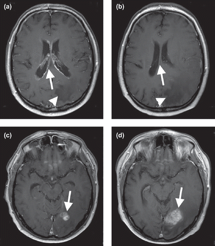
We describe the case of a 50-year-old male, Karnofsky Performance Status 80% who was diagnosed with melanoma of the trunk at level of lumbar vertebral body 2 in November 2003. The patient received multiple therapies for systemic manifestations (local excision, radiotherapy, interferon 2b, cisplatin, carmustin, DTIC, peptide vaccination) of thoracic soft tissue, lung, and the adrenal gland. Retrosternal lymphonodal metastasis was treated with seed-implantation. In January 2008, he presented to our department with headache, vertigo and lumbar radicular pain. Magnetic resonance imaging (MRI) of the whole neuroaxis revealed cerebral metastases of the occipital lobe and next to third ventricle and widespread intraventricular lesions () and affection of the conus medullaris (at level of lumbar vertebral body 3) and lumbar nerve roots. Initial cytological analysis revealed normal cell count, but elevated protein (727 mg/l) and lactate (2.63 mmol/l) levels in the cranial spinal fluid (CSF). Neuropathologic analysis identified melanoma cells. The patient received radiation therapy of the lumbar vertebrae and spinal cord (lumbar vertebral bodies 2–4 with 10 × 3 Gy), whole brain radiation with (20 × 2 Gy) combined with temozolomide (TMZ, 75 mg/m2) and an additional stereotactic boost of the occipital metastasis up to a local dose of 50 Gy. The radiochemotherapy was followed by eight cycles of adjuvant TMZ (200 mg/m2 on 5 of 28 days) and 22 cycles of intrathecal treatment with DepoCyte (50 mg biweekly). The therapy was well tolerated without hematotoxic or any other side effects. Upon therapy, the patient showed a complete clinical response and MRI revealed a significant regression of leptomeningeal lesions in April 2008. Due to an increase of liver metastasis detected by regular sonographic control the systemic therapy was changed after 10 months to carboplatin and etoposid. The contrast-enhanced cranial MRI was stable at that time. The intrathecal therapy with liposomal cytarabine was continued. The follow-up 12 months after diagnosis of LM showed a stable clinical-neurological examination, a clear regression of leptomeningeal lesions on MRI while the cerebral parenchymal metastases increased in size (). Interestingly, every cytological analysis of CSF revealed malignant cells as well elevated levels of cell count, protein and lactate. The patient died 14 months after diagnosis of cerebral and spinal metastasis due to diffuse systemic metastasis and multi-organ failure.
Figure 1a and c. Axial T1-weighted contrast-enhanced scans (3,0T, TR 550, TE 13) after radiochemotherapy but before treatment with adjuvant TMZ and intrathecal DepoCyte showing widespread subependymal enhancement (a, arrow), parenchymal metastases of the occipital lobe (c, arrow) with perifocal edema (a, arrowhead). b and d. Corresponding images (3,0T, TR 550, TE 13) 12 months later without evidence of leptomeningeal or subependymal lesions (b, arrow) while the cerebral parenchymal metastases (d, arrow) and the perifocal edema (b, arrowhead) increased in size.

It is interesting in our case that combination therapy including continuous IT with liposomal cytarabine was associated with prolonged clinical and MRI response of LM despite of a persistently positive CSF cytology. Considering the generally very short prognosis of LM, the combination therapy with radiotherapy, temozolomide and liposomal cytarabine was highly effective in our patient surviving more than one year. It is important to note that while CNS parenchymal and systemic metastases progressed over time, leptomeningeal metastases as documented on MRI, regressed and did not reappear. This could be an indication that intrathecal therapy with liposomal cytarabine was effective in this patient. The course of LM in our patient is encouraging to further explore combination therapy in general and intrathecal therapy with liposomal cytarabine in particular in patients with LM.
Acknowledgements
U. Herrlinger and M. Glas have received honoraria from Mundipharma (manufacturer of DepoCyte) in the past. All remaining authors have declared no conflict of interest.
References
- Davies MA, Liu P, McIntyre S, Kim KB, Papadopoulos N, Hwu WJ, . Prognostic factors for survival in melanoma patients with brain metastases. Cancer 2011;117:1687–96.
- Harstad L, Hess KR, Groves M. Prognostic factors and outcomes in patients with leptomenigeal melanomatosis. Neuro Oncol 2008;10:1010–8.
- Glantz MJ, Van Horn A, Fisher R, Chamberlain MC. Route of intracerebrospinal fluid chemotherapy administration and efficacy of therapy in neoplastic meningitis. Cancer 2010; 116:1947–52.
