Abstract
Background. Locally advanced cervical cancer is commonly treated with external radiation therapy combined with local brachytherapy. The brachytherapy is traditionally given based on standard dose planning with prescription of dose to point A. Dosimetric aspects when changing from former standard treatment to individualized treatment plans based on computed tomography (CT) images are here investigated. Material and methods. Brachytherapy data from 19 patients with a total of 72 individual treatment fractions were retrospectively reviewed. Standard library plans were analyzed with respect to doses to organs at risk (OARs), and the result was compared to corresponding delivered individualized plans. The theoretical potential of further optimization based on prescription to target volumes was investigated. The treatments were performed with a Fletcher applicator. Results. For standard treatment planning, the tolerance dose limits were exceeded in the bladder, rectum and sigmoid in 26%, 4% and 15% of the plans, respectively. This was observed most often for the smallest target volumes. The individualized planning of the delivered treatment gave the possibility of controlling the dose to critical organs to below certain limits. The dose was still prescribed to point A. An increase in target dose coverage was achieved when additional individual optimization was performed, while still keeping the dose to the OARs below predefined limits. Relatively low average target coverage, especially for the largest volumes was however seen. Conclusion. The individualized delivered treatment plans ensured that doses to OARs were within acceptable limits. This was not the case in 42% of the corresponding standard plans. Further optimized treatment plans were found to give an overall better dose coverage. In lack of MR capacity, it may be favorable to use CT for planning due to possible protection of OARs. The CT based target volumes were, however, not equivalent to the volumes described in magnetic resonance imaging (MRI) based recommendations. Prescription and assessment of dose, when introducing such target volumes, should be evaluated and preferably compared to well known treatment regimens.
Chemoradiation is the standard treatment for patients with locally advanced cervical carcinoma. The five-year survival rate ranges from 70–85% for stage IB to 5–15% for stage IV [Citation1]. The radiotherapy is often delivered as a combination of external irradiation and intracavitary brachytherapy. The bladder as well as the rectum and sigmoid are situated close to the cervix, and radiotherapy related long-term side effects from these organs may occur.
The brachytherapy has traditionally been performed by prescribing the radiation dose to a geometrical point A [Citation2]. For this type of treatment the individual size of the tumor is not considered, nor are the positions and shapes of the surrounding organs at risk.
To improve the treatment, the gynaecological (GYN) GEC-ESTRO working group has introduced recommendations for image-guided brachytherapy of cervical cancer [Citation3–5]. These comprehensive guidelines address delineations of target volumes on magnetic resonance (MR) images, as well as reporting of dose parameters from three dimensional (3D) treatment planning. They differentiate between a high risk (HR) clinical target volume (CTV), including the whole cervix and the presumed extracervical tumor extension at the time of brachytherapy with no safety margins, and an intermediate risk (IR) CTV encompassing the HR CTV with a safety margin of 5–15 mm. The total prescribed dose should be appropriate to eradicate the macroscopic disease in HR CTV and significant microscopic disease in IR CTV.
Whereas MR capacity is still often a limited resource, more radiotherapy departments have access to CT facilities. A US survey shows that while an increasing number of clinics perform CT prior to brachytherapy, 76% continue to prescribe the dose to point A rather than to a 3D based volume, and 52% use ICRU points instead of values from dose volume histogram when evaluating OARs [Citation6]. Our institution has made a stepwise approach from the use of standard library brachytherapy treatment plans, based on prescribed dose to point A, towards the GYN GEC-ESTRO recommendations.
The aims of this retrospective study were to evaluate dosimetric aspects when changing from standard library plans to individualized treatment plans based on CT images. The delivered treatment was mainly based on prescription of dose to point A, while taking doses to OARs into consideration. A further evaluation of the theoretical potential of individualized optimization was done by introducing prescription of dose to target volumes. Analyses were performed with respect to target coverage and dose to OARs. The dose to point A was in this case recorded, but not used for prescription.
Material and methods
Patient group and treatment delivery
The patients in this study have received treatment for locally advanced cervical cancer as a combination of external radiotherapy, weekly distribution of Cisplatin and brachytherapy. They were the first patients in our hospital to receive brachytherapy treatment with individualized 3D planning based on CT images, following 15 years of treatment with standard library plans based on dose to point A [Citation7].
Twenty one consecutive patients treated with individually planned brachytherapy between September 2005 and February 2007 constituted the basis for this study, and 19 patients accepted to participate. The study was approved by the Regional Committee for Medical and Health Research Ethics.
CT of the thorax/abdomen and magnetic resonance imaging (MRI) of the pelvis were performed as part of the diagnostics prior to treatment start. The patient characteristics are summarized in . The group of patients included in the study covers a wide range of tumor sizes and stages of the disease at the time of diagnosis. Follow-up evaluations based on MRI and clinical examinations occurred at 10–12 weeks, and every three months the first two years after treatment completion. Additional information regarding patient outcome was obtained autumn 2010, from 3.5 to 5 years after treatment completion.
The external radiation treatment was based on 3D planning in CT images. Standard treatment was delivered with the fractionation of 2 Gy × 25 with an external boost to enlarged lymph nodes of 2 Gy × 3. Other fractionation regimens were given to some patients (see ).
The brachytherapy treatment was carried out using the GammaMed 12i afterloading equipment Varian (Palo Alto, USA) with an Ir192 stepping source. A Fletcher type applicator (Sauerwein Isotopen Tecknik 926500) with standard colpostat segment of the ovoids (20 mm diameter) was used. The length of the intrauterine probe and the distance between the colpostats are adjustable for too some degree adapt to the patients anatomy. Gauze packs were used to stabilize the applicator and push the bladder and the rectum away from the tumor. CT images (2 mm thick, Siemens Somatom Emotion CT (Siemens, Munich, Germany) spiral-CT) were acquired prior to every brachytherapy fraction. Before both CT survey and treatment, the bladder was drained, and then filled with 100 ml 0.9% NaCl. Individualized plans were made for every treatment (see details below). The prescribed standard brachytherapy dose was 5 Gy × 4, given twice weekly during the two last weeks of the external radiation treatment. No external radiation was given on the day of brachytherapy. Other fractionation regimens were given to some patients (see ). The 19 patients received a total of 76 individual brachytherapy fractions. Four of these fractions were excluded from the retrospective study due to the use of a different type of Fletcher applicator.
Delineation of volumes of interest
The delineation of structures on CT images was done in Oncentra Masterplan (Nucletron, Veenendaal, The Netherlands) version 1.5 (Nucletron).
Clinical target volumes, CTV, were retrospectively drawn, including tumor, cervix and extracervical tumor extension. Delineation was always done with additional information from the diagnostic MR images and the clinical examination performed prior to the brachytherapy treatment. The tumor volume at the time of diagnosis was used as a base for the delineation. When obvious tumor shrinkage had occurred, the CTV was modified accordingly to better represent the actual extent of the disease. The GYN GEC-ESTRO recommendations were tentatively used.
The non-tissue material including the lower parts of the catheters, ovoids and the vaginal packing were excluded from the target volumes ().
Figure 1. Delineation of volumes of interest in CT images in FIGO stage IIIB cervical cancer, second brachytherapy fraction. Left: Axial view. Right: Sagittal view.
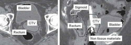
Table I. Patient, tumor and treatment characteristics.
The OARs, that is bladder, rectum and sigmoid colon, were drawn according to published guidelines [Citation4]. Every volume in this study was outlined and approved by the same experienced physician.
Treatment planning
Treatment planning was performed using the Plato Brachytherapy Planning System v.14.2.6 (Nucletron, Veenendaal, The Netherlands). Three different plans were selected or made for each treatment fraction as described below:
Standard treatment plans. Standard plans were originally constructed to treat volumes containing the cervix and the entire endometrium, providing the prescribed dose of 5 Gy to point A and the prescription isodose to enclose the colpostat segments with a margin of 2 mm. These plans gave a ‘pear’-shaped dose distribution in the frontal plane and a ‘banana’-shaped distribution in the sagittal plane of the patient. In our department there is a library of standard plans based on different adjustments of the applicator geometry.
The standard plans used in the study were chosen based on the actual applicator geometry measurements on sagittal and coronal projections of the CT images. The resulting dose distributions were analyzed for CTV dose coverage as well as doses to OARs.
Delivered treatment plans; a semi optimization of the standard treatment. The actual patient treatments were initially based on traditionally planning, prescribing the dose to point A. These plans were however modified to not exceed the dose limits for the OARs (see below). For some patients with tumor volumes extending beyond point A, the dose to point A was allowed to exceed the prescribed dose to provide better visual dose coverage, while still keeping the dose to the OARs within the specified limits. A total of 57 treatment fractions were delivered based on such planning and were eligible for further study. In the remaining 15 fractions no individualization was attempted at the time of brachytherapy and standard plans were used for patient treatment.
Optimized treatment plans. A set of equally spaced dose points on the surface of each CTV were defined, and the dose point optimization method in Plato was employed. This optimization method provides a treatment plan where the average dose to the target surface equals the prescribed dose, without the ability to set any other dose constraints to OARs or normal tissue. Manual graphical optimization was then performed to make sure that the dose to any of the OARs did not exceed the given tolerance limits. The dose to point A was not taken into account during the optimization.
Dose to organs at risk. The recommended dose limit for bladder is a total dose of 90 Gy to the most exposed 2 cm3 (D2cc). For rectum and sigmoid the corresponding limit is 75 Gy. The underlying assumption is that the total dose may be expressed in fractions of 2 Gy using α/β = 3 [Citation4]. For our standard radiation regimen, assuming 2 Gy × 25 given externally and 5 Gy × 4 given as brachytherapy, the tolerance limits per brachytherapy fraction are then 5.7 Gy for bladder and 4.3 Gy for both rectum and sigmoid.
Conformity indices and parameters for comparing treatment plans
The conformal index COIN [Citation8] was used as a tool to define the quality of the treatment plans, taking into consideration both target coverage and the degree of normal tissue irradiation. The definitions of target coverage (TC), conformity index (CI) and COIN index are:

V100: Volume (cm3) of target receiving 100% or more of the prescription dose.
Vtot: Total volume (cm3) of target.
VExt100: Volume (cm3) within the external contour receiving 100% or more of the prescription dose.
Other parameters presented are:
Vnnxx: Volume (cm3) enclosed by the xx% isodose for structure nn.
D90: Minimum dose to 90% of target volume.
Dose and volume data were extracted from the dose-volume histograms for each treatment plan.
Statistical analyses
The dose and volume data were statistically analyzed using SPSS. The Wilcoxon Signed Rank Test was used to establish whether the distributions of the paired dose and volume data for two types of treatment plans were the same. This is a test that can be used when the variables are not normally distributed, as was the case in our material. A 95% confidence interval was used.
Results
Patient follow-up
None of the patients had severe complications during or immediately after completion of treatment, and there were no treatment delays. None of the patients had pelvis failure as the first recurrence. By follow-up 3.5 to 5 years from treatment completion, five patients were dead due to progression of the disease outside the pelvis area. Three of these patients had enlarged para-aortic lymph nodes at the time of diagnosis, one patient developed enlarged para-aortic lymph nodes eight weeks after completion of the treatment and one patient had brain metastases at the time of diagnosis and underwent palliative radiation treatment. One patient was alive with progression outside the external radiation field and one patient was dead due to another disease. The rest of the patients (12) showed no sign of recurrence and had no major complications that can be related to the treatment.
Target volumes
The sizes of the CTV were in general found to increase with the stage of the disease. The average size of the volumes for the different stages were 93 ± 44 cm3 (stage I), 113 ± 54 cm3 (stage II) and 117 ± 35 cm3 (stage III and IV). The initial tumors diameters from the diagnostic MRI were compared to measurement of diameters of the delineated CTVs in the first brachytherapy fraction. The calculated average volumes of the CTVs were fairly equal to the original tumor volumes. Such measurements are however encumbered with uncertainties due to the presence of the applicator at treatment. The average CTV decreased from 124 cm3 in the first brachytherapy fraction to 100 cm3 in the last fraction.
Standard plans – dose to OARs
When employing standard plans to the patients, 26%, 4% and 15% of the plans were found to cause dose distributions that exceeded the tolerance limits for the bladder, rectum and sigmoid, respectively. In total, 42% of the standard plans were observed to exceed one or two of the tolerance limits. Furthermore, for 8 of the 19 patients the same organ would have received a dose that was higher than the tolerance dose in two fractions or more if treated with the chosen standard plan. The minimum dose to the most exposed 2 cm3 volumes of the OARs were generally found to decrease with increasing target volumes for the standard plans as seen in , and the dose limits were more often exceeded for the smaller target volumes as seen in .
Figure 2. Target coverage against volume of CTV (linear regressions) for optimized plans (—) and standard (– –) plans. The individual fractions are shown for standard plans below OAR limits (○), or with bladder (▴), rectum (•) or sigmoid (♦) exceeding tolerance limits.
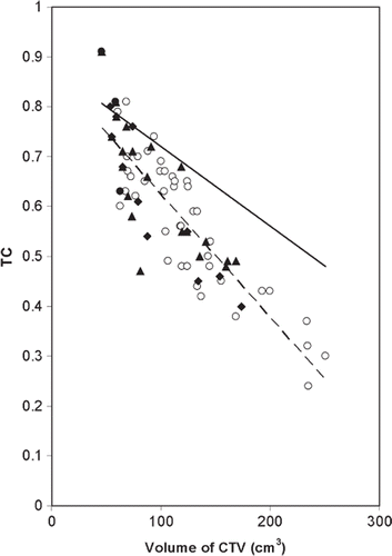
Table II. Mean values of dose and volume data for optimized and standard treatment plans.
Comparison of delivered and standard treatment plans
Comparing the delivered plans to the corresponding standard plans revealed no significant difference for the mean dose to point A (average of point A1 and A2). Although a higher dose was given to point A for the largest tumor volumes for the delivered treatment plans (data not shown), the standard deviation of dose to point A obtained for the brachytherapy plans was relatively low, reflecting our careful transition from the use of standard plans to 3D image based planning. The mean value of VExt100, which represents the volume treated to the prescription dose or more, was quite similar in the two treatment plans, as seen in and .
Comparison of optimized and standard treatment plans
Data for optimized versus standard plans based on analyses for CTV are given in . The table includes averages of the complete amount of data, with columns in which the data are split into subgroups representing the smallest and the largest target volumes, respectively. Dose data are presented as EQD2Gy for total treatment calculated as if each brachytherapy fraction was given as 5 Gy × 4 in addition to external radiation 2 Gy × 25, using σ/β = 10 for tumor and σ/β = 3 for OARs.
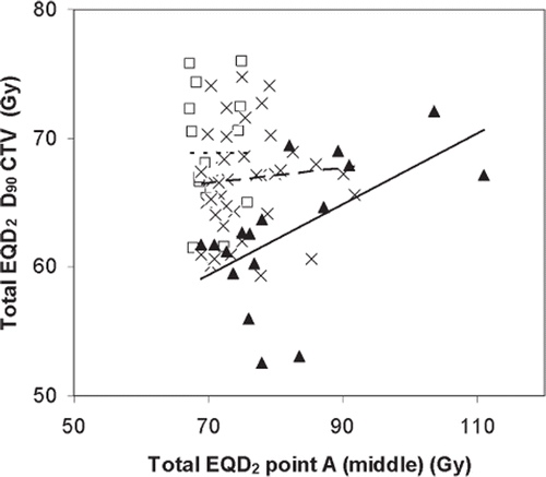
The mean total D90 value was found to be 3.3 Gy higher for the optimized than for the standard treatment plans. When considering only the brachytherapy treatment, this additional dose represents an increase of 26%. It was further seen that the D90 value decreased with increasing target volumes. shows the D90 value versus dose to point A for the optimized plans. No clear correlation was found between the values for small and intermediate target volumes, whereas such a correlation might be seen for the largest volumes.
Figure 3. Optimized plans: EQD2Gy for CTV D90 plotted against dose to point A for different ranges of CTV sizes. CTV < 70 cm3 (- - ○), 70 cm3 < CTV < 140 cm3 (- - x), CTV > 140 cm3 (— ▴). The data are presented for total treatment calculated as if each brachytherapy fraction was given equally four times in addition to standard external radiation (2 Gy × 25) using σ/β = 10 for tumor.
The mean dose to the A points (average of point A1 and A2) for the optimized plans was significantly lower than the corresponding mean dose to point A for standard plans for the smallest target volumes and significantly higher for the largest volumes (). It was seen that the treatment plans based on standard loading did not result in a mean dose to point A exactly equal to the prescribed dose (5 Gy). This is due to individual, sometimes non-symmetrical placements of the ovoids in the patients that were not adjusted for when employing the standard plans.
For the optimized treatment plans the mean value of TC was found to be 19% higher than that of the standard plans and was further seen to decrease with increasing target volume. This effect of target volume on the value of TC was, however, more pronounced for the standard plans (). The mean total volume receiving the reference dose, VExt100, was significantly larger (30%) for the optimized plans than for the standard plans. As a consequence, the corresponding mean CI value for the optimized plans was 6% lower than for the standard plans. This means that more normal tissue was irradiated when employing the optimized plans compared to the standard plans. The mean COIN value was however improved for the optimized plans, most clearly seen for the largest volumes ().
Treatment plans having COIN value > 0.5 and doses within OAR tolerances were considered to be acceptable plans. 52% of the optimized plans were in accordance to these criteria. This was an improvement compared to standard treatment for which only 23% were considered to be acceptable.
Doses to OARs were the limiting factors when making optimized treatment plans, aiming for 100% coverage of the target volume (CTV). The bladder dose was seen to be the main limiting factor in 64% of the cases and the doses to sigmoid and rectum appeared to be the limiting factor in 21% and 14% of the cases, respectively.
For the optimized plans the doses to the OARs were increased for the largest volumes compared to the standard plans (). For both types of treatment plans, a minor volume of tissue outside the CTV received twice the reference dose, 1.8 cm3 for the optimized plans and 1.3 cm3 for the standard plans.
Discussion
It is recommended that delineation of brachytherapy target volumes is based on MR images due to superior soft tissue contrast compared to CT [Citation4]. A few studies based on 3D CT brachytherapy and the new recommendations are published. Viswanathan et al. [Citation9] have shown that CT images may overestimate the tumor width compared to MR based contours, especially in the lateral direction. Based on a material consisting of 10 patients, they developed so called standardized CT contours, which gave a mean HR CTV of 48 cm3 and IR CTV of 118 cm3. Kang et al. [Citation10] investigated local control in cervical cancer patients where target volumes delineated in CT images were reported as ‘close to’ HR CTV. However, their volumes included fixed margins and information from additional MRI performed at the time of brachytherapy. In a Japanese study based on CT planning [Citation11], MRI was also used prior to the brachytherapy sessions for most patients, reporting a mean HR CTV of 33 cm3.
Mean volumes of HR CTV based on MRI are reported to vary between 34 cm3 and 44 cm3 in similar studies [Citation12–15], while corresponding volumes of IR CTV vary between 90 cm3 and 112 cm3.
We believe that our target volumes, defined in CT images, can not be directly compared to the HR CTV or the IR CTV as described by GYN GEC-ESTRO [Citation3]. The median volume of CTV in our study was observed to be 109 cm3. Thus, when considering only the size of the volume, CTV is more similar to the IR CTV than to the HR CTV. The fear of missing malignant tissue (tumor or so called “grey zones” not clearly seen on CT), may have led to an overestimation of the CTV compared to HR CTV. This is supported by measurements of the width of the CTV compared to MRI measurements. Tumor shrinkage during the weeks of treatment is more difficult to identify in CT images than in MRI, but the CTVs were clearly smaller at the end of the treatment. The patients in our study generally had a higher stage of disease (45% in stage III and IV) compared to the other studies (14–40% in stage III and IV). This will most probably affect the size of the target volumes.
As mentioned above, target volumes delineated in the present study, based on CT images, seem to represent an overestimation of the MRI based HR CTV. Dose values from our study can therefore not be directly compared to values in the literature based on HR CTV. Dimopoulos et al. [Citation16,Citation17] have demonstrated that local tumor control rates of >90% may be expected at D90 doses >86 Gy for HR CTV. An equivalent dose of 86 Gyα/β = 10 would in our case require a brachytherapy boost of 6.5 Gy × 4, a dose actually represented by the 130% isodose in our treatment plans with the standard fractionation regimen described earlier. The mean Vext130 for the delivered treatment plans was found to have an average value of 45 cm3 which is comparable to reported volumes of HR CTV [Citation12–15,Citation18]. This may explain the fact that our treatment has been successful for local control despite of apparently low target coverage. A fraction of our relatively big target volume represents the ‘true’ HR CTV and will receive an amount of irradiation that is sufficient to eradicate the malignant tissue.
The prescribed total dose used in our institution equals 75 Gyα/β = 10 (2 Gy × 25 external radiation and 5 Gy × 4 with brachytherapy). This place us among the other comparable institutions prescribing a total dose of 71.4–82.0 Gyα/β = 10 to point A [Citation12–13,Citation15,Citation18–19]. The average CTV D90 was found to be 65.8 Gy for the optimized treatment plans. According to GYN GEC-ESTRO it is recommended to cover the IR CTV with at least 60 Gy [Citation4]. We believe that our CTV was not necessarily equal to IR CTV, which ultimately is based on MR images, but this comparison supports that this approach towards individualized planning was apparently safe regarding dose prescription. The recommendation was also met for the average CTV D90 when providing the standard library plans.
Further increase in conformity was obtained by optimizing the treatment plans, however relatively low values of TC and COIN were generally obtained for these optimized plans, especially for the largest volumes. This corresponds with data from studies based on MRI and CT brachytherapy, getting the best optimization for the smallest tumors [Citation16,Citation20].
When prescribing the dose to a target volume (as was the basis for the optimized treatment plans) instead of to point A (being the basis for the standard treatment plans), no significant change in the mean dose to point A was observed (). Similar findings are described by Tanderup et al. [Citation21] in optimized treatment plans based on MR images. They point out that dose optimization will not necessarily change the average dose given to the patient population; it will rather add conformity and provide a higher therapeutic ratio.
There was no correlation between D90 and the dose to point A for small and intermediate target volumes for optimized plans (). The dose to point A does therefore not provide any valuable information about the actual dose to the target volume on an individual basis.
CT and MR images provide basically similar quality for discrimination of the OARs [Citation4]. The total biological dose limits used in the present study regarding the OARs were therefore in accordance with the guidelines [Citation4]. The better target coverage for standard plans for some of the smallest target volumes was at the expense of exceeding tolerance doses to OARs (). This is in compliance with other institutions that have analyzed typical standard loading [Citation10,Citation22]. The main improvement of individualized treatment planning was the opportunity of keeping doses to OARs within limits.
Attention should be paid to the high doses obtained close to the applicator, represented by the volume receiving twice the reference dose, VExt200. Our results revealed no significant change in this volume between the optimized and the standard treatment plans when considering the average of all the individual plans (). For the largest target volumes a significant increase in VExt200 was recorded for the optimized plans, and small parts of these volumes consist of normal tissue outside the CTV. A contiguous high dose volume is assumed clinically unfavorable compared to a more distributed high dose. Data from DVHs is however of limited use for analyzing hot spots as they provide no information regarding the distribution of the high dose.
As D90 and TC are considered useful parameters to evaluate dose to target volumes, other indices include additional information regarding healthy tissue and OARs. The conformal index COIN [Citation8] defined for brachytherapy reflects the quality of the treatment plans by taking target coverage and surrounding irradiated normal tissue equally into account. As dose to radiation sensitive organs is handled separately in our study, one may argue that target coverage may clinically be of greater importance than the amount of irradiated normal tissue. Weighting factors for such indices have been suggested [Citation23]. It would be of interest to define suitable weighting factors to make the COIN index more appropriate.
Conclusions
Our study suggests that in lack of MR capacity, it is favorable to use CT for brachytherapy planning due to possible protection of OARs. Individual 3D treatment planning, based on prescription to point A, ensured that the doses to OARs were within acceptable limits for the treatment delivered to the patients. This was not the case in 42% of the corresponding standard plans, most often seen for the smallest target volumes. It is possible to gain important benefits from use of CT imaging without having to change the prescription protocols in the first place.
An increase in target dose coverage and COIN was attained when further individual optimization based on prescription to target volumes was performed. This was achievable without exceeding tolerance limits for the OARs.
MR is assumed to be the gold standard for brachytherapy planning of cervical cancer. This study showed that it is feasible to perform planning also on CT based volumes. Prescription and evaluation of target dose should however be performed carefully as target volumes delineated on CT are not necessarily equal to the volumes defined in the GYN GEC-ESTRO recommendations. More knowledge and guidelines may be needed to encourage institutions to use 3D brachytherapy.
Acknowledgements
The technical support from Thorbjørn Tveit and Eva Susanne Oddvik, St. Olavs Hospital is greatly acknowledged. The authors alone are responsible for the content and writing of the paper.
Declaration of interest: The authors report no conflicts of interest. The authors alone are responsible for the content and writing of the paper.
References
- Waggoner SE. Cervical cancer. Lancet 2003;361:2217–25.
- Gerbaulet A, Pötter R, Mazeron JJ, Meertens H, Van Limbergen E. The GEC ESTRO Handbook of Brachyterapy. Brussels: European Society of Therapeutic Radiology and Oncology; 2002.
- Haie-Meder C, Pötter R, Van Limbergen E, Briot E, De Brabandere M, Dimopoulos J, . Recommendations from Gynecological (GYN) GEC-ESTRO Working Group (I): Concepts and terms in 3D image based 3D treatment planning in cervix cancer brachytherapy with emphasis on MRI assessment of GTV and CTV. Radiother Oncol 2005;74:235–45.
- Pötter R, Haie-Meder C, Van Limbergen E, Barillot I, De Brabandere M, Dimopoulos J, . Recommendations from Gynecological (GYN) GEC-ESTRO Working Group (II): Concepts and terms in 3D image-based treatment planning in cervix cancer brachytherapy – 3D dose volume parameters and aspects of 3D image-based anatomy, radiation physics, radiobiology. Radiother Oncol 2006;78:67–77.
- Hellebust TP, Kirisits C, Berger B, Pérez-Calatayud J, De Brabandere M, De Leeuw A, . Recommendations from Gynaecol (GYN) GEC-ESTRO Working Group: Considerations and pitfalls in commissioning and applicator reconstruction in 3D image-based treatment planning of cervix cancer brachytherapy. Radiother Oncol 2010;96: 153–60.
- Viswanathan AN, Erickson BA. Three-dimensional imaging in gynecologic brachytherapy: A survey of the American Brachytherapy Society. Int J Radiat Oncol Biol Phys 2010; 76:104–9.
- Lorenz E, Strickert T, Hagen B. Radiation therapy in cervical carcinoma fifteen years experience in a Norwegian health region. Eur J Gynaecol Oncol 2009;30:20–4.
- Baltas D, Kolotas C, Geramani K, Mould RF, Ioannidis G, Kekchidi M, . A conformal index (COIN) to evaluate implant quality and dose specification in brachytherapy. Int J Radiat Oncol Biol Phys 1998;40:515–24.
- Viswanathan AN, Dimopoulos J, Kirisits C, Berger D, Pötter R. Computed tomography versus magnetic resonance imaging-based contouring in cervical cancer brachytherapy: Results of a prospective trial and preliminary guidelines for standardized contours. Int J Radiat Oncol Biol Phys 2006;68:491–8.
- Kang H-C, Shin KH, Park S-Y, Kim J-Y. 3D CT-based high-dose-rate brachytherapy for cervical cancer: Clinical impact on late rectal bleeding and local control. Radiother Oncol 2010;97:507–13.
- Wadasaki K, Monzen Y, Kurose T, Okazaki H, Mito M. Computed tomography-based three-dimensional dosimetry of intracavitary brachytherapy for cervical cancer. Jpn J Radiol 2010;28:740–5.
- Lindegaard J, Tanderup K, Nielsen SK, Haack S, Gelineck J. MRI-guided 3D optimization significantly improves DVH parameters of pulsed-dose-rate brachytherapy in locally advanced cervical cancer. Int J Radiat Oncol Biol Phys 2008;71:756–64.
- Kiristis C, Pötter R, Lang S, Dimopoulus J, Wachter-Gerstner N, Georg D. Dose and volume parameters for MRI-based treatment planning in intracavitary brachytherapy for cervical cancer. Int J Radiat Oncol Biol Phys 2005;62:901–11.
- Kiristis C, Lang S, Dimopoulus J, Berger D, Georg D. Pötter R. The Vienna applicator for combined intracavitary and interstitial brachytherapy of cervical cancer: Design, application, treatment planning, and dosimetric results. Int J Radiat Oncol Biol Phys 2006;65:624–30.
- Chargari C, Magne N, Dumas I, Messai T, Vicenzi L, Gillion N, . Physics contributions and clinical outcome with 3D-MRI-based pulsed-dose-rate intracavitary brachytherapy in cervical cancer patients. Int J Radiat Oncol Biol Phys 2009;74:133–9.
- Dimopoulos JC, Lang S, Kirisits C, Fidarova, EF, Berger D, Georg P, . Dose–volume histogram parameters and local tumor control in magnetic resonance image-guided cervical cancer brachytherapy. Int J Radiat Oncol Biol Phys 2009;75:56–63.
- Dimopoulos JC, Potter R, Lang S, Fidarova, E, Georg P, Dörr W, . Dose-effect relationship for local control of cervical cancer by magnetic resonance image-guided brachytherapy. Radiother Oncol 2009;93:311–5.
- Shin KH, Kim TH, Cho JK, Kim JY, Park SY, Park SY, . CT-guided intracavitary radiotherapy for cervical cancer: Comparison of conventional point A plan with clinical target volume-based three-dimensional plan using dose-volume parameters. Int J Radiat Oncol Biol Phys 2006;64:197–204.
- De Brabandere M, Mousa AG, Nulens A, Swinnen A, Van Limbergen E. Potential of dose optimisation in MRI based PDR brachytherapy of cervix carcinoma. Radiother Oncol 2008;88:217–26.
- Zwahlen D, Jezioranski J, Chan P, Haider MA, Cho YB, Yeung I, . Magnetic resonance image-guided intracavitary brachytherapy for cancer of the cervix. Int J Radiat Oncol Biol Phys 2009;74:1157–64.
- Tanderup K, Nielsen SB, Nyvang, G-B, Pedersen EM, Røhl L, Aagaard T, . From point A to the sculpted pear: MR image guidance significantly improves tumour dose and sparing of organs at risk in brachytherapy of cervical cancer. Radiother Oncol 2010;94:173–80.
- Onal C, Arslan G, Topkan E, Pehlivan B, Yavuz M, Oymak E, . Comparison of convential and CT-based planning for intracavitary brachytherapy for cervical cancer: Target volume coverage and organs at risk doses. J Exp Clin Cancer Res 2009;28:95.
- Oozeer R, Chauvet B, Garcia R, Berger C, Felix-Faure C, Reboul F. Évaluation dosimetrique d'une radiothérapie conformationnelle: Le facteur de conformation. Cancer Radiother 2000;4:207–16.