Abstract
Background. The prevalence of esophageal cancer accompanied by hypopharyngeal cancer (HPC) is high and increasing rapidly in Asia. The purpose of this prospective study was to evaluate the prevalence of esophageal cancer during the pretreatment of HPC patients who were routinely examined using esophagogastroduodenoscopy (EGD) and 18F-fluorodeoxyglucose/computed tomography (FDG-PET/CT) and to discuss the utility of these examinations. Material and methods. Between September 2005 and September 2010, 33 patients with newly diagnosed HPC (all with squamous cell carcinoma) underwent EGD (after a conventional endoscopy, iodine staining was performed) and FDG-PET/CT examinations. We evaluated the prevalence of esophageal cancer among HPC patients according to the EGD findings and determined the sensitivity of FDG-PET/CT for the detection of esophageal primary tumors for each clinical T classification. Results. In 17 of the 33 patients (51.5%), 29 biopsy-proven esophageal squamous cell carcinomas were diagnosed using EGD. In eight of the 17 (47.1%) patients, two or more esophageal cancer lesions were diagnosed. Twenty-four of the 29 (82.8%) lesions were superficial esophageal cancers, and the remaining five (17.2%) lesions were advanced esophageal cancers. In six of the 29 (20.7%) esophageal cancer lesions that were detected using FDG-PET/CT, only one of the 29 (3.4%) lesions was evaluated as being equivocal; the remaining 22 (75.9%) lesions were not detected. The distribution of the clinical T classifications detected using FDG-PET/CT was as follows: T1a, 0/21 (0%); T1b, 1/3 (33%); and T3, 5/5 (100%). Conclusions. The prevalence of esophageal cancer during the pretreatment of HPC patients was 51.5%; this prevalence was higher than that in previous reports. We believe that the increasing proportion of superficial lesions (82.8%) detected using iodine staining and EGD may have led to the relatively high prevalence. FDG-PET/CT detected only 20.7% of the esophageal cancers, although FDG-PET/CT is capable of detecting unexpected primary malignant tumors other than esophageal cancer.
Hypopharyngeal cancer (HPC) is difficult to diagnose at an early stage. The overall five-year survival rate is relatively poor compared with those for other head and neck cancers (HNC) [Citation1]. HPC initially presents at an advanced stage (III or IV), even in developed countries, accounting for a five-year survival prognosis of only 20–40% [Citation2]. Moreover, the detection of multi-primary cancer (MPC) is quite important because the presence of this condition affects the prognosis. Patient survival after the diagnosis of cancer varies according to the site of the secondary cancer, with esophagus or lung cancer offering the worst prognoses [Citation3]. The frequency of MPC among HPC patients is approximately 5–22% [Citation4,Citation5]. The prevalence of esophageal cancer accompanied by HNC is known to be high and to be increasing rapidly in Asia [Citation6]. In particular, esophageal cancer among HPC patients is common.
In Japan, the incidence of HNC according to the International Classification of Disease (ICD-10: C00-14) was 2.0 per 100 000 person-years (world population) in 1975, whereas in 2006 the incidence of HNC was 4.7 per 100 000 person-years (world population); the incidence was 7.3 per 100 000 person-years among men and 2.2 per 100 000 person-years among women [Citation7]. The incidence of HPC in Japan was not clear, although among HNC, the frequency of HPC (18.0%) was the third highest after oral cancer (31.8%) and laryngeal cancer (23.8%) [Citation8]. The incidence of esophageal cancer (ICD-10: C15) was 4.7 per 100 000 person-years (world population) in 1975, whereas the incidence of esophageal cancer in 2006 was 6.3 per 100 000 person-years (world population); the incidence was 11.7 per 100 000 person-years among men and 1.6 per 100 000 person-years among women [Citation7]. Thus, the incidences of HNC and esophageal cancer have gradually increased in Japan.
Among secondary cancers in the digestive tract, including the esophagus, stomach, colon, and rectum, the esophagus is the only site of secondary cancer associated with an excess risk in patients with oral or pharyngeal carcinoma. Interestingly, the risk increases with the proximity of the primary index tumor to the esophagus, with a descending sequence of hypopharynx > oropharynx > oral cavity [Citation6]. Therefore, the prevalence of esophageal cancer among HPC patients is a great concern. Nevertheless, the recent prevalence of esophageal cancer detected during the pretreatment of patients with HPC remains unclear. A number of clinical factors have been recorded that have a predictive association in patients who may or may not develop a second primary malignancy. Tobacco smoking and alcohol drinking have been identified as major risk factors. Moreover, patients who present with an early-stage primary tumor are said to have a higher risk of developing a secondary primary tumor (SPT) than are patients who present with an advanced tumor, probably because of the anticipated longer survival of such patients [Citation5]. Generally, for the detection of esophageal cancer, an esophagogastroduodenoscopy (EGD) is performed as a standard examination, although an EGD can only evaluate focal sites. Therefore, other examinations, such as computed tomography (CT) and magnetic resonance imaging (MRI), are often performed for staging. Additionally, positron emission tomography using 18F-fluorodeoxyglucose (FDG-PET) has also shown good results for the staging and detection of local recurrences of HNC [Citation9,Citation10]. Recently, combined functional-anatomic imaging with fused FDG-PET/CT has emerged as a further development of the PET technique. FDG-PET/CT enables a whole-body examination during only one imaging session. Moreover, FDG-PET/CT is a less-invasive technique. If FDG-PET/CT were to demonstrate an ability to detect esophageal cancer in HPC patients equal to that of EGD, FDG-PET/CT would be likely to take the place of EGD.
The clinical role of FDG-PET/CT for the detection of esophageal cancer during the pretreatment of HPC patients is unclear. The purpose of this study was to evaluate the prevalence of esophageal cancer during the pretreatment of HPC patients based on routinely performed EGD and FDG-PET/CT examinations and to discuss the utility of these examinations.
Material and methods
Patients
This is a prospective study evaluating the prevalence of esophageal cancer in pretreatment HPC patients. From September 2005 to September 2010, 33 patients with newly diagnosed HPC (29 men, four women; average age, 65 years; range, 40–79 years) underwent EGD and FDG-PET/CT. For the laryngeal findings, we had already examined the larynx at the time of the diagnosis of HPC, and none of the patients had simultaneous laryngeal cancer. Informed consent was obtained from all the patients for the EGD and the FDG-PET/CT examinations.
The criteria in this study were as follows: (1) the patients were newly diagnosed as having HPC based on histopathological findings; (2) the patients had no medical history of esophageal cancer before the HPC diagnosis; (3) FDG-PET/CT was performed before treatment for the HPC; (4) EGD was performed before treatment for the HPC; and (5) FDG-PET/CT was performed within one month before or after the EGD. The clinical staging of HPC and esophageal cancer was according to the International Union Against Cancer (UICC) [Citation11].
EGD
The endoscopy examination was performed using an Advancia or Sapientia system (Fujifilm, Tokyo, Japan). The endoscope was inserted into the pharynx, and a careful examination was performed while suctioning any secretions. After advancing the endoscope beyond the upper esophageal sphincter, the esophageal mucosa was flushed with 40–80 ml of water through the biopsy port. After conventional endoscopy from the esophagus down to the duodenum, 10–15 ml of a 1.0–1.5% iodine solution was performed until the normal mucosa was evenly stained. If unstained lesions were detected, biopsy specimens were taken for histopathological examination. If esophageal cancer lesions were diagnosed histopathologically, the number of lesions and each T classification (depth of invasion) were recorded. We evaluated the prevalence of esophageal cancer during the pretreatment of HPC patients based on these results.
FDG-PET/CT imaging
After fasting for at least four hours, the patients received an intravenous injection of 18F-FDG (3.7 MBq/kg). PET-CT examination was performed with an Aquiduo machine (Toshiba, Tokyo, Japan). First, CT images were acquired from head to the upper thigh. After CT, PET scanning of the same region was performed. The final FDG-PET/CT findings (positive, equivocal, or negative) were analyzed as follows. The criterion for a positive finding of malignancy was a visible, focally increased FDG uptake, compared with the surrounding structures and tissues. When an abnormal FDG uptake was found in the esophageal regions, circular regions of interest (ROI) were manually placed over the entire area of abnormal uptake to measure the maximum standardized uptake value (SUVmax). We analyzed the sensitivity of detecting esophageal cancer lesions at each T classification. The clinical T classification of esophageal cancer was decided by referring to the EGD findings; if advanced esophageal cancer was detected, the contrast-enhanced CT findings were referred to.
Treatment for esophageal cancer
We also recorded what treatments were used for the esophageal cancer in each patient, such as endoscopic mucosal resection (EMR), operation, radiation therapy, chemotherapy, or chemoradiotherapy (CRT), etc.
Results
Patients characteristics
All 33 patients underwent EGD and FDG-PET/CT examinations. The characteristics of the 33 patients are shown in . The histopathological findings for all the HPC patients were squamous cell carcinoma (SCC). The distribution of the clinical stages of HPC was as follows: stage I, four patients; stage II, six patients; stage III, seven patients; stage IVA, 14 patients; stage IVB, one patient; and stage IVC, one patient.
Table I. Characteristics and study results of 33 HPC patients.
EGD findings
In 17 of the 33 patients (51.5%), 29 biopsy-proven esophageal SCC were diagnosed using EGD. The distribution of the clinical T classifications was as follows: T1a, 21; T1b, 3; and T3, 5. Twenty-four of the 29 (82.8%) lesions were superficial cancers, and the remaining 5 (17.2%) lesions were advanced esophageal cancers. In eight of the 17 (47.1%) patients, two or more esophageal cancer lesions were diagnosed. In Case 9, four esophageal cancer lesions were diagnosed, which was the largest number in any case. Four of five (80%) patients who were diagnosed as having advanced esophageal cancer were also diagnosed as having superficial esophageal cancer.
FDG-PET/CT findings
Six of the 33 patients (18.2%) were diagnosed as having esophageal cancer based on FDG-PET/CT findings. In six of the 29 (20.7%) esophageal cancer lesions that were detected, only one of the 29 (3.4%) lesions was evaluated as being equivocal; the remaining 22 (75.9%) were not detected. The range of the SUVmax of the six positive cases was 4.4–33.2. Of these six cases, five cases were advanced esophageal cancer (T3) (). The remaining one (Case 17) was a superficial esophageal cancer (T1b). In another example (Case 14), diffuse (rather than focal) FDG uptake (SUV max, 3.8) was confirmed (). This case was diagnosed as equivocal, though an inflammatory change, such as reflux esophagitis, was considered. The case was ultimately diagnosed as T1b esophageal cancer based on the EGD findings. The sensitivity of FDG-PET/CT at each T classification was as follows (): T1a, 0/21 (0%); T1b, 1/3 (33%); and T3, 5/5 (100%).
Figure 1. A. Maximum intensity projection (MIP). B,C. Transaxial PET/CT. A 79-year-old man (Case 11). An FDG-PET/CT image shows focal FDG uptake on the right side of the hypopharynx (SUVmax, 13.4). An additional FDG uptake is observed from the lower thoracic to abdominal esophagus (SUVmax, 33.2); this uptake was confirmed to represent an esophageal squamous cell carcinoma (T3).
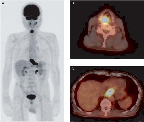
Figure 2. A. Maximum intensity projection (MIP). B,C. Transaxial PET/CT. A 68-year-old man (Case 14). An FDG-PET/CT image shows focal FDG uptake on the left side of the hypopharynx (SUVmax, 4.4), bilateral neck lymph nodes (SUVmax, 8.2), and the left lower jaw (SUVmax, 18.1), which was already identified as a mandibular neoplasm. The additional detection of FDG uptake (arrow) was observed from the upper to middle thoracic esophagus (SUVmax, 3.8). This case was considered to possibly reflect an inflammatory change, such as reflux esophagitis, because the distribution of the FDG uptake was diffuse, rather than focal; thus, we diagnosed the FDG-PET/CT image as equivocal. The uptake was subsequently confirmed to represent an esophageal cancer (T1b) based on the EGD findings.
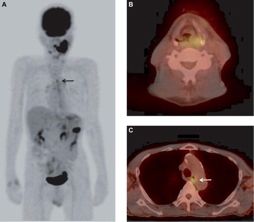
Table II. The distribution of the clinical T classifications of esophageal cancer lesions.
Clinical stages of esophageal cancer
The distribution of the clinical stages of esophageal cancer was as follows: stage IA, 11 patients; stage IIA two patients; stage IIB, one patient; stage IIIA, one patient; and stage IIIB, two patients.
Treatment for esophageal cancer
EMR was performed in eight patients with stage IA esophageal cancer. An operation was performed in three patients (stage IIA, one patient; stage IIIA, one patient; stage IIIB, one patient). Chemotheraphy was performed in one patients (Stage IA) CRT was performed in two patients (stage IIA, one patient; stage IIB, one patient). In two of the three patients (stage IA, one patient; stage IIIB, one patient) were transferred to another hospital; therefore, the details of their treatment were unknown, the remaining one patient did not undergo any treatment because of the advanced stage of HPC.
Discussion
In our study the prevalence of esophageal cancers in HPC patients was 51.5%. In previous studies using conventional esophageal endoscopy, the reported incidence varied regionally. In Taiwan, Wang et al. reported six esophageal cancers in 27 HPC patients (22%) [Citation12]. In France, Petit et al. examined patients treated for HNC and reported 50 of 1560 (3.2%) esophageal cancers were detected. According to the initial HNC anatomic site, 2.6% of the patients with an oral cavity tumor, 5.7% of the patients with an oropharynx tumor, 2.3% of the patients with a hypopharynx tumor, and 1.7% of the patients with a larynx tumor had metachronous esophageal cancer [Citation13]. Guardiola et al. examined 487 HNC patients and detected 10 (2.0%) esophageal cancers. Esophageal cancer was observed in 1.3% of the patients with an oropharyngeal tumor, 2.0% of the patients with a laryngeal tumor, none of the patients with a tumor of the oral cavity, and 9.2% of the patients with a hypopharyngeal tumor [Citation14]. These results suggest that the incidence of esophageal cancer among HPC or HNC patients is relatively high in Asia.
Our present result was higher than that in previous reports. The reason for the high prevalence was firstly, EGD with iodine staining screening was performed. Makuuchi et al. performed endoscopic screening using iodine staining in 788 patients with HNC [Citation15]. Among them, 93 cases (11.8%) of esophageal cancers were detected; moreover 72 of these 93 cases (77.4%) had superficial esophageal cancers that were limited to the submucosal layer [Citation15]. Our study, the prevalence of superficial esophageal cancers was 24/29 (82.8%). Our present result is similar to those of the previous reports. EGD with iodine staining screening enabled the efficient detection of superficial esophageal cancer lesions. Secondly, our population was limited to HPC patients, instead of HNC patients. Su et al. detected 15 esophageal cancers in 293 HNC patients (5.1%), and the prevalence was 15.9% (7/44) among patients with HPC, which was significantly higher than a prevalence of 8.3% (2/24) for laryngeal cancer, 7.1% (3/42) for oropharyngeal cancer, and 1.6% (3/183) for oral cancer [Citation16]. The incidence of esophageal cancer in HPC patients was high, compared with other head and neck tumors.
Though the etiological factors for the development of subsequent esophageal cancer in HPC patients remains to be defined. According to the concept of field cancerization, patients with esophageal SCC or HNSCC are at a high risk for the development of multiple SCCs [Citation17]. Field cancerization may be attributed to the development of multiple tumors by common risk factors, where the carcinogenic effects of alcohol and tobacco may simultaneously act on the entire mucosa of the mouth, pharynx and upper aerodigestive tract to trigger the development of multiple cancers. In recent epidemiological studies, the chewing of betel quid was reported to be a common habit prevalent in India, Taiwan and Southeast Asia. Whether betel quid chewing is an independent risk factor for HNC and esophageal cancer remains controversial [Citation18,Citation19]. In Japan, an Asian country where betel quid chewing is not a common habit, aldehydrogenase 2 (ALDH2) deficiency is closely associated with second primary esophageal cancers in patients with HNSCC [Citation20,Citation21]. Further genetic and epidemiologic studies are crucial to clarify the carcinogenic effect of these factors and to prevent esophagus and head and neck tumors.
If superficial esophageal cancer is detected, a complete cure can be expected after minimally invasive EMR [Citation22]. Morimoto et al. reported that the five-year overall survival rates of HPC patients with esophageal cancer, according to the esophageal cancer staging system, were as follows: stage 0 (83%), stage I (47%), stage IIA–IVB (0%) [Citation23]. Following Morimoto's data, the early detection of esophageal cancer has strong relation to lengthen the survival period. We believe that EGD screening using iodine staining should be performed during the pretreatment of HPC patients, although how long the screening should be continued following treatment for HPC is uncertain based on the results of the present analysis. Lee et al. reported that the risk of developing a second esophageal cancer peaked during the first year of follow-up care [Citation6].
However, iodine staining has some shortcomings; it can cause coughing and heartburn, and it is not suitable for patients who are allergic to iodine. For the sites of esophagus entrance, the neck-esophagus and the hypopharynx, iodine staining is not suitable. The narrow band imaging (NBI) system does not have the shortcomings listed above [Citation24]. The NBI system is an optical technique that applies narrow-band spectrum filters to enhance the visualization of mucosal and submucosal microvascular patterns, based on the principle of the different penetration depths of light according to its wavelength. NBI filters select the blue and green light with wavelengths of 415 and 540 nm, respectively, corresponding to the peaks of absorption of hemoglobin [Citation25]. Muto et al. reported that NBI detected superficial esophageal cancer more frequently than did white light imaging (WLI). The sensitivity of NBI was significantly higher than that of WLI (97.2% vs. 55.2%; p < 0.001), and the accuracy was also higher for NBI than for WLI (88.9% vs. 56.5%; p < 0.001) [Citation26]. The combination of NBI and iodine staining should improve the diagnostic rate of early esophageal cancer and precancerous lesions [Citation24].
Except for esophageal cancer, lung cancer is troublesome in HNC patients because of the poor prognosis. Strobel et al. reported that the majority of SPT in HNSCC patients was the lung (46%) [Citation27]. Therefore, the necessity of evaluating the respiratory tract using a bronchoscopy should be considered, in addition to EGD. Since a bronchoscopy is generally uncomfortable and invasive for most patients, we did not routinely perform a bronchoscopy. If abnormal findings suggesting the possibility of primary lung cancer or lung metastasis were observed in the trachea or lung using FDG-PET/CT or other modalities (such as chest x-ray or chest CT), a bronchoscopy should be performed for further examination. In our study, abnormal findings in the trachea or lung were not confirmed from the initial FDG-PET/CT findings; therefore, a bronchoscopy was not performed during the pretreatment of HPC patients.
FDG-PET/CT is capable of detecting a considerable number of MPCs during the initial staging of HNC [Citation27]. The detection of MPC during an early stage is quite important, as the presence of this condition affects the prognosis. Kaida et al. reported that esophageal cancer was present in 10 of 70 HPC patients (14.3%) based on FDG-PET/CT examinations [Citation4]. In our study, esophageal cancer was present in six of 33 HPC patients (18.2%) based on FDG-PET/CT examinations, these results are similar to those of the previous reports. Regarding the ability of FDG-PET to detect primary esophageal cancer, Kato et al. reported that the mean sensitivity for the detection of all stages was 78%, for stage T1b or greater, the mean sensitivity was 85% [Citation28]. However, the ability to detect a superficial T1a lesion was only 25%. As the depth of invasion increased, the FDG uptake also increased (p < 0.05). A significant correlation was observed between the tumor dimensions and the SUV (p < 0.01) [Citation28]. Himeno et al. reported a 0% ability to detect lesions confined to the mucosa (Tis or T1a) using FDG-PET [Citation29]. This result was thought to have occurred because the detection by PET requires a certain minimum tumor volume. In our study, the sensitivity of detecting T1a esophageal cancer was 0% and 33% for T1b. These results are similar to those of the previous reports.
In Case 17, a T1b esophageal cancer was detected using FDG-PET/CT (SUV max 4.4). In this case, the possibility of a false-positive result arising from an inflammatory change caused by a biopsy was suspected. However, the FDG-PET/CT examination was performed three weeks after the esophageal biopsy in this case, so the possibility of inflammatory change was thought to be relatively low. In Case 14, a T1b esophageal cancer was diagnosed using EGD. However, a FDG-PET/CT examination showed a diffuse uptake. In such cases, the possibility of an inflammatory change, such as reflux esophagitis, should be considered. Therefore, if a superficial esophageal cancer with a slight and focal FDG uptake coexists with inflammatory changes, the cancer may be overlooked.
FDG-PET/CT cannot replace EGD because of its limited ability to detect superficial esophageal cancer, whereas FDG-PET/CT is capable of detecting unexpected primary malignant tumors other than esophageal cancer. Ishimori et al. reported that FDG-PET/CT detected new unexpected primary malignant tumors in at least 1.2% of patients with cancer [Citation30]. Kim et al. reported that the prevalence of SPT in HNC patients is 4–17% based on FDG-PET findings [Citation31]. Strobel et al. performed FDG-PET/CT in 589 HNSCC patients, and the prevalence of SPT was 9.5%, of which 84% were detected using FDG-PET/CT. In 80% of the patients, the therapy was changed because of the detection of SPT [Citation27]. In our study, an unexpected prostate cancer was detected in one of the 33 patients during the initial FDG-PET/CT examinations. Moreover, unexpected lung cancers were detected in two patients during the follow-up FDG-PET/CT examinations and bronchoscopy was performed. If FDG-PET/CT examinations detect two or more unexpected primary malignant tumors, the presence of these other tumors may affect the treatment planning and patient prognosis.
Conclusion
The prevalence of esophageal cancer during the pretreatment of HPC patients was 51.5%, based on EGD screening with iodine staining. This prevalence was higher than those mentioned in previous reports. We believe that the higher percentage of superficial lesions (82.8%) detected by iodine staining and EGD may have led to the high prevalence of esophageal cancer. EGD is the preferred technique for the pretreatment examination of HPC patients, since the depth of invasion of the esophageal cancer detected in the HPC patients was T1a in most cases; therefore, a complete cure can be expected after minimally invasive EMR. FDG-PET/CT examinations detected only 20.7% of the esophageal cancers, although FDG-PET/CT is capable of detecting unexpected primary malignant tumors other than esophageal cancer.
Declaration of interest: The authors report no conflicts of interest. The authors alone are responsible for the content and writing of the paper.
References
- Wycliffe ND, Grover RS, Kim PD. Hypopharyngeal cancer. Top Magn Reson Imaging 2007;18:243–58.
- Vandenbrouck C, Eschwege F, De la Rochefordiere A, Sicot H, Mamelle G, Le Ridant AM, . Squamous cell carcinoma of the pyriform sinus: Retrospective study of 351 cases treated at the Institut Gustave-Roussy. Head Neck Surg 1987;10:4–13.
- Schwartz LH, Ozsahin M, Zhang GN, Touboul E, De Vataire F, Andolenko P, . Synchronous and metachronous head and neck carcinomas. Cancer 1994;74:1933–8.
- Kaida H, Ishibashi M, Kurata S. The utility of FDG-PET for detecting multiple primary cancers in hypopharyngeal cancer patients. Nuklearmedizin 2009;48:179–84.
- Raghavan U, Quraishi S, Bradley PJ. Multiple primary tumors in patients diagnosed with hypopharyngeal cancer. Otolaryngol Head Neck Surg 2003;128:419–25.
- Lee KD, Lu CH, Chen PT, Chan CH, Lin JT, Huang CE, . The incidence and risk of developing a second primary esophageal cancer in patients with oral and pharyngeal carcinoma: A population-based study in Taiwan over a 25 year period. BMC Cancer 2009;9:373.
- Matsuda T, Marugame T, Kamo KI, Katanoda K, Ajiki W, Sobue T; The Japan Cancer Surveillance Research Group. Cancer incidence and incidence rates in Japan in 2005: Based on data from 12 population-based cancer registries in the monitoring of cancer incidence in Japan (MCIJ) Project. Jpn J Clin Oncol 2011;41:139–47.
- Japan Society for Head and Neck Cancer, Cancer Registry Committee: Report of Head and Neck Cancer Registry in Japan. Clinical Statistics of Registered Patients, 2002. Jpn J Head Neck Cancer 2006;32(Suppl):1–98.
- Stokkel MP, ten Broek FW, Hordijk GJ. Preoperative evaluation of patients with primary head and neck cancer using dual-head 18fluorodeoxyglucose positron emission tomography. Ann Surg 2000;231:229–34.
- Stokkel MP, Terhaard CH, Hordijk GJ. The detection of local recurrent head and neck cancer with fluorine-18 fluorodeoxyglucose dual-head positron emission tomography. Eur J Nucl Med 1999;26:767–73.
- Sobin LH, Gospodarowicz MK, Wittekind C. TNM classification of malignant tumors. 7th ed. New York: Wiley-Blackwell; 2009. p. 30–8, 66–72.
- Wang CP, Lee YC, Yang TL, Lou PJ, Ko JY. Application of unsedated transnasal esophagogastroduodenoscopy in the diagnosis of hypopharyngeal cancer. Head Neck 2009;31:153–7.
- Petit T, Georges C, Jung GM, Borel C, Bronner G, Flesch H, . Systematic esophageal endoscopy screening in patients previously treated for head and neck squamous-cell carcinoma. Ann Oncol 2001;12:643–6.
- Guardiola E, Pivot X, Dassonville O, Poissonnet G, Marcy PY, Otto J, . Is routine triple endoscopy for head and neck carcinoma patients necessary in light of a negative chest computed tomography scan? Cancer 2004; 101:2028–33.
- Makuuchi H, Machimura T, Shimada H. Endoscopic screening for esophageal cancer in 788 patients with head and neck cancers. Tokai J Exp Clin Med 1996;21:139–45.
- Su YY, Fang FM, Chuang HC, Luo SD, Chien CY. Detection of metachronous esophageal squamous carcinoma in patients with head and neck cancer with use of transnasal esophagoscopy. Head Neck 2010;32:780–5.
- Slaughter DP, Southwick HW, Smejkal W. Field cancerization in oral stratified squamous epithelium; clinical implications of multicentric origin. Cancer 1953;6:963–8.
- Lee CH, Wu DC, Lee JM, Wu IC, Goan YG, Kao EL, . Anatomical subsite discrepancy in relation to the impact of the consumption of alcohol, tobacco and betel quid on esophageal cancer. Int J Cancer 2007;120:1755–62.
- Wang WL, Lee CT, Lee YC, Hwang TZ, Wang CC, Hwang JC, . Risk factors for developing synchronous esophageal neoplasia in patients with head and neck cancer. Head Neck 2011;33:77–81.
- Muto M, Takahashi M, Ohtsu A, Ebihara S, Yoshida S, Esumi H. Risk of multiple squamous cell carcinomas both in the esophagus and the head and neck region. Carcinogenesis 2005;26:1008–12.
- Yokoyama A, Omori T. Genetic polymorphisms of alcohol and aldehyde dehydrogenases and risk for esophageal and head and neck cancers. Jpn J Clin Oncol 2003;33:111–21.
- Makuuchi H. Endoscopic mucosal resection for early esophageal cancer indication and techniques. Dig Endosc 1996;8:175–9.
- Morimoto M, Nishiyama K, Nakamura O, Kawaguchi Y, Nakajima A, . Significance of endoscopic screening and endoscopic resection for esophageal cancer in patients with hypopharyngeal cancer. Jpn J Clin Oncol 2010;40:938–43.
- Huang LY, Cui J, Wu CR, Liu YX, Xu N. Narrow-band imaging in the diagnosis of early esophageal cancer and precancerous lesions. Chin Med J 2009;122:776–80.
- Piazza C, Cocco D, Del Bon F, Mangili S, Nicolai P, Peretti G. Narrow Band Imaging and High Definition Television in the endoscopic evaluation of upper aero-digestive tract cancer. Acta Otorhinolaryngol Ital 2011;31:70–5.
- Muto M, Minashi K, Yano T, Saito Y, Oda I, Nonaka S, . Early detection of superficial squamous cell carcinoma in the head and neck region and esophagus by narrow band imaging: A multicenter randomized controlled trial. J Clin Oncol 2010;28:1566–72.
- Strobel K, Haerle SK, Stoeckli SJ. Head and neck squamous cell carcinoma (HNSCC) – detection of synchronous primaries with (18)F-FDG-PET/CT. Eur J Nucl Med Mol Imaging 2009;36:919–27.
- Kato H, Kuwano H, Nakajima M. Comparison between positron emission tomography and computed tomography in the use of the assessment of esophageal carcinoma. Cancer 2002;94:921–8.
- Himeno S, Yasuda S, Shimada H. Evaluation of esophageal cancer by positron emission tomography. Jpn J Clin Oncol 2002;32:340–6.
- Ishimori T, Patel PV, Wahl RL. Detection of unexpected additional primary malignancies with PET/CT. J Nucl Med 2005;46:752–7.
- Kim SY, Roh JL, Yeo NK. Combined 18F-fluorodeoxyglucose-positron emission tomography and computed tomography as a primary screening method for detecting second primary cancers and distant metastases in patients with head and neck cancer. Ann Oncol 2007;18:1698–703.