To the Editor,
The most recent edition of the World Health Organization (WHO) “Classification of Tumors of Hematopoietic and Lymphoid Tissues” included a new category of lymphomas with histologic, phenotypic, and genetic features similar to both Burkitt lymphoma (BL) and diffuse large B-cell lymphoma (DLBCL), and described them as “B-cell lymphoma, unclassifiable, with features intermediate between diffuse large B-cell lymphoma and Burkitt lymphoma (iDLBCL/BL)”. This type of neoplasm, which was previously described as a Burkitt-like lymphoma, shares some of the morphological features of BL, but some cells are larger than those that are typical of BL [Citation1]. A classical DLBCL shows rearrangements that involve the immunoglobulin heavy chain (IGH) locus (14q32) with different genes such as B-cell lymphoma 2 protein (BCL2) (18q21). On the contrary, a classical BL presents translocations joining c-MYC (MYC) (8q24) and immunoglobulin genes, usually IGH. In some of these cases, concurrent IGH-BCL2 and MYC rearrangement occurs, and these are called double-hit lymphoma (DHL). DHLs are more aggressive and have a higher incidence of central nervous system (CNS) involvement than either BL or DLBCL [Citation2].
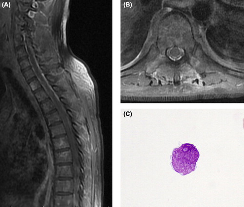
A 30-year-old man was admitted to our hospital with an enlarged right cervical lymph node. Computed tomography (CT) scans showed lymphadenopathy of the right cervical, para-aortic, and left inguinal regions. Histological examination of the right cervical lymph node biopsy showed monotonous proliferation of medium-sized lymphocytes accompanied by scattered tangible body macrophages, which gave the tumor a starry-sky appearance (). Immunohistochemical analyses showed that the tumor cells had membrane staining for CD20, CD79a, and CD10, cytoplasmic staining for BCL2 (124; Dako, Glostrup, Denmark) (), and nuclear staining for MYC (C19; Santa Cruz Biotechnology, Santa Cruz, CA, USA) (), but were negative for CD3, CD5, and CD56. Lymphoma cells demonstrated cytoplasmic staining for IL-6 (10C12; Novocastra, Newcastle Upon Tyne, UK) (), and TNF-α (52B83; Santa Cruz Biotechnology) (), and membrane staining for the IL-6 receptor (IL-6R; Deciphargen Biotechnology, Cheshire, CT, USA) (), TNF-α receptor-1 (TNF-R1; H-271; Santa Cruz Biotechnology) (), TNF-R2 (L-20; Santa Cruz Biotechnology) (), and CC chemokine receptor 7 (CCR7) (Lifespan Bioscience, Seattle, WA, USA) (). The proliferation index assessed by Ki67 was 90%. Fluorescence in situ hybridization (FISH) analysis revealed fusion between the heavy chain of immunoglobulin H (IgH) and the MYC gene (IgH/MYC). FISH analysis also revealed fusion between the IgH and BCL2 gene (IgH/BCL2). A bone marrow aspiration and biopsy showed infiltration of lymphoma cells. These findings were consistent with a new category of iDLBCL/BL, stage IVA, and the International Prognostic Index was categorized as low–intermediate [Citation1]. Following treatment with three courses of R-hyper-CVAD therapy (a regimen of rituximab, hyperfractionated cyclophosphamide, doxorubicin, vincristine, and dexamethasone alternating with a high dose of methotrexate and cytarabine), the patient's clinical symptoms and lymphoma lesions disappeared. However, after the fourth course of chemotherapy, he presented with bone pain. Observation of the bone marrow revealed hypercellular marrow with 86.8% lymphoma cells showing the disease to be progressive. Salvage chemotherapy with fludarabine did not improve the patient's clinical symptoms and the patient became refractory to chemotherapy. Thereafter, he developed progressive paresthesia in his chest, back, and lower limb and weakness of the lower limbs. MRI disclosed remarkable enhancement of the meninges, including the dura mater and leptomeninges, extending from C5 to T3 and T9–L2 ( and ). Cerebrospinal fluid (CSF) examination revealed pleocytosis, mainly including abnormal lymphocytes () (7 cells/mm3; normal: < 5 cells/mm3), normal glucose (64 mg/dL; normal: 50–80 mg/dL), and elevated protein levels (312.5 mg/dL; normal: 15–45 mg/dL). These clinical findings showed CNS infiltration. He was treated with an infusion of 50 mg etoposide and intrathecal injections of 15 mg methotrexate, 40 mg cytarabine, and 10 mg prednisolone. However, the patient died because the disease was progressive.
Figure 1. (A) Histological examination showing monotonous proliferation of medium-sized lymphocytes accompanied by a starry-sky appearance (hematoxylin-eosin stain; original magnification, 40×). (B) Lymphoma cells were positive for BCL2 (objective magnification, 40×). (C) Lymphoma cells were positive for MYC (objective magnification, 40×). (D) Lymphoma cells were positive for IL-6 (objective magnification, 40×). (E) Lymphoma cells were positive for TNF-α (objective magnification, 40×). (F) Lymphoma cells were positive for IL-6R (objective magnification, 40×). (G) Lymphoma cells were positive for TNFR1 (objective magnification, 40×). (H) Lymphoma cells were positive for TNFR2 (objective magnification, 40×). (I) Lymphoma cells were positive for CCR7 (objective magnification, 40×).
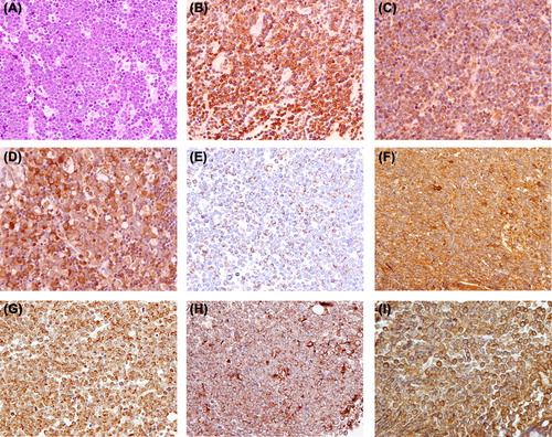
Figure 2. MRI disclosed remarkable enhancement of the meninges in a T1-weighted sequence. (A) Sagittal view section. (B) Axial view section. (C) CSF samples showing lymphoma cells with irregular nuclei with moderately dispersed chromatin and conspicuous nucleoli (May-Giemsa stain, objective magnification 100 ×).
The 18q21.3/BCL2 gene was initially observed by cloning of the chromosomal breakpoint in cases with t(14;18) translocations, which are presumed to result from an error during VDJ rearrangement of the IG gene. Neoplastic B-lymphocytes with this translocation constantly express the BCL2 protein, an apoptosis inhibitor, whereas in normal B-cell differentiation, the BCL2 protein is not expressed in the germinal center (GC). Neoplastic B-lymphocytes with BCL2 overexpression by t(14;18) translocation become apoptosis-resistant and proliferate in the GC [Citation3]. BCL2 was also promoted by TNF-α (3) and IL-6 [Citation4]. The 8q24/MYC (c-MYC) gene was discovered in the analysis of the chromosomal breakpoint in cases with BL. The MYC gene has been reported to be amplified in various types of cancer, and MYC aberrations are likely to confer a powerful growth advantage to lymphoma cells [Citation5]. MYC is thought to be an oncogene that encodes the MYC protein which is involved in cell proliferation, apoptosis, and cell cycle control. In cases with translocation between the MYC and IG genes, the MYC protein is constantly expressed in all stages of cell turnover. In addition, MYC expression is stimulated by IL-6 [Citation6] and TNF-α [Citation7]. The basis for the extremely aggressive clinical behavior of DHL is likely to be related to the combination of the MYC-induced growth promotion and the anti-apoptotic effects conferred by BCL2 overexpression [Citation8].
CCR7 is an attractive candidate for recruiting lymphoma cells to CNS, because it is a known regulator of lymphocyte migration, and it has been suggested to be important for the trafficking of lymphocytes participating in CNS immunosurveillance. The single chemokine-receptor interaction acting as a CNS entry signal and the importance of CCR7-mediated T-cell acute lymphoblastic leukemia cell recruitment to the CNS have been shown [Citation9]. However, there have been no reports of CCR7-expressing DH iDLBCL/BL lymphoma with CNS infiltration which may indicate CCR7-mediated DH iDLBCL/BL lymphoma cell recruitment to the CNS. Analysis of the CCR7 promoter sequence revealed two potential binding sites for NFκB [Citation10] and CCR7 up-regulation was shown to be mediated by constitutive NFκB activity [Citation11]. NFκB is a ubiquitously expressed transcription factor that is involved in the activation of genes associated with inflammation and cell adhesion. NFκB activity can be induced by BCL2 and proinflammatory cytokines, such as IL-6 and TNF-α [Citation12,Citation13]. In this case, BCL2 and the cytokines produced by lymphoma cells may promote the aberrant expression of CCR7, resulting in CNS infiltration.
To the best of our knowledge, this is the first case report on the CNS infiltration of multiple cytokine-, multiple cytokine receptor-, and chemokine receptor-expressing DH iDLBCL/BL that showed the immunohistological expression of BCL2, MYC, IL-6, IL-6R, TNF-α, TNFRs, and CCR7 in lymphoma cells. These observations suggest that IL-6- and TNF-α-producing lymphoma cells with IL-6 and TNF-α promoted BCL2 and MYC expression and those with BCL2, IL-6, and TNF-α promoted CCR7 expression, and may show CCR7-mediated efficient migration toward CNS infiltration. DH iDLBCL/BL produce multiple cytokines which may result in the aberrant expression of BCL2, MYC, and CCR7, thereby playing a role in the initiation and enhancement of the recruitment of lymphoma cells to the CNS. These cytokines and the chemokine receptor may play a key role in CNS infiltration in some DH iDLBCL/BL lymphomas.
Declaration of interest: The authors report no conflicts of interest. The authors alone are responsible for the content and writing of the paper.
References
- Swerdlow S, Campo E, Harris NL, Jaffe ES, Pileri SA, Stein H, et al, editors. World Health Organization classification of tumors. Pathology & genetics, tumors of hematopoietic and lymphoid tissues. Lyon, France: IARC Press; 2008.
- Snuderl M, Kolman OK, Chen YB, Hsu JJ, Ackerman AM, Dal Cin P, et al. B-cell lymphomas with concurrent IGH-BCL2 and MYC rearrangements are aggressive neoplasms with clinical and pathologic features distinct from Burkitt lymphoma and diffuse large B-cell lymphoma. Am J Surg Pathol 2010;34:327–40.
- Esche C, Shurin GV, Kirkwood JM, Wang GQ, Rabinowich H, Pirtskhalaishvili G, et al. Tumor necrosis factor-alpha-promoted expression of Bcl-2 and inhibition of mitochondrial cytochrome c release mediate resistance of mature dendritic cells to melanoma-induced apoptosis. Clin Cancer Res 2001;7:974s–9s.
- Steiner MK, Syrkina OL, Kolliputi N, Mark EJ, Hales CA, Waxman AB. Interleukin-6 overexpression induces pulmonary hypertension. Circ Res 2009;104:236–44.
- Klapper W, Stoecklein H, Zeynalova S, Ott G, Kosari F, Rosenwald A, et al. Structural aberrations affecting the MYC locus indicate a poor prognosis independent of clinical risk factors in diffuse large B-cell lymphomas treated within randomized trials of the German High-Grade Non-Hodgkin’s Lymphoma Study Group (DSHNHL). Leukemia 2008; 22:2226–9.
- Shi Y, Frost P, Hoang B, Benavides A, Gera J, Lichtenstein A. IL-6-induced enhancement of c-Myc translation in multiple myeloma cells: Critical role of cytoplasmic localization of the RNA-binding protein hnRNP A1. J Biol Chem 2011; 286:67–78.
- Tselepis C, Perry I, Dawson C, Hardy R, Darnton SJ, McConkey C, et al. Tumour necrosis factor-alpha in Barrett’s oesophagus: A potential novel mechanism of action. Oncogene 2002;21:6071–81.
- Lin P, Medeiros LJ. High-grade B-cell lymphoma/leukemia associated with t(14;18) and 8q24/MYC rearrangement: A neoplasm of germinal center immunophenotype with poor prognosis. Haematologica 2007;92:1297–301.
- Buonamici S, Trimarchi T, Ruocco MG, Reavie L, Cathelin S, Mar BG, et al. CCR7 signalling as an essential regulator of CNS infiltration in T-cell leukaemia. Nature 2009;459: 1000–4.
- Schweickart VL, Raport CJ, Godiska R, Byers MG, Eddy RL Jr, Shows TB, et al. Cloning of human and mouse EBI1, a lymphoid-specific G-protein-coupled receptor encoded on human chromosome 17q12-q21.2. Genomics 1994;23:643–50.
- Höpken UE, Foss HD, Meyer D, Hinz M, Leder K, Stein H, et al. Up-regulation of the chemokine receptor CCR7 in classical but not in lymphocyte-predominant Hodgkin disease correlates with distinct dissemination of neoplastic cells in lymphoid organs. Blood 2002;99:1109–16.
- de Moissac D, Mustapha S, Greenberg AH, Kirshenbaum LA. Bcl-2 activates the transcription factor NFkappaB through the degradation of the cytoplasmic inhibitor IkappaBalpha. J Biol Chem 1998;273:23946–51.
- Wang L, Walia B, Evans J, Gewirtz AT, Merlin D, Sitaraman SV. IL-6 induces NF-kappa B activation in the intestinal epithelia. J Immunol 2003;171:3194–201.