Abstract
Purpose. To dosimetrically evaluate a margin-of-the-day (MoD) online adaptive intensity-modulated radiotherapy (IMRT) strategy for cervical cancer patients. The strategy is based on a single planning computed tomography (CT) scan and a pretreatment constructed IMRT plan library with incremental clinical target volumes (CTV)-to-planning target volumes (PTV) margins. Material and methods. For 14 patients, 9–10 variable bladder filling CT scans acquired at pretreatment and after 40 Gy were available. Bladder volume variability during the treatment course was recorded by twice-weekly US bladder-volume measurements. A MoD strategy that selects the best IMRT plan of the day from a library of plans with incremental margins in steps of 5 mm was compared with a clinically recommended population-based margin (15 mm). To compare the strategies, for each fraction that had a recorded US bladder-volume measurement, the CT scan with the nearest bladder volume was selected from the pretreatment CT series and from the CT series acquired after 40 Gy. A frequency-weighted average of the dose-volume histograms (DVH) parameters calculated for the two selected CT scans was used to estimate the DVH parameters of the fraction of interest. Results. The 15-mm recommended margin resulted in cervix-uterus underdosage in six of 14 patients. Compared with the 15-mm margin, the MoD strategy resulted in significantly better cervix-uterus coverage (p = 0.008) without a significant difference in the sparing of rectum, bladder, and small bowel. For each patient, 3–8 (median 5) plans were needed in the library of plans for the MoD strategy. The required range of the MoD was 5–45 mm (median 15 mm). Twenty-five percent of all fractions could be treated with a MoD of 5 mm and 81% of all fractions could be treated with a MoD up to 25 mm. Conclusions. Compared with a clinically recommended margin, a simple online adaptive strategy resulted in better cervix-uterus coverage without compromising organs at risk sparing.
Introduction
The use of intensity-modulated radiation therapy (IMRT) for cervical cancer patients is challenged by the large inter- and intra-patient variability in the extent and type of cervix and uterus motion [Citation1]. Most of the published studies show that bladder filling variations have a large impact on the shape and position of the cervix and uterus [Citation1–5]. Unfortunately, drinking instructions, a common approach to limit bladder filling variations, demonstrate limited efficacy [Citation6]. Generous population-based margins (24–40 mm) can be used to accommodate the extent of motion [Citation7,Citation8], but the use will come at the cost of increased irradiation of normal tissues.
Alternatively, our group demonstrated that individualized IMRT strategies accounting for the patient-specific cervix-uterus motion can provide adequate coverage and better organ at risk (OAR) sparing than population-based margins [Citation8]. However, these individualized strategies require a minimum of two pretreatment computed tomography (CT) scans with a full and empty bladder and sophisticated non-rigid registration and shape modeling tools [Citation8–10].
The aim of this study was to quantify the potential benefit of a straightforward online margin-of-the-day (MoD) adaptive treatment strategy based on a single planning CT scan. The strategy adapts the treatment online by selecting the best plan-of-the-day from a library of plans with incremental uniform clinical target volumes (CTV)-to-planning target volumes (PTV) margins.
The efficacy of the MoD strategy was benchmarked against the clinically recommended population-based margin [Citation11]. The comparison was performed by simulating treatment courses using US bladder volume data that was recorded twice weekly and CT data series of variable bladder filling acquired before the start of the treatment and after the delivery of 40 Gy. In the discussion section we propose practical solutions for the clinical implementation of the MoD strategy.
Material and methods
Patient data
Fourteen locally advanced cervical cancer patients were included, after providing written consent, in this local ethics committee approved study. For 13 patients, EBRT was delivered in 23 daily fractions of 2 Gy and for one patient in 28 fractions of 1.8 Gy. Patients were treated prone (lying on a small bowel displacement system).
Imaging and delineation
Each patient underwent two series of variable bladder filling CT scans in treatment position: One before the start of the treatment and one after 40 Gy [Citation1]. For each series, patients were asked to drink 500 ml of water 1 hour prior to each series. The first scan was acquired immediately at the start of a series with a full bladder. After acquisition of this scan, the patient was asked to go to the toilet to empty her bladder, and then drink another 300 ml. This was followed by acquisition of the other 3–4 CT scans on the belly board, acquired every 20 minutes while the bladder filled naturally [Citation1]. Immediately after each acquisition, the bladder volume was measured using a portable three-dimensional (3D) bladder US scanner (see below). The planning CT scan was the first CT scan of the first series and had a full bladder. In an attempt to achieve day-to-day reproducibility in having a full bladder, the patients were asked to drink 500 ml of water 1 hour prior to each treatment fraction [Citation6]. For each patient, 3–4 repeat CT scans of the first series and 4–5 CT scans of the second series were aligned to the planning CT in order to remove the translations and rotations of the bony anatomy in the pelvic region.
For each patient, the nodal CTV and the primary CTV (cervix, uterus, superior part of the vagina, and parametria) were delineated in the planning CT. The cervix-uterus structure (cervix, uterus, and superior part of the vagina), bowel, bladder, and rectum were delineated in all CT scans.
Bladder volume data during the treatment course
Twice-weekly US bladder volume measurements were available for this study. These were performed immediately after the delivery of a treatment fraction with a portable 3D bladder US scanner (Mobile BladderScan® BVI 6400) [Citation6]. For each patient 6–10 (median 8.5) measurements were available, summing up to a grand total of 120 [Citation1], (60 for the first half of the treatment and 60 for the second half). The fraction number at which the measurement was performed was recorded. These data were used to evaluate the MoD strategy and the use of the clinically recommended population-based margin.
Treatment planning
To construct the library of plans, first three IMRT plans with a 5, 10, and 15 mm CTV-to-PTV margin around the cervix-uterus were created for each patient using the planning CT scan. Then, for each of the 8–9 subsequent aligned CT scans we investigated whether at least 99% of the cervix-uterus volume received at least 95% (43.7 Gy) of the prescription dose (46 Gy). If for a repeat CT scan, the 15 mm margin was not sufficient to meet this requirement, additional library plans were made by increasing the margins in steps of 5 mm until the requirement was met. For all plans, the margin around the nodal CTV was set to 10 mm. This procedure guaranteed that for each patient an IMRT plan that provided sufficient coverage was available for each CT scan, without having to make, for each patient, IMRT plans up to the maximum margin required for this population.
Treatment plans were generated by a single experienced dosimetrist (P.V), using the commercial inverse planning software, Monaco TPS, version 2.04 (Elekta-CMS). All calculations were done using 10-MV photons with a grid spacing of 3 mm for an Elekta Synergy (Elekta MLCi, Elekta Oncology Systems, Crawley, UK) linear accelerator with 40 multileaf collimator leaf pairs, and leaf width at the isocenter of 10 mm. The dose prescribed to the PTV was 46 Gy, given in 23 fractions (five fractions weekly). We ensured that at least 99.5% of the PTV received at least 95% of the prescribed dose. To ensure a homogeneous dose in the PTV, a planning constraint was used that allowed a maximum dose of 110% of the prescribed dose in 0.2% of the PTV.
Construction of the margin-of-the-day strategy
We tested an online adaptive strategy in which the patients are treated with the tightest fitting MoD plan from a library of IMRT plans generated at pretreatment. In clinical practice, the daily cervix-uterus shape and position could be determined by a direct visualization of the cervix-uterus in a daily cone beam CT (CBCT) image prior to delivery of each treatment fraction [Citation12]. In this study, the tightest fitting MoD IMRT plan was selected by the criterion that at least 99% of the cervix-uterus volume received at least 95% of the prescription dose. The MoD plan selection was performed for each of the 8–9 repeat CT scans of the variable bladder filling CT scan series. Here, we assumed the repeat CT scans to be representative for actual treatment fractions. To evaluate the MoD and the population-based strategy for realistic bladder-volume patterns during the treatment course, the US bladder-volume data obtained twice weekly was used (see next section).
Evaluation of the margin-of-the-day strategy and the population-based strategy
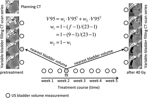
For each fraction that had a recorded US bladder-volume measurement, the CT scan with the nearest bladder volume was selected from the pretreatment CT scan series and from the CT scan series acquired after 40 Gy. The planning CT scan was not included herein. By including the CT scan series after 40 Gy anatomy variations due to bowel displacement, rectal filling changes, and tumor regression were included in the dosimetric evaluation. A frequency-weighted average of the dose-volume histograms (DVH) parameters calculated for the two selected CT scans was used to estimate the DVH parameters of the fraction of interest. The frequency weighting emphasized the pretreatment CT scan for the first half of the treatment and emphasized the after 40 Gy CT scan for the second half of the treatment. See for a schematic example of calculating bladder V95 for a certain treatment fraction.
Figure 1. Schematic representation of the calculation of the DVH parameters during the treatment course. This example illustrates the calculation of bladder V95 for fraction 9 (of 23 fractions) as a weighted sum of the bladder V95 of the pretreatment (V951) CT scan and the after 40 Gy CT scan (V952) that had the nearest bladder volume to the bladder volume measured by US at fraction 9.

The MoD strategy was compared with a standard population-based margin strategy. A uniform population-based margin of 15 mm around the cervix-uterus in the planning CT was selected based on previous recommendations [Citation11]. The DVH parameters for the population-based margin strategy were calculated as described above by replacing the MoD plan with the 15-mm margin plan.
Comparison between margin-of-the-day and population-based margin
We calculated DVH parameters that correlated with normal tissue toxicity [Citation13] and that were previously used for comparative studies of IMRT strategies for cervical cancer [Citation14,Citation15]. The following parameters were calculated: The percentage (%) of the cervix-uterus volume that received 95% (43.7 Gy) of the prescribed dose (V95), the volume of the small bowel that received 99% (45.0 Gy) of the prescribed dose (V99), and the percentage of the bladder and rectum volume that received 90% (41.4 Gy) of the dose (V90), respectively. The averages per patient of the DVH parameters obtained by the two strategies were compared using the signed rank Wilcoxon test. Furthermore, we compared the volumes of the PTV for the two approaches. The calculated volumes included both the nodal and primary part of the PTV. In addition, the performance of the two strategies was related to the maximum extent of cervix-uterus motion observed during treatment and the extent of motion observed in the pretreatment CT series. The extent of motion was expressed as the displacement of the tip of uterus (ToU) which, was marked in all CT scans using axial, sagittal, and coronal views [Citation1]. For each patient, the magnitude of the ToU displacement vector and of the absolute displacement in LR, AP, and CC directions between the planning CT scan and the subsequent CT scans was previously calculated and reported [Citation1]. For each patient, the maximum ToU extent of motion (vector, LR, AP, or CC) was defined as the maximum of ToU displacements between the planning and all subsequent CT scans acquired at pretreatment and at after 40 Gy. The pretreatment ToU extent of motion for a patient was defined as the ToU displacement between the planning and the empty bladder pretreatment CT scan.
Results
Simulation of the margin-of-the-day strategy
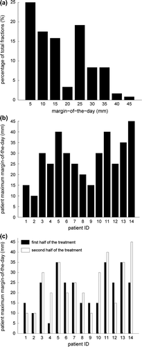
A total of 71 plans were generated. For each patient, 3–8 (median 5) plans were needed to generate the library of plans for the MoD strategy. The distribution of the MoD needed to adapt the treatment at the treatment fractions for all patients is summarized in . The range of the MoD was 5–45 mm (median 15 mm). The patient maximum value of the MoD over all fractions ranged from 10 to 45 mm (). The large variation in the maximum value illustrates the inter-patient variability in type and extent of cervix-uterus motion. The patient maximum MoD value for the first half of the treatment () was significantly correlated with the patient maximum MoD value for the second half of the treatment (R = 0.67, p = 0.008 Pearson's correlation analysis).
Figure 2. a) Distribution of the margin-of-the-day. b) Maximum margin-of-the-day per patient. c) Maximum of the margin-of-the-day for each treatment half.

A strong correlation was found between the patient maximum MoD and the maximum ToU vector and AP displacement (R = 0.9, p < 0.001, R = 0.79, p < 0.001), respectively. The maximum MoD correlated moderately with the pretreatment ToU AP displacement (R = 0.57, p = 0.03).
Comparison between margin-of-the-day and population-based margin
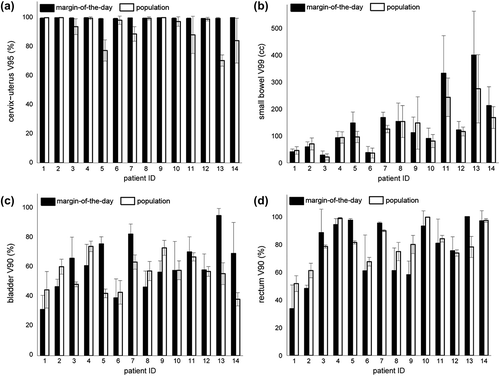
Applying a population-based margin of 15 mm around the cervix-uterus resulted in a cervix-uterus underdosage in six of 14 patients (the average cervix-uterus V95 was 92.66 ± 9.5%, the average per patient ranged between 70% and 100%, see ). Compared with the population-based margin, the MoD strategy resulted in significantly better cervix-uterus coverage (p = 0.008, average cervix-uterus V95 was 99.5 ± 0.2%, the average per patient ranged between 99.1% and 99.8%, see ). shows the volume of the PTV (including the nodal target volume) for the population-based margin and the frequency-weighted volume of the PTV for the MoD strategy. For the eight patients that had a sufficient coverage using the population-based margin, the MoD strategy reduced the irradiated volume by on average −11% (range −24% to 9%; a negative sign denotes a volume reduction if the MoD strategy is applied). Obviously, for the other six patients the irradiated volume of the MoD strategy was larger compared to the population-based margin in order to achieve sufficient CTV coverage. For all patients together the volume of the PTV for the MoD strategy was not significantly larger than for the population-based approach (p-value 0.1), despite that the MoD strategy provided better CTV coverage.
Figure 3. DVH parameters of the margin-of-the-day strategy and the population-based strategy. a) Percentage of cervix-uterus that received 95% of the prescribed dose b) Absolute volume of small bowel that received 99% of the dose and percentage of c) bladder and d) rectum that received 90% of the dose. The error bar denotes ± 1SD variation around the mean.

Table I. Volume of the PTV using the population-based margin and the frequency-weighted volume of the PTV of the MoD strategy. An * in the last column points out the patients for whom the population-based margin did not provide sufficient coverage of the CTV.
The improved coverage of the MoD strategy did not result in a significant increase in the average per patient of bowel V99, and bladder and rectum V90 values (p-value 0.06–0.5, ). The increase in cervix-uterus coverage and the difference in bladder, rectum and small bowel sparing with respect to population-based margins were significantly correlated with the maximum ToU (R = 0.66, p = 0.009; R = −0.75, p = 0.001; R = −0.75, p < 0.001; R = −0.74, p = 0.002) and pretreatment ToU vector displacement (R = 0.69, p = 0.006; R = −0.73, p = 0.002; R = −0.85 p < 0.001; R = −0.6, p = 0.01), respectively.
Discussion
In this study we evaluated a straightforward online adaptive strategy for locally advanced cervical cancer. The strategy adapts the treatment to the daily cervix-uterus position by selecting the plan with the tightest daily margin from a library of plans based on a single planning CT scan and incremental CTV-to-PTV margins. The strategy was dosimetrically benchmarked against a clinically recommended population-based margin of 15 mm [Citation11]. Concurrent with a previous study [Citation8], the 15-mm margin was insufficient to accommodate the cervix-uterus motion and resulted in underdosage for 40% of the patients. The proposed MoD strategy resulted in adequate cervix-uterus coverage without compromising OARs sparing.
To ensure coverage for patients with large cervix-uterus motion, the MoD was for some treatment fractions larger than the 15-mm margin. However, our study demonstrated that 25% of the fractions could be treated with a margin of 5 mm, 58% with a margin up to 15 mm, and 81% with a daily margin up to 25 mm. Note that in this study the set-up errors were assumed zero. Similarly, Tyagi et al. [Citation12] demonstrated that a margin of 15 mm was insufficient to encompass the CTV in 32% of the fractions of 10 cervical cancer patients. However, in that study no dosimetric analysis was performed and no solution to improve the coverage was evaluated.
Compared to the previously proposed ITV-based adaptive strategy [Citation8], the MoD approach might require less workload at pretreatment since it uses only one CT scan. For example, for the adaptive ITV-based strategy, two pretreatment CT scans need to be acquired and delineated and 2–3 model-based ITVs need to be generated based on non-rigid registration tools. However, the MoD strategy might require more treatment planning workload since the MoD library will contain up to five plans whereas the ITV-based library contains 2–3 plans. While the MoD approach is using a homogeneous margin, the ITV-based approach takes advantage of the individualized motion model of the cervix-uterus. Consequently, the target volume is expanded only in the direction in which the cervix-uterus is expected to move. Regarding tissue sparing, we demonstrated that compared with a population-based margin the ITV-based approach significantly reduced the irradiation of rectum at the cost of a reduced bladder sparing whereas for the MoD approach there was no significant difference in OAR sparing.
Our study showed that it is not possible to accurately predict before the start of the treatment the number of the plans in the library needed for a patient. For example, although the largest MoD correlated strongly with the maximum cervix-uterus displacement during treatment, a weaker correlation with the displacement of the uterus tip at pretreatment was found (R = 0.57, p = 0.03). Therefore, an additional pretreatment empty bladder CT scan might not provide a clinically reliable indicator of the required number of plans. Similarly, although our study showed that the largest MoD needed for the first half of the treatment was correlated with the one needed for the second treatment half, the strength of the correlation (R = 0.67) is not sufficient to make a reliable prediction (see, e.g. the results for Patients 4 and 10, ).
Therefore, to construct the library, we recommend generating at pretreatment five plans with margins ranging from 5 to 25 mm. To reduce workload, one plan could be generated manually and the subsequent plans could be automatically optimized by using automated plan generation methods [Citation15,Citation16]. Since there might be little or no benefit of IMRT versus 3DCRT for large IMRT margins, to save treatment planning and delivery time the department might decide to deliver the fractions that would require margins beyond 25 mm by using a 3DCRT plan. The plan-of-the-day could be selected based on a direct visualization of the cervix-uterus in a daily acquired CBCT scan. For example, by overlaying the PTVs corresponding to different margins on the daily image, the plan of the day could be selected by finding the smallest PTV that encompasses the daily cervix-uterus position. Alternatively, future research will investigate the potential use of deformable registration or segmentation methods to automatically identify the shape and position of the CTV and OARs in a daily CBCT. These approaches could support computer-aided plan selection and open up the possibility for dose-guided adaptive radiotherapy [Citation17]. For example, Thor et al. [Citation18] demonstrated promising deformable results on pelvic CBCT data.
We would not recommend selecting the plan-of-the-day based on a US bladder volume measurement, as the relationship between the cervix-uterus and the volume of the bladder might change during the treatment course [Citation1,Citation8,Citation9]. In this study, the US measurements were used only for selecting CT scans from the two variable bladder filling CT scan series in order to evaluate both strategies for a representative set of bladder volumes during the treatment course.
A limitation of this study is that the US measurements were linked to the CT scan series that were made at only two time points, i.e. at pretreatment and after 40 Gy. Therefore, we may not have evaluated all possible internal anatomies. However, by acquiring the second CT scan series nearly at the end of the treatment course, we believe to have captured most of the gradual changes during the treatment such as tumor regression and a decrease in rectal filling. Another limitation of this study is that the dose to the CTV and OARs was not accumulated using deformable registration. However, we would like to point out the uncertainties related to dose accumulation that still remain, especially in the pelvic region [Citation19].
In conclusion, a simple online adaptive IMRT treatment strategy was designed that adapts the treatment to the daily cervix-uterus shape and position. In this strategy, the plan with the smallest margin encompassing the daily cervix-uterus is selected from a pretreatment-generated library of plans with incremental CTV-to-PTV margins. Compared with a recommended margin of 15 mm, the margin-of-the-day strategy resulted in better cervix-uterus coverage without compromising normal tissue sparing.
Declaration of interest: The authors report no conflicts of interest. The authors alone are responsible for the content and writing of the paper.
Rozilawati Ahmad was funded by University Kebangsaan Malaysia (grant number UKM-DLP-2011-078). Luiza Bondar was funded by the Dutch Cancer Society (grant number 2007-3777). The authors thank Laura Velema for contouring work.
References
- Ahmad R, Hoogeman MS, Bondar M, Dhawtal V, Quint S, de Pree I, et al. Increasing treatment accuracy for cervical cancer patients using correlations between bladder-filling change and cervix-uterus displacements: Proof of principle. Radiother Oncol 2011;98:340–6.
- Chan P, Dinniwell R, Haider MA, Cho YB, Jaffray D, Lockwood G, et al. Inter- and intrafractional tumor and organ movement in patients with cervical cancer undergoing radiotherapy: A cinematic-MRI point-of-interest study. Int J Radiat Oncol Biol Phys 2008;70:1507–15.
- Taylor A, Powell M, An assessment of interfractional uterine and cervical motion: Implications for radiotherapy target volume definition in gynaecological cancer. Radiother Oncol 2010;88:250–7.
- Beadle B, Jhingran A, Salehpour A, Sam M, Iyer R, Eifel P, Cervix regression and motion during the course of external beam chemoradiation for cervical cancer. Int J Radiat Oncol Biol Phys 2009;73:235–41.
- Buchali A, Koswig S, Dinges S, Rosenthal P, Salk P, Lackner L, et al. Impact of the filling status of the bladder and rectum on their integral dose distribution and the movement of the uterus in the treatment planning of gynaecological cancer. Radiother Oncol 1999;52:29–34.
- Ahmad R, Hoogeman MS, Quint S, Mens JW, de Pree I, Heijmen BJM. Inter-fraction bladder filling variations and time trends for cervical cancer patients assessed with a portable 3-dimensional ultrasound bladder scanner. Radiother Oncol 2008;89:172–9.
- van de Bunt L, Jürgenliemk-Schulz IM, de Kort GAP, Roesink JM, Tersteeg RJ, van der Heide UA. Motion and deformation of the target volumes during IMRT for cervical cancer: What margins do we need?. Radiother Oncol 2008;88:233–40.
- Bondar ML, Hoogeman MS, Mens JW, Quint S, Ahmad R, Dhawtal G, et al. Individualized nonadaptive and online-adaptive IMRT strategies for cervical cancer patients based on pretreatment acquired variable bladder filling CT-scans. Int J Radiat Oncol Biol Phys 2012;83:1617–23.
- Bondar L, Hoogeman M, Mens JW, Dhawtal G, de Pree I, Ahmad R, et al. Toward an individualized target motion management for IMRT of cervical cancer based on model-predicted cervix-uterus shape and position. Radiother Oncol 2011;99: 240–5.
- Bondar L, Hoogeman M, Vásquez Osorio E, Heijmen B. A symmetric non-rigid registration method to handle large organ deformations in cervical cancer patients. Med Phys 2010;37:3760–72.
- Lim K, Small W Jr, Portelance L, Creutzberg C, Jürgenliemk-Schulz IM, Mundt A, et al. Consensus guidelines for delineation of clinical target volume for intensity-modulated pelvic radiotherapy for the definitive treatment of cervix cancer. Int J Radiat Oncol Biol Phys 2011;79:348–55.
- Tyagi N, Lewis JH, Yashar CM, Vo D, Jiang SB, Mundt A, et al. Daily online cone beam computed tomography to assess interfractional motion in patients with intact cervical cancer. Int J Radiat Oncol Biol Phys 2011;80:273–80.
- Roeske J, Bonta D, Mell L, Lujan AE, Mundt AJ. A dosimetric analysis of accute gastrointestinal toxicity in women receiving intensity-modulated whole-pelvic radiation therapy. Radiother Oncol 2003;69:201–7.
- Lim K, Kelly V, Stewart J, Xie J, Cho YB, Moseley J, et al. Pelvic radiotherapy for cancer of the cervix: Is what you plan actually what you deliver?. Int J Radiat Oncol Biol Phys 2009;74:304–12.
- Stewart J, Lim K, Kelly V, Xie J, Brock KK, Moseley J, et al. Automated weekly replanning for intensity-modulated radiotherapy of cervix cancer. Int J Radiat Oncol Biol Phys 2010; 78:350–8.
- Breedveld S, Storchi PR, Voet PW, Heijmen BJ. iCycle: Integrated, multicriterial beam angle, and profile optimization for generation of coplanar and noncoplanar IMRT plans. Med Phys 2012;39:951–63.
- Elstrøm UV, Wysocka BA, Muren LP, Petersen JB, Grau C. Daily kV cone-beam CT and deformable image registration as a method for studying dosimetric consequences of anatomic changes in adaptive IMRT of head and neck cancer. Acta Oncol 2010;49:1101–8.
- Thor M, Petersen JB, Bentzen L, Høyer M, Muren LP. Deformable image registration for contour propagation from CT to cone-beam CT scans in radiotherapy of prostate cancer. Acta Oncol 2011;50:918–25.
- Zhong H, Kim J, Chetty IJ. Analysis of deformable image registration accuracy using computational modeling. Med Phys 2010;37:970–9.
