To the Editor,
Different cone beam computed tomography (CBCT) systems are commercially available for radiotherapy (RT). The image quality of these systems differs as a consequence of different hardware and software. However, image quality assessment based on images of phantoms and related physical measures usually cannot tell us whether we should prefer one system over the other, in a specific clinical setting. On the other hand, subjective, non-quantitative evaluations based on the preferences of individual observers are often not sufficient either. Therefore, a quantitative evaluation method based on clinically relevant features of images of actual patients is called for.
A method called visual grading characteristics (VGC) analysis has been developed by Båth and colleagues [Citation1,Citation2]. The VGC is a non-parametric, rank-invariant statistical analysis which can be used for quantitative evaluation of the difference in image quality between two imaging systems. To our knowledge, VGC has never been applied in the field of RT.
The aim of this study was to test VGC analysis as a tool to evaluate clinically relevant image quality differences between CT-based imaging systems used in RT. Furthermore, the aim was to give a preliminary indication of whether one of two specific CBCT systems should be preferred when used for visualization of the bladder in RT [Citation3].
Material and methods
Both home clinics of the authors of the present paper have two different treatment units with CBCT capabilities, i.e. Varian iX (CBCTiX) [Citation4] and Varian TrueBeam (CBCTTb). The CBCT systems on these two different treatment unit types differ and the image quality may differ as well. In short, the CBCTiX system uses a standard phantom-based normalization in the image reconstruction to account for scatter and beam hardening effects. By contrast, the CBCTTb employs a patient-specific scatter correction algorithm as well as an improved analytical beam hardening correction.
The patient data used in this retrospective study were obtained from Aarhus University Hospital. Four male patients were included. All patients had scans obtained with CBCTiX and CBCTTb, as well as a planning CT (pCT) (Supplementary Appendix, to be found online at http://informahealthcare.com/doi/abs/10.3109/0284186X.2013.818252).
Based on European guidelines provided by the Commission of the European Communities (CEC) [Citation5,Citation6] and in cooperation with a physician (physician 1, HL), eight image quality criteria in relation to bladder on CBCT were formulated. For every CBCT and pCT, the fulfillment of the criteria was graded using an ordinal scale from 1 to 5; with 1 and 5 corresponding to “Confident that the criterion is fulfilled” and “Confident that the criterion is not fulfilled”, respectively. The grading was performed by four observers: physicians 1 and 2, and RTTs 1 and 2 (see Supplementary Appendix to be found online at http://informahealthcare.com/doi/abs/10.3109/0284186X.2013.818252).
As in several other studies, e.g. [Citation7–9], the results were pooled for all criteria, however, not for the observers. Thus, individual VGC curves for the observers were created using the software DBM MRMC 2.32 Build 3 [Citation10–17]. The DBM MRMC is a suitable receiver operating characteristic (ROC) software and can be used for VGC analysis [Citation1]. By using ANOVA provided by the software, the areas under the curve (AUCs) and the 95% CIs were determined.
The smallest number of CBCTiX was three (for Patient III). Hence, for each patient, three CBCTTb and three CBCTiX were randomly selected using Matlab R2011b. This ensured that all patients contributed equally to the analysis. The images were given a random number between 1 and 28. Thus, the observers were blinded when performing the analysis. The order in which the observers examined the images was also random. The randomization was performed to avoid reading order effects [Citation18]. The results were analyzed in terms of VGC analysis of pCT compared to CBCTiX and CBCTTb compared to CBCTiX.
Results
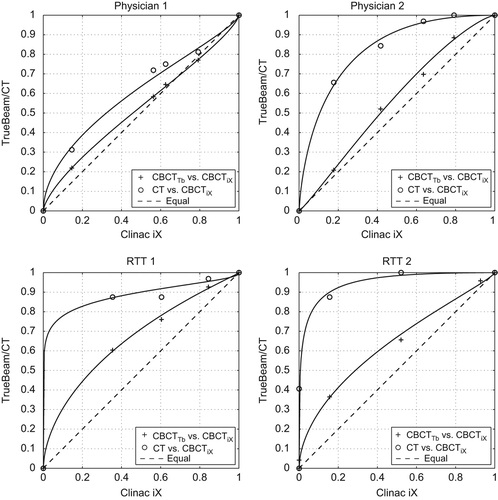
The AUCs for all pCT vs. CBCTiX curves were above 0.5, and 0.5 was not included in the 95% CIs except for physician 1 where 0.5 was just within the 95% CI ( and ). For CBCTTb versus CBCTiX the results were less clear. There was a tendency that CBCTTb was superior to CBCTiX with 95% CIs shifted towards values higher than 0.5. However, it was only for the two RTTs that 0.5 was not included in the CIs.
Figure 1. VGC analysis for physician 1, physician 2, RTT1, and RTT2, respectively. pCT performs better than CBCTiX with all points above the “equal line”. The equal line corresponds to no difference between the two modalities. In general, CBCTTb seems to be superior to CBCTiX, however, one point is below the equal line (for physician 1) and 0.5 is within the CIs for the physicians ().

Table I. The results of the VGC analysis. AUCs for pCT compared to CBCTiX and CBCTTb compared to CBCTiX as well as the 95% CIs.
Discussion
The superiority in image quality of CT over CBCT is well known [Citation22–24]. Several methods to improve the image quality of CBCT images have been suggested and investigated, e.g. scatter corrections based on either measurements, Monte Carlo simulations or other algorithms or improvement of the CBCT image quality by using the information from a standard CT scan [Citation23–28]. The result of this study that CT seems to provide better image quality than CBCT was therefore expected from the basic differences in the two acquisition techniques. As opposed to CT, CBCT acquisition takes approximately 60 seconds of scan time with the entire anatomical region in the field of view. Thus, CBCT images are more prone to artifacts from movement of interfaces between different densities during acquisition. Furthermore, scatter and beam hardening effects will deteriorate the soft tissue contrast centrally in the patient where the bladder is located. The contribution from the latter has been decreased in the CBCT reconstruction on the TrueBeam platform compared to iX which is reflected in the results of this study.
Other studies have compared image quality of different CBCT systems for RT or different operating modes or software solutions of the same CBCT system, however not using the VGC method used in this study [Citation28–31]. The VGC curves () give a clear and quick illustration of the trend in results, but they cannot stand alone as the CIs are needed in order to clarify whether the results are statistical significant or not. However, the CIs and AUCs do not tell the whole story either.
The calculations of the CIs are based on the assumption that the AUC can be treated as a normally distributed variable. This assumption is valid in most cases if AUC is not close to 1 and if the number of cases is > 50 [Citation1]. In the present study, the number of cases is < 50 and the assumption of normality may not hold. Therefore, even though, e.g. CT is superior to CBCT for the tested cases as evaluated by the observers of this study, the current study does not have the statistical power to generalize this.
VGC analysis is well suited for comparing different CT-based image modalities used in RT. The results of VGC analysis can be based on clinically relevant differences in image quality of actual patient images. The method thus overcomes the potential problems that images of phantoms and related physical measures may have little relevance in the daily clinical use of the imaging systems.
Supplementary Appendix, Supplementary Figure 1 and Supplementary Tables I–IV
Download PDF (60.3 KB)Acknowledgments
Thanks to physician Camilla Kjær Lønkvist, and RTTs Camilla Lee Dann and Dorrit Larsen from Herlev Hospital for their time and participation in the VGC analysis.
Declaration of interest: The authors report no conflicts of interest. The authors alone are responsible for the content and writing of the paper.
References
- Båth M, Månsson LG. Visual grading characteristics (VGC) analysis: A non-parametric rank-invariant statistical method for image quality evaluation. Br J Radiol 2007;80:169–76.
- Båth M, Zachrisson S, Månsson LG. VGC analysis: Application of the ROC methodology to visual grading tasks. In: Proc SPIE 2008;6917:69170X–1–9.
- Vestergaard A, Søndergaard J, Petersen JB, Høyer M, Muren LP. A comparison of three different adaptive strategies in image-guided radiotherapy of bladder cancer. Acta Oncol 2010;49:1069–76.
- Sjöström D, Bjelkengren U, Ottosson W, Behrens CF. A beam-matching concept for medical linear accelerators. Acta Oncol 2009;48:192–200.
- European Commission. European guidelines on quality criteria for diagnostic radiographic images. Report EUR 16260 EN; 1996.
- European Commission. European guidelines on quality criteria for computed tomography. Report EUR 16262 EN; 1999.
- Zachrisson S, Hansson J, Cederblad Å, Geterud K, Båth M. Optimisation of tube voltage for conventional urography using a Gd2O2S:Tb flat panel detector. Radiat Prot Dosimetry 2010;139:86–91.
- Leander P, Söderberg M, Fält T, Gunnarsson M, Albertsson I. Post-processing image filtration enabling dose reduction in standard abdominal CT. Radiat Prot Dosimetry 2010;139: 180–5.
- Burmeister HP, Baltzer PAT, Möslein C, Bitter T, Gudziol H, Dietzel M, et al. Visual grading characteristics (VGC) analysis of diagnostic image quality for high resolution 3 tesla MRI volumetry of the olfactory bulb. Acad Radiol 2011; 18:634–9.
- DBM MRMC; 2013. Available from: http://perception.radiology.uiowa.edu/Software/tabid/109/Default.aspx, Free download for registered users. [cited 2013 April 20].
- Dorfman DD, Berbaum KS, Lenth RV, Chen YF, Donaghy BA. Monte Carlo validation of a multireader method for receiver operating characteristic discrete rating data: Factorial experimental design. Acad Radiol 1998;5: 591–602.
- Dorfman DD, Berbaum KS, Metz CE. Receiver operating characteristic rating analysis: Generalization to the population of readers and patients with the jackknife method. Invest Radiol 1992;27:723–31.
- Hillis SL. A comparison of denominator degrees of freedom methods for multiple observer ROC analysis. Stat Med 2007;26:596–619.
- Hillis SL, Berbaum KS. Monte Carlo validation of the Dorfman-Berbaum-Metz method using normalized pseudovalues and less data-based model simplification. Acad Radiol 2005;12:1534–41.
- Hillis SL, Berbaum KS. Power estimation for the Dorfman-Berbaum-Metz method. Acad Radiol 2004;11:1260–73.
- Hillis SL, Berbaum KS, Metz CE. Recent developments in the Dorfman-Berbaum-Metz procedure for multireader ROC study analysis. Acad Radiol 2008;15:647–61.
- Hillis SL, Obuchowski NA, Schartz KM, Berbaum KS. A comparison of the Dorfman-Berbaum-Metz and Obuchowski-Rockette methods for receiver operating characteristic (ROC) data. Stat Med 2005;24:1579–607.
- Metz CE. Some practical issues of experimental design and data analysis in radiological ROC studies. Invest Radiol 1989;24:234–45.
- Lanhede B, Båth M, Kheddache S, Sund P, Björneld L, Widell M, et al. The influence of different technique factors on image quality of chest radiographs as evaluated by modified CEC image quality criteria. Br J Radiol 2002;75: 38–49.
- Månsson L. Methods for the evaluation of image quality: A review. Radiat Prot Dosimetry 2000;90:89–99.
- Van Erkel AR, Pattynama PMT. Receiver operating characteristic (ROC) analysis: Basic principles and applications in radiology. Eur J Radiol 1998;27:88–94.
- Stock M, Pasler M, Birkfellner W, Homolka P, Poetter R, Georg D. Image quality and stability of image-guided radiotherapy (IGRT) devices: A comparative study. Radiother Oncol 2009;93:1–7.
- Lou Y, Niu T, Jia X, Vela PA, Zhu L, Tannenbaum AR.Joint CT/CBCT deformable registration and CBCT enhancement for cancer radiotherapy. Med Image Anal 2013;17:387–400.
- Niu T, Al-Basheer A, Zhu L. Quantitative cone-beam CT imaging in radiation therapy using planning CT as a prior: First patient studies. Med Phys 2012;39:1991–2000.
- Poludniowski G, Evans PM, Hansen VN, Webb S. An efficient Monte Carlo-based algorithm for scatter correction in keV cone-beam CT. Phys Med Biol 2009;54:3847–64.
- Mainegra-Hing E, Kawrakow I. Variance reduction techniques for fast Monte Carlo CBCT scatter correction calculations. Phys Med Biol 2010;55:4495–507.
- Jina JY, Ren L, Liu Q, Kim J, Wen N, Guan H, et al. Combining scatter reduction and correction to improve image quality in cone-beam computed tomography (CBCT). Med Phys 2010;37:5634–44.
- Qiu W, Pengpan T, Smith ND, Soleimani M. Evaluating iterative algebraic algorithms in terms of convergence and image quality for cone beam CT. Comput Methods Programs Biomed 2013;109:313–22.
- Kamath S, Song W, Chvetsov A, Ozawa S, Lu H, Samant S, et al. An image quality comparison study between XVI and OBI CBCT systems. J Appl Clin Med Phys 2011;12: 376–90.
- Kim S, Yoo S, Yin FF, Samei E, Yoshizumi T. Kilovoltage cone-beam CT: Comparative dose and image quality evaluations in partial and full-angle scan protocols. Med Phys 2010;37:3648–59.
- Elstrøm UV, Muren LP, Petersen JB, Grau C. Evaluation of image quality for different kV cone-beam CT acquisition and reconstruction methods in the head and neck region. Acta Oncol 2011;50:908–17.
