
Abstract
Background. In a selective group of patients accelerated partial breast irradiation (APBI) might be applied after conservative breast surgery to reduce the amount of irradiated healthy tissue. The role of volumetric modulated arc therapy (VMAT) and voluntary moderately deep inspiration breath-hold (vmDIBH) techniques in further reducing irradiated healthy – especially heart – tissue is investigated.
Material and methods. For 37 partial breast planning target volumes (PTVs), three-dimensional conformal radiotherapy (3D-CRT) (3–5 coplanar or non-coplanar 6 and/or 10 MV beams) and VMAT (two partial 6 MV arcs) plans were made on CTs acquired in free-breathing (FB) and/or in vmDIBH. Dose-volume parameters for the PTV, heart, lungs, and breasts were compared.
Results. Better dose conformity was achieved with VMAT compared to 3D-CRT (conformity index 1.24 ± 0.09 vs. 1.49 ± 0.20). Non-PTV ipsilateral breast receiving ≥ 50% of the prescribed dose was on average reduced by 28% in VMAT plans compared to 3D-CRT plans. Mean heart dose (MHD) reduced from 2.0 (0.1–5.1) Gy in 3D-CRT(FB) to 0.6 (0.1–1.6) Gy in VMAT(vmDIBH). VMAT is beneficial for MHD reduction if MHD with 3D-CRT exceeds 0.5Gy. Cardiac dose reduction as a result of VMAT increases with increasing initial MHD, and adding vmDIBH reduces the cardiac dose further. Mean dose to the ipsilateral lung decreased from 3.7 (0.7–8.7) to 1.8 (0.5–4.0) Gy with VMAT(vmDIBH) compared to 3D-CRT(FB). VMAT resulted in a slight increase in the contralateral breast dose (DMean) always remaining < 1.9 Gy).
Conclusions. For APBI patients, VMAT improves PTV dose conformity and delivers lower doses to the ipsilateral breast and lung compared to 3D-CRT. This goes at the cost of a slight but acceptable increase of the contralateral breast dose. VMAT reduces cardiac dose if MHD exceeds 0.5 Gy for 3D-CRT. Adding vmDIBH results in a further reduction of heart and ipsilateral lung dose.
Breast conserving therapy (BCT) is a well-established procedure for early stage breast cancer. It consists of resection of the primary breast tumor followed by whole breast irradiation (WBI) [Citation1]. Radiation therapy (RT) is an essential component of BCT as it reduces the risk of future ipsilateral breast cancer events [Citation2]. Long-term follow-up of randomized trials concluded that the survival and local control rates among women treated with BCT is equivalent to those of patients treated with mastectomy [Citation3,Citation4].
However, some authors suggest that RT may be withheld in a selected group of elderly patients after conservative surgery, without exposing these patients to an increased risk of distant recurrences. Moreover, 44 − 86% of all local recurrences occur close to the primary tumor bed [Citation3,Citation4]. Therefore, WBI following tumor resection has been challenged as being possibly an over-treatment for low-risk patients.
Accelerated partial breast irradiation (APBI) has emerged in the recent years as a treatment option for selected patients with early stage breast cancer [Citation5,Citation6]. APBI offers several advantages over WBI, including a reduced overall treatment time and a decrease in the dose delivered to the uninvolved portions of the ipsilateral breast and adjacent organs (heart and ipsilateral lung). Several randomized clinical trials have been initiated to compare the effectiveness and safety of APBI [Citation7]. Even as we await the long-term results of these trials, APBI is increasingly being employed routinely in the management of early stage breast cancers. This led both the American Society for Radiation Oncology (ASTRO) and the Groupe Européen de Curiethérapie- European SocieTy for Radiotherapy & Oncology (GEC-ESTRO) to develop consensus statements regarding patient selection criteria and best practices for the use of APBI outside the context of a clinical trial [Citation6,Citation8].
External beam radiation therapy (EBRT) for APBI has many potential advantages over other APBI techniques such as brachytherapy and intraoperative RT [Citation9]. These include its non-invasive nature, widespread availability, and familiarity of technical demands and quality assurance issues in most RT centers.
For patients with left-sided breast tumors, the benefits of radiation therapy may be offset by the increase in morbidity and mortality from late cardiovascular damage that are associated with WBI [Citation10,Citation11]. This is assumed to be a consequence of the inclusion of a portion of the heart into the RT fields. APBI with conventional three-dimensional conformal radiation therapy (3D-CRT) techniques might also deliver a significant dose to the cardiac structures.
Multiple studies have confirmed the effectiveness of breath-hold techniques in reducing cardiac radiation dose for left-sided breast cancer treated with WBI and with locoregional RT [Citation12,Citation13]. Moreover, several recent studies have focused on the reduction of the dose to the heart and other organs at risk (OAR) using intensity-modulated radiation therapy (IMRT) and volumetric modulated arc therapy (VMAT) [Citation14,Citation15]. To the best of our knowledge, there is currently no study published comparing both 3D-CRT and VMAT in free-breathing and in breath-hold for APBI treatments.
In our four arm planning study, we systematically investigate and compare the dosimetric results of APBI planned with 3D-CRT and with VMAT, both in free-breathing (FB) and in voluntary moderately deep inspiration breath-hold (vmDIBH). We also aim to suggest guidelines for the optimum treatment method based on patient characteristics, e.g. the tumor location within the breast or the initial (i.e. for the 3D-CRT technique in FB) mean heart dose (MHD).
Material and methods
Patient data
A total of 37 patients with left-sided breast cancer were retrospectively included in this study. For 21 patients originally treated with whole breast radiotherapy, two CT scans were acquired before irradiation, one in FB and the other in vmDIBH. The vmDIBH [Citation16] scans were acquired using audio-couching (recorded voice, both during CT scan as well as during irradiation, asking the patient to “quietly breath in, breath out, breath in, breath out, take a deep breath and hold your breath”) and verified using breathing signals from the Real-Time Position Management (RPM) respiratory gating system (Varian Medical Systems, Palo Alto, California, USA). Using the RPM, we can verify that the patient holds her breath in the same amplitude during the treatment scan, the RPM system was also used on the treatment machine, as well as kV images. The CT slice thickness was 2.5 mm. For this APBI planning study, the clinical target volume (CTV) consisted of the lumpectomy cavity with a margin of 15 mm, modified to stay within the glandular breast tissue. The planning target volume (PTV) was constructed by adding a 5 mm margin to the CTV and then restricting the PTV to the tissue inside the first 5 mm of skin and superficial to the chest wall [Citation9]. In addition, for 16 patients originally treated with 3D-CRT APBI (13 in FB and 3 in BH) in the framework of the IRMA trial [Citation17], VMAT plans were acquired as well. In this way, we could compare 3D-CRT and VMAT plans for 37 patients: 10 patients with a centrally (C) located tumor, and nine in the upper-inner (UI), six in the upper-outer (UO), four in the lower-inner (LI), and eight in the lower-outer (LO) quadrant. The following structures were contoured as well: body contour, ipsilateral non-PTV breast, contralateral breast, heart, left lung and right lung. For all treatment plans, we applied the IRMA fractionation scheme with a prescribed dose of 38.5 Gy given in 10 fractions (3.85 Gy per fraction), twice a day (a total of 5 days) [Citation17].
Treatment planning
Treatment plans were generated in the Eclipse≥ treatment planning system (TPS) (Varian Medical systems, Acuros version 10.0). For 21 patient PTVs, four treatment plans were generated: 3D-CRT(FB); 3D-CRT(vmDIBH); VMAT(FB); VMAT(vmDIBH). For 13 patients, for whom only a FB scan was available, a 3D-CRT(FB) and a VMAT(FB) plan were made, and for three patients with only a vmDIBH scan, we calculated 3D-CRT(vmDIBH) and VMAT(vmDIBH) plans. The prescription was to obtain coverage of ≥ 95% of the PTV by ≥ 95% of the prescribed dose of 38.5 Gy.
For 3D-CRT plans a mixture of 6 MV and/or 10 MV (if necessary non-coplanar, maximum couch rotation 25°) photon beams was used (minimum of three beams per plan). Beam arrangements were made to cover the PTV according to the above prescription while avoiding beams directions including cardiac structures as much as possible. Wedges, MLCs and beam weights were individualized to optimize the dose distribution. The typical VMAT technique consists of two 6MV photon partial arcs (start and stop angles between 300° and 320° and between 150° and 170° depending on the PTV location and avoiding as much as possible direct inclusion of the lungs and the contra-lateral breast. As a starting point, PTV minimum and maximum dose objectives were 100% and 102% of the prescribed dose, respectively, with a somewhat higher weight than the weights for the constraints of the organs at risk. Dose-volume and mean dose constraints are used for the heart (mostly, a mean heart dose constraint is sufficient, and the weight can be increased to reduce the heart dose, if necessary), contralateral breast, lungs and sometimes ipsilateral non-PTV breast. In this study, no monitor units (MU) constraint was used to allow a maximum sparing of all OARs.
Dosimetric evaluation parameters
The following was analyzed for the 21 patients with four treatment plans: For PTV the fraction of the volume receiving ≥ 95% of the prescribed dose (V95%), the near-maximum dose (D1%) which represents the highest dose received by 1% of the volume, and the mean dose (DMean) were analyzed. In addition, the conformity index (CI), which is defined as TV95%/ PTV, where TV95% is the volume of the 95% isodose and PTV is the planning target volume, was calculated. For organs at risk (OAR), mean and maximum doses as well as relevant dose-volume parameters were compared. For ipsilateral (non-PTV) breast, the fraction receiving 50% of the prescription dose (V50%) and V5Gy were analyzed. For the heart, the volume receiving ≥ 5Gy (V5Gy) as well as DMean (MHD) and DMax (maximum dose) are reported. Contralateral breast and contralateral lung DMax and DMean values were compared, as well as ipsilateral lung V5Gy and DMean. Statistical analysis was made using a Wilcoxon signed-rank test, two-tailed for each evaluation parameter at a significance level of ≤ 0.05.
The 16 extra patients, with only one CT scan, were not included in the statistical analysis, but were included to determine the difference in MHD between 3D-CRT and VMAT for each individual (in total 37) patient.
Results
lists dosimetric parameters for the 21 PTVs with four treatment plans (2 CTs). PTV coverage and conformity was slightly better for VMAT than for 3D-CRT plans, while the near-maximum dose, (D1%), was slightly worse. An overview of the doses received by the OARs using the different APBI treatment techniques for these 21 patients is presented in . Up to 30% reduction in the ipsilateral (non-PTV) breast V50% was observed with VMAT plans compared to 3D-CRT plans. For all 21 patients, ipsilateral lung dose parameters were significantly improved with VMAT compared to 3D-CRT. An overall increase in contralateral breast maximum dose was observed in VMAT plans compared to 3D-CRT plans. Despite this increase in the maximum dose, the mean contralateral breast dose remained always less than 1.9 Gy. Similarly, the contralateral lung maximum doses were slightly increased in VMAT plans while the mean dose remained very low. The MHD is significantly reduced by applying either vmDIBH or VMAT compared to 3D-CRT(FB), but there is no significant difference in MHD between the 3D-CRT(vmDIBH) and VMAT(vmDIBH) plans, Also, applying VMAT in vmDIBH does not improve the MHD significantly for the total group of patients. However, applying both vmDIBH and VMAT does result in a cumulative reduction in V5Gy and Dmax for the heart.
Table I. Comparison of population averaged PTV dose parameters from different planning techniques (3D-CRT and VMAT) in free-breathing (FB) and in vmDIBH (voluntary moderate deep inspiration breath hold) for the 21 patients with 2 CT scans and 4 different plans.
Table II. Comparison of number of monitor units (MU), ipsilateral lung volumes, and organs at risk dose parameters from different planning techniques (3D-CRT and VMAT) in free-breathing (FB) and breath-hold (vmDIBH), averaged for the 21 patients with 2 CT scans and four different plans.
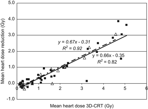
and show MHD data for individual patients. shows the reduction in MHD for the three other techniques compared to the conventional 3D-CRT(FB) technique for the 21 patients with four plans. The benefit of each technique increases with increasing initial MHD, and vmDIBH and VMAT show a small cumulative benefit on MHD reduction. There are a few outliers, e.g. the patients with an initial MHD of 4.8 and 5.1 Gy. These patients have a very unfavorable geometry, with the PTV in the center of the breast, immediately adjacent to the heart. Although vmDIBH causes a lung volume increase of a factor 1.6 and 2, respectively, the PTV is still very close to the heart in vmDIBH. For these patients, VMAT is the treatment of choice for a sharp dose fall-off around the PTV. In , VMAT is compared to 3D-CRT, both in FB and vmDIBH, for different tumor locations and for all 37 patients (including the 16 clinical patients with only one CT scan). There is no clear relation between tumor location and initial MHD in 3D-CRT or between tumor location and reduction in MHD as a result of VMAT. shows the MHD reduction if VMAT is used instead of 3D-CRT for both FB and vmDIBH situations. VMAT results in MHD reduction if MHD in the 3D-CRT plan exceeds 0.5 Gy. VMAT can reduce the MHD by 0.67 Gy per Gy above 0.5 Gy. If it is lower than 0.5 Gy, then VMAT might result in similar MHD, or a slightly higher MHD, with a maximum increase of 0.3 Gy found for this patient group.
Figure 1. Mean heart dose reduction compared with the 3D-CRT(FB) technique as a result of 3D-CRT(vmDIBH) (×), VMAT(FB) (×) and VMAT(vmDIBH) (×) as a function of the mean heart dose for 3D-CRT(FB) for the 21 patients with 2 CT scans and 4 treatment plans.
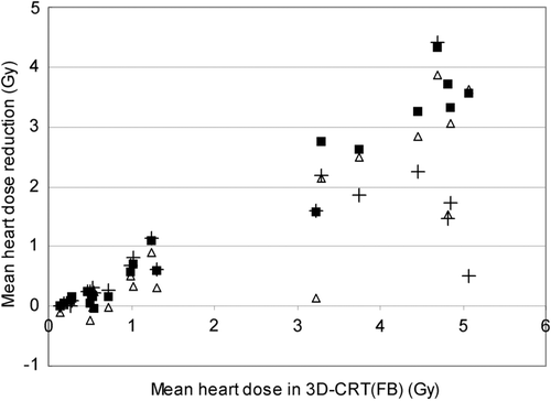
Figure 2. Mean heart dose reduction of the VMAT plans compared with the with 3D-CRT plans, for centrally located tumors (×), and tumors located in the upper inner(×), lower inner (×), upper outer (×) and lower outer (×) quadrant of the breast, for all 37 patients in FB and in vmDIBH.
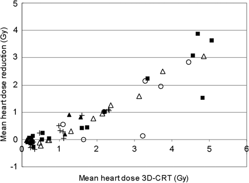
Figure 3. Mean heart dose reduction with VMAT compared with the 3D-CRT plans, in FB (×, solid trendline with right equation) and in vmDIBH (×, dashed trendline with left equation), for all 37 patients in FB and in vmDIBH.
Discussion
For all patients in this study, treatment planning with VMAT provided significantly improved target coverage and dose conformity in both FB and vmDIBH, as was already shown in the literature [Citation15]. A significant reduction in irradiated non-PTV tissue could be achieved using VMAT, although the dose to non-PTV breast tissue was also acceptable for 3D-CRT plans, since Jagsi et al. [Citation18] recommended limiting the proportion of the non-PTV breast tissue receiving 50% of prescribed dose to < 40% in order to reduce the risk of toxicity. This was based on their report on the outcomes of APBI using IMRT with active breathing control, where they reported unacceptable cosmesis in about 22% of the treated cases [Citation18]. Therefore, as far as PTV coverage and non-PTV ipsilateral breast sparing is concerned, our data suggest that VMAT is, in FB as well as in breath-hold, the treatment method of choice for external beam APBI. However, a very important issue in breast radiotherapy is the heart dose. According to the literature, any reduction of the dose delivered to the cardiac structures might be clinically relevant, probably even more with the increased use of systematic therapies with cardiotoxic agents [Citation10,Citation11,Citation19]. Therefore, the goal of treatment planning optimization is to minimize the irradiated heart volume as much as possibly achievable without compromising target volume coverage. Our study showed that VMAT is able to reduce the heart dose if the mean heart dose in 3D-CRT is above 0.5 Gy. Therefore, we suggest applying VMAT for patients where the mean heart dose without VMAT exceeds 0.5 Gy. As we also demonstrated that the use of vmDIBH (alone or with VMAT) always results in lower or equal heart and lung dose values compared to FB, we suggest to always apply vmDIBH, being a very patient friendly and easy way of respiratory regulation, proven to be feasible in our clinic in 95% of the patients. Many recent studies also showed that DIBH is even more useful for heart dose reduction if the whole breast, with or without regional nodes, is irradiated [e.g. 20,21]. vmDIBH also results in a very reproducible and stable (intrafraction) patient setup. In June 2012, image-guided vmDIBH RT was introduced at our department as part of the standard approach for all left-sided breast cancer patients. In a pilot study of 20 vmDIBH candidates, the accuracy of the online setup procedure including the reproducibility and stability of breath-hold was tested and the residual errors were quantified. Using a standard margin formula CTV-PTV margins of 4.8 mm were calculated, justifying the 5 mm CTV-PTV margin used in this study (results not published). A recent study by Damkjær et al. [Citation22] also showed very good reproducibility of DIBH (SD = 0.4 mm).
As expected, contralateral breast and contralateral lung received significant but still low higher maximum doses with VMAT compared to 3D-CRT, as reported by other authors before [e.g. 14]. Despite this increase in the maximum dose to the contralateral breast, the risk of a secondary tumor is known to be low for patients above 40 years old and for doses below 5 Gy [Citation23]. As one of the patient selection criteria for APBI is based on age, we think that the choice of treatment technique should not be affected by the small increase in contralateral breast dose. In hypofractionation settings, Smith et al. recommends limiting V30% to ≤ 15% for the ipsilateral lung and V5% to ≤ 15% for the contralateral lung [Citation6]. The highest ipsilateral lung dose observed in our study was with 3D-CRT, but even then the dose parameters were well below these recommended values. Significant sparing of ipsilateral lung was observed in VMAT plans compared to 3D-CRT plans ().
Several other groups have investigated the use of modern RT techniques including IMRT [Citation24] and VMAT [Citation15] for APBI. Compared to 3D-CRT, these techniques were reported to improve the dose conformity, and to reduce the dose to the ipsilateral breast and to the ipsilateral lung at the cost of a small increase of the dose to the contralateral lung and breast. In their study, Qiu et al. [Citation15] showed average heart dose(-volume) data that were lower for VMAT than for 3D-CRT, and in the same range as our results, but because of the small number of patients (only four left-sided breast cancer patients), these differences were not significant. Due to the number of patients is much larger in our study, statistical significance of 3D-CRT or VMAT for heart dose reduction could be shown. In agreement with our study, Moran et al. [Citation24] showed no significant reduction in heart dose as a result of IMRT, for patients irradiated with DIBH (10 patients). However, in contrast to our study, they did not show results for individual patients.
The total number of MU in our VMAT plans was on average 843 in vmDIBH and 862 in FB plans. For the 3D-CRT plans, the average number of MU is 525. In a recent pre-clinical study, Nicolini et al. [Citation25] showed the efficiency of VMAT delivery under respiratory-gated conditions. Film measurements conducted at our department show that the dose distribution of an uninterrupted and an interrupted arc are exactly the same. Therefore, combining vmDIBH with VMAT could be done by simply using the “beam on” button to interrupt and restart the arc in 4–5 breath-hold cycles. So, even though the number of MU is higher for VMAT than for 3D-CRT, no wedges, additional gantry or couch rotations are necessary, which will result in the same or even a slightly shorter treatment time compared to a conventional technique.
Based on our findings, we conclude vmDIBH has equal or reduced MHD and lung dose compared to FB, with the benefit increasing with initial MHD. The VMAT treatment technique is beneficial for left-sided APBI patients where the MHD with 3D-CRT exceeds 0.5 Gy, because of the reduced heart dose. Moreover, VMAT results in improved target volume coverage and dose conformity, a decreased dose to the uninvolved ipsilateral breast and a lower volume of the ipsilateral lung treated to a higher dose. The dose to the contralateral breast is slightly increased but below an acceptable level for the patient age group that might be a suitable candidate for APBI. Based on the results of our investigation, we suggest to always apply vmDIBH and to combine this with VMAT if MHD without VMAT exceeds 0.5 Gy and VMAT is able to provide equal or reduced heart dose.
Declaration of interest: The authors report no conflicts of interest. The authors alone are responsible for the content and writing of the paper.
References
- Early Breast Cancer Trialists’ Collaborative Group. Favourable and unfavourable effects on long-term survival of radiotherapy for early breast cancer: An overview of the randomised trials. Lancet 2000;355:1757–70.
- Clarke M, Collins R, Darby S, Davies C, Elphinstone P, Evans E, et al. Effects of radiotherapy and of differences in the extent of surgery for early breast cancer on local recurrence and 15-year survival: An overview of the randomised trials. Lancet 2006;366:2087–106.
- Fisher B, Anderson S, Bryant J, Margolese RG, Deutsch M, Fisher ER, et al. Twenty-year follow-up of a randomized trial comparing total mastectomy, lumpectomy, and lumpectomy plus irradiation for the treatment of invasive breast cancer. N Engl J Med 2002;347:1233–41.
- Veronesi U, Cascinelli N, Mariani L, Greco M, Saccozzi R, Luini A, et al. Twenty-year follow-up of a randomized study comparing breast-conserving surgery with radical mastectomy for early breast cancer. N Engl J Med 2002;347: 1227–32.
- Njeh CF, Saunders MW, Langton CM. Accelerated partial breast irradiation using external beam conformal radiation therapy: A review. Crit Rev Oncol Hematol 2012;81:1–20.
- Smith BD, Arthur DW, Buchholz TA, Haffty BG, Hahn CA, Hardenbergh PH, et al. Accelerated partial breast irradiation consensus statement from the American Society for Radiation Oncology (ASTRO). Int J Radiat Oncol Biol Phys 2009;74:987–1001.
- Mannino M, Yarnold J. Accelerated partial breast irradiation trials: Diversity in rationale and design. Radiother Oncol 2009;91:16–22.
- Polgár C, Van Limbergen E, Pötter R, Kovács G, Polo A, Lyczek J, et al. GEC-ESTRO breast cancer working group. Patient selection for accelerated partial-breast irradiation (APBI) after breast-conserving surgery: Recommendations of the Groupe Européen de Curiethérapie-European Society for Therapeutic Radiology and Oncology (GEC-ESTRO) breast cancer working group based on clinical evidence (2009). Radiother Oncol 2010;94:264–73.
- Offersen BV, Overgaard M, Kroman N, Overgaard J. Accelerated partial breast irradiation as part of breast conserving therapy of early breast carcinoma: A systematic review. Radiother Oncol 2009;90:1–13.
- Gutt R, Correa CR, Hwang WT, Solin LJ, Litt HI, Ferrari VA, et al. Cardiac morbidity and mortality after breast conservation treatment in patients with early-stage breast cancer and preexisting cardiac disease. Clin Breast Cancer 2008;8:443–8.
- Darby SC, Ewertz M, McGale P, Bennet AM, Blom- Goldman U, Brønnum D, et al. Risk of ischemic heart disease in women after radiotherapy for breast cancer. N Engl J Med 2013;368:987–98.
- Remouchamps VM, Vicini FA, Sharpe MB, Kestin LL, Martinez AA, Wong JW. Significant reductions in heart and lung doses using deep inspiration breath hold with active breathing control and intensity-modulated radiation therapy for patients treated with locoregional breast irradiation. Int J Radiat Oncol Biol Phys 2003;5:392–406.
- Nissen HD, Appelt AL. Improved heart, lung and target dose with deep inspiration breath hold in a large clinical series of breast cancer patients. Radiother Oncol 2013;106:28–32.
- Johansen S, Cozzi L, Olsen DR. A planning comparison of dose patterns in organs at risk and predicted risk for radiation induced malignancy in the contralateral breast following radiation therapy of primary breast using conventional, IMRT and volumetric modulated arc treatment techniques. Acta Oncol 2009;48:495–503.
- Qiu JJ, Chang Z, Wu QJ, Yoo S, Horton J, Yin FF. Impact of volumetric modulated arc therapy technique on treatment with partial breast irradiation. Int J Radiat Oncol Biol Phys 2010;78:288–96.
- Borst GR, Sonke JJ, den Hollander S, Betgen A, Remeijer P, van Giersbergen A, et al. Clinical results of image-guided deep inspiration breath hold breast irradiation. Int J Radiat Oncol Biol Phys 2013;78:1345–51.
- Study protocol IRMA, [cited 2007 May 1]. Available from: http://groups.eortc.be/radio/res/irma/synopsis_trial_irma1.pdf
- Jagsi R, Ben-David MA, Moran JM, Marsh RB, Griffith KA, Hayman JA, et al. Unacceptable cosmesis in a protocol investigating intensity-modulated radiotherapy with active breathing control for accelerated partial-breast irradiation. Int J Radiat Oncol Biol Phys 2010;76:71–8.
- Sardaro A, Petruzzelli MF, D’Errico MP, Grimaldi L, Pili G, Portaluri M. Radiation-induced cardiac damage in early left breast cancer patients: Risk factors, biological mechanisms, radiobiology, and dosimetric constraints. Radiother Oncol 2012;103:133–42.
- Vikström J, Hjelstuen MH, Mjaaland I, Dybvik KI. Cardiac and pulmonary dose reduction for tangentially irradiated breast cancer, utilizing deep inspiration breath-hold with audio-visual guidance, without compromising target coverage. Acta Oncol 2011;50:42–50.
- Hjelstuen MH, Mjaaland I, Vikström J, Dybvik KI. Radiation during deep inspiration allows loco-regional treatment of left breast and axillary-, supraclavicular- and internal mammary lymph nodes without compromising target coverage or dose restrictions to organs at risk. Acta Oncol 2012;51:333–44.
- Damkjær SM, Aznar MC, Pedersen AN, Vogelius IR, Bangsgaard JP, Josipovic M. Reduced lung dose and improved inspiration level reproducibility in visually guided DIBH compared to audio coached EIG radiotherapy for breast cancer patients. Acta Oncol 2013;52:1458–63.
- Ibrahim EM, Abouelkhair KM, Kazkaz GA, Elmasri OA, Al-Foheidi M. Risk of second breast cancer in female Hodgkin's lymphoma survivors: A meta-analysis. BMC Cancer 2012;12:197.
- Moran JM, Ben-David MA, Marsh RB, Balter JM, Griffith KA, Hayman JA, et al. Accelerated partial breast irradiation: What is dosimetric effect of advanced technology approaches?Int J Radiat Oncol Biol Phys 2009;75:294–301.
- Nicolini G, Vanetti E, Clivio A, Fogliata A, Cozzi L. Pre-clinical evaluation of respiratory-gated delivery of volumetric modulated arc therapy with RapidArc. Phys Med Biol 2010;55:N347–57.