
Abstract
Background. Improvement in radiotherapy during the past decades has made the risk of developing a radiation-induced secondary cancer as a result of dose to normal tissue a highly relevant survivorship issue. Important factors expected to influence secondary cancer risk include dose level and dose heterogeneity, as well as gender and type of tissue irradiated. The elevated radio-sensitivity in children calls for models particularly tailored to paediatric cancer patients.
Material and methods. Treatment plans of six paediatric medulloblastoma patients were analysed with respect to secondary cancer risk following cranio-spinal irradiation (CSI), using either: 1) electrons and photons combined; 2) conformal photons; 3) double-scattering (DS) protons; or 4) intensity-modulated proton therapy (IMPT). The relative organ equivalent dose (OED) concept was applied in three dose-risk scenarios: a linear response model, a plateau response and an organ specific linear-exponential response. Life attributable risk (LAR) was calculated based on the BEIR VII committee's preferred models for estimating age- and site-specific solid cancer incidence. Uncertainties in the model input parameters were evaluated by error propagation using a Monte Carlo sampling procedure.
Results. Both DS protons and IMPT achieved a significantly better dose conformity compared to the photon and electron irradiation techniques resulting in a six times lower overall risk of radiation-induced cancer. Secondary cancer risk in the thyroid and lungs contributed most to the overall risk in all compared modalities, while no significant difference was observed for the bones. Variations between DS protons and IMPT were small, as were differences between electrons and photons.
Conclusion. Regardless of technique, using protons decreases the estimated risk of secondary cancer following paediatric CSI compared to conventional photon and electron techniques. Substantial uncertainties in the LAR estimates support relative risk comparisons by OED.
Cranio-spinal irradiation (CSI) remains an important radiotherapy (RT) technique in the management of cancer of the central nervous system (CNS). CNS malignancies account for 20–30% of cancer diseases in paediatric patients. A main CNS tumour entity treated with CSI is medulloblastoma, with incidence peaking at ages 5–8 years [Citation1]. Surgery, chemotherapy as well as RT are all important components in the current treatment strategy of this indication with a prognosis for five-year survival in the order of 80% [Citation2]. Since the target volumes are overlapping with or are located close to several critical organs, side effects following treatment may be severe. These may include hearing loss, cardiac disease, hypothyroidism and neurocognitive deficits; in addition there is a risk of inducing secondary cancers [Citation3–5].
A significant increase in the risk of developing a subsequent cancer after RT has been found for medulloblastoma patients, not only at the primary tumour locations but also for the thyroid and the digestive system [Citation6,Citation7]. These patients are also at high risk of having subsequent development of bone cancer, although the relation to previous RT is somewhat uncertain [Citation6]. Patient follow-up data stems from a range of RT regimens and modalities. Due to lack of biological and consistent epidemiologic data for the various scenarios of RT, the dose-response relationship of cancer-induction following RT is at present not adequately described.
Our most reliable data on radiation-induced cancer is based on exposures to low levels of low-LET ionising radiation, demonstrating a linear dose-risk relationship with variations in age, gender and type of tissue involved [Citation8]. Individuals having undergone RT receive doses that by far exceed the dose levels of the referred statistics and several scenarios are conceivable including a continued linear risk as well as a risk that levels-off or eventually is reduced with dose [Citation9–12]. For RT patients also the homogeneity of the dose and the choice of fractionation have to be accounted for [Citation11].
Due to the very large and complex-shaped target volume, CSI is one of the most technically challenging RT planning and delivery procedures. Conventional CSI strategies usually involve photon fields with static beam configurations and several abutting radiation fields combined with a junction shift strategy in order to avoid under- and over-dosage in the field matching areas. In some cases the shallower depth dose distribution of electrons is exploited for the spinal fields [Citation13]. Non-conventional photon techniques for CSI, including intensity-modulated RT (IMRT) and volumetric modulated arc therapy (VMAT), have also been found applicable for these patients [Citation14,Citation15]. Proton beams delivered using the passive double scattering (DS) mode have also been used for CSI and has shown potential in improving dose conformity with decreased volumes with low dose compared to photon/electron-based techniques [Citation16–18]. Spot scanning intensity-modulated proton therapy (IMPT) to CSI patients has so far been sparsely applied in clinics, however, this technique has been proven feasible for treatment [Citation19].
In the current investigation we have compared CSI using a range of RT modalities, including photons, electrons and photons combined, standardised DS protons as well as IMPT. The risk of radiation-induced cancers has been evaluated for organs either in or near the spinal fields where the dose-response scenarios are expected to have much impact.
Material and methods
Six paediatric patients aged 5–11 years, three girls and three boys, having undergone CSI as part of treatment of medulloblastoma were selected for this study. All structures applied in the analysis were individually delineated by an experienced oncologist. The primary clinical target volume (CTV) included the brain and the spinal canal with caudal restrictions set at the level of the S2/S3 vertebral junction. The CTV was used as basis for further modality-specific margins and planning target volumes (PTVs). For all techniques investigated in this study, a common field configuration was employed by combining two spinal fields and two oblique opposing cranial fields with the patients in prone position. For patients younger than about 15 years of age, bone growth of the skeleton is still ongoing and age-specific target volumes including the entire vertebrae were defined for the proton plans in order to prevent asymmetric growth due to sharp dose gradients in the vertebral body [Citation17].
A radiation dose of 23.4 Gy administered in 13 fractions was applied to the CTV, this being the conventional RT prescription when treating standard risk medulloblastoma. For the proton plans, a biologically weighted dose of 23.4 Gy (RBE) was used, obtained from expanding the reduced physical dose by a factor of 1.1 [Citation20].
The cranial fields were defined by blocks or multi-leaf collimators (MLCs) with the exception of when applying the IMPT technique, in which the scanning fields were equipped with appropriate margins for target coverage. For all techniques the effective target margins were designed to achieve equivalent dose coverage to ensure a just plan comparison, with the exception of the additional inclusion of the vertebrae in the target volume for the proton plans. All plans were generated in the Eclipse treatment planning system (Varian Medical Systems, Palo Alto, CA, USA).
Conventional electron/photon technique
These plans consisted of 6 MV cranial photon fields and 15–22 MeV spinal electron fields. The electron energy was selected based on an assessment of the ability of the electron beam to adequately cover the distal end of the target volume. For some of the patients a bolus was applied for dose adjustment. Margins were defined by expanding the CTV by 5 mm both in the distal and proximal depth with respect to the beam, and laterally by a 2 cm adjustment of the beam shaping blocks. In accordance with treatment protocols, three junction shifts of about 2 cm each were distributed between the fractions in order to avoid compromising the target volume coverage or increasing hot spots on the borderline of two patched fields. The electron Monte Carlo 8908 algorithm was applied for dose calculation.
Conventional photon technique
For the photon plans, 6 MV photon beams shaped by the MLCs were applied for both cranial fields and spinal fields. The lower spinal fields were directed at an angle of 5–10° with respect to the beam axis in order to achieve an optimal patch between the adjacent fields. Junction shifts as well as margins were applied in the same manner as for the electron treatment. The dose calculation model used for the photon plans was the Analytical Anisotropic Algorithm 11.0.31.
Passive DS protons
For the passive DS protons, the treatment planning procedure described in Giebeler et al. was applied [Citation17]. The spinal fields including margins were fitted to the target volume by brass blocks. The same junction shifts as with the previous techniques were applied. For the spinal fields, the distal end of the target volume was shaped by a range compensator for depth modulation, while no compensators were used for the cranial fields. The nominal beam energies were 100–110 MeV for the spinal fields and 140–150 MeV for the cranial fields. The proton beam algorithm Proton Dose Calculation 10.0.28 was used in the dose calculations. The DS proton beams were generated from beam data based on a generic IBA machine.
Intensity-modulated proton therapy
As no standardised treatment protocol for CSI with scanning proton beams was available, the IMPT plans were generated based on information from treatment planning studies [e.g. 19–21]. The results of such studies are highly dependent on the target volume definition, and our approach was to achieve target dose coverage consistent with the passive scattering proton technique applied. The aim was to have the same 95% dose coverage of the target volumes including margins, however, with reduced dose in the entrance channel as this can be achieved using scanning beams. With respect to the incident beam, the age-specific CTV was effectively expanded by 5 mm laterally in addition to 4 mm proximally to account for range uncertainties as described in [Citation17]. The distal end of the age-specific CTV was not expanded further; nevertheless it was cropped to the lung and oesophagus by 2 mm. The calculation model used was the Proton Dose Calculation 11.0.31 - 90 MeV, with dose objectives applied to the thyroid and oesophagus, in addition to an optimisation volume used for lowering the dose to the normal tissue surrounding the spinal target. Inherent range shifters in the (virtual) machine were applied in order to spread out the nominal energies of 135–150 MeV for the spinal fields and 175–190 MeV for cranial fields. The beam spot spacing in the scanning direction was set to 5 mm. The scanning proton beams were generated from generic beam data based on the first generation Varian ProBeam machine.
Secondary cancer risk analysis
We applied both relative and absolute secondary cancer risk estimates for the different treatment techniques. Furthermore, we performed a quantitative analysis of the confidence levels of the estimates based on error propagation of the input parameters of the models. All secondary cancer risks were estimated for each patient based on the analysis of individual dosimetric data. Nevertheless, we present average doses and mean dose-volume distributions in order to illustrate the overall dosimetric characteristics of each treatment technique.
Three dose-response relationships were included in the analysis in order to cover a range of possible risk scenarios. The dose-volume distributions for each organ were evaluated using the following models: a Linear No Threshold (LNT) risk, a plateau response with no further increase in risk above a threshold of 4–5 Gy [Citation10] and an organ-specific linear-exponential response obtained from fits to Hodgkin's patient statistics [Citation11]. From the latter publication, the organ equivalent dose (OED) concept was introduced. This method states that a dose-volume distribution can be converted into a single measure, a weighting factor in units of Gy, representing the same imposed risk as a uniform (organ) dose of the same quantity would impose. This weighting factor can further be applied in plan comparison on a relative scale. We have adapted this method for all of the dose-response scenarios; and from here on, we refer to this (truncated) measure entity as OEDs in all applied models as explained in Supplementary Appendix A available online at http://informahealthcare.com/doi/abs/10.3109/0284186X.2014.928420.
For absolute risk calculations the lifetime attributable risk (LAR) estimates for age- and sex- dependent site-specific solid cancer incidence from the Biological Effects of Ionizing Radiation (BEIR) VII report was used [Citation22]. The LARs are obtained as combined estimates based on relative and absolute risk transport and have been adjusted by a dose to dose-rate effectiveness factor (DDREF) of 1.5. For each individual patient and type of tissue, we used a risk coefficient R as listed in and subsequently combined the risk with the model dependent OEDs:
Table I. Patient characteristics and patient- and organ- specific risk coefficients estimated from the BEIR VII report. Risk coefficients for a male and female of 50 years of age are shown for reference.
The LAR results are presented for each patient per organ, as well as the nominal risk values of all organs combined as the average across age and gender of all patients. Since medulloblastoma incidence is more frequent in boys than in girls with a ratio of about 2:1 [Citation23], the average risk was weighted accordingly.
Uncertainty estimates for the results are presented as 95% confidence intervals derived for both the OEDs and LARs by error propagation using a Monte Carlo sampling procedure of the model input parameters. Since typically only the 95% confidence interval for the input parameters were known, we randomly sampled the lower and upper interval values, and then applied the full OED and LAR calculation to each sample. The minimum and maximum of the resultant distributions were taken as the 95% confidence interval for the OEDs and LARs. These intervals were further adjusted to take into account systematic uncertainties arising from the calculation methods used. The details are described in Supplementary Appendix A available online at http://informahealthcare.com/doi/abs/10.3109/0284186X.2014.928420
Results
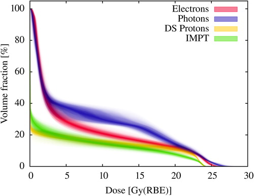
The resulting dose distributions for one of the cases illustrate some of the general differences between the techniques investigated (). Distinct variations were seen in the region beyond the distal end of the spinal target volume, where little or almost no dose was deposited when applying protons. The dose to the organs at risk varied considerably depending on treatment technique with distinctively lower doses to all organs at risk for the proton techniques (). This was also reflected by the mean dose-volume distributions for the normal tissue (including the vertebrae) showing the integral dose to the patient outside the primary CTV (). Similar dose distributions for different organs may be inspected in Supplementary Appendix B available online at http://informahealthcare.com/doi/abs/10.3109/0284186X.2014.928420. The volumes receiving doses below 5 Gy was significantly reduced with the proton techniques, and also moderately reduced in the volumes receiving doses between 5 and 20 Gy. In the latter dose region the difference between the photons and electrons was most pronounced with larger volumes receiving these doses from the photon treatment. Between 20 and 24 Gy there was little difference between all techniques, to some extent expected due to the inclusion of the vertebrae in the age-specific CTV for protons. When applying photons and electrons, a volume fraction also received doses above the prescribed target dose. This was difficult to avoid due to the higher entrance dose when using a single photon or electron beam.
Figure 1. Dose distributions for an 11-year-old male patient from CSI technique applying electrons, photons, DS protons and spot scanning IMPT.
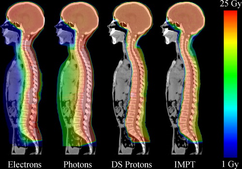
Figure 2. Average of mean doses to all patients for selected organs at risk. The error bars presents the 95% confidence interval of the bootstrapped mean dose for the six patients.
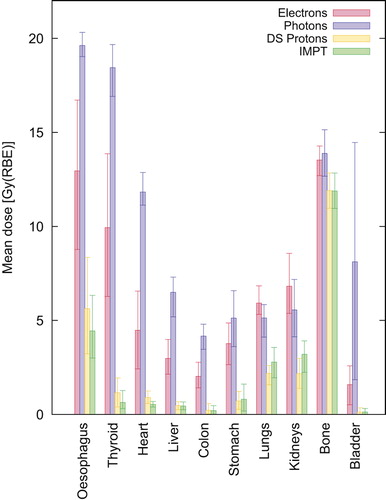
Figure 3. Mean dose-volume distribution of all patients for the dose to normal tissue (including vertebrae). The shading of each distribution extends out to the 95% confidence interval of the bootstrapped mean distribution.
Relative cancer risks based on OED
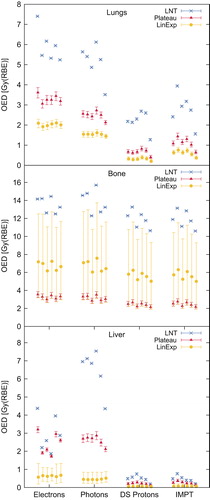
Representative OED results for each individual patient are shown for selected organs in . Other organs exhibited similar behaviour and these results are presented in Supplementary Appendix C available online at http://informahealthcare.com/doi/abs/10.3109/0284186X.2014.928420. The highest risks were found with the linear dose-risk relationship, and for all patients and most organs, substantially lower risks were predicted from the linear-exponential and plateau model. OED results for the proton techniques were in general significantly lower than for the phonton and electron techniques, typically 4–8 times lower. An exception was seen for the bones () with the OED only slightly lower with the proton techniques. Variations between the electron and photon treatments were also observed; and the OEDs for the lungs from the electron technique was higher using all models. For the liver, colon and thyroid however, the OEDLinear for the photons was larger compared to the electrons. Nontheless, the risk for these organs were similar for both techniques using the non-linear models. With respect to the proton techniques, the inherent variations in OEDs were less pronounced. An exception was for the lungs where the IMPT technique resulted in slightly higher doses in the range up to 10 Gy and therefore higher OEDs. Differences in OED between patients were small relative to the variations between the treatment techniques and the models applied. However, this was not the case for the bladder, where rather large dose variations were seen between patiens for the conventional photons.
Figure 4. Organ equivalent doses for the lungs, bone and liver grouped by technique. Results for each individual patient in sequence from left to right, female: 5 years, 7 years, 8 years, male: 8 years, 8 years, 11 years.
Absolute cancer risks
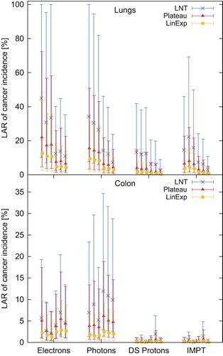
Overall, the nominal values compared in favour of the proton techniques for all patients and all organs included in the LAR analysis: lungs, stomach, colon, liver, thyroid and bladder ( and Supplementary Appendix D available online at http://informahealthcare.com/doi/abs/10.3109/0284186X.2014.928420). Taking the confidence levels into account, our results show that patients have a significant risk of radiation-induced cancer in the lung, thyroid, colon and partially the stomach after electron and photon treatments. However, for the liver and bladder this was not so evident from the results of the plateau and linear-exponential response models. Regarding the proton techniques, the lower threshold of most of the 95% confidence intervals approached zero risk with the exception of the lung for the female patients being treated with IMPT. The difference in the risk of colon cancer between males and females was also observable in the LAR estimations for both the electron and the photon technique. The electron technique resulted in the highest risk for the lungs, which in the case of the 8-year-old female patient ranged from 2% to 36% according to the linear-exponential model and from 8% to 100% from the linear model. The DS proton technique resulted in the lowest estimates for the lungs in this patient with corresponding risks of 0–7% and 3–40% for linear-exponential and linear model, respectively. In contrast to the LAR estimated for the lungs, the risk was lower for the colon; with all patients and all models included, the LARs for the colon ranged up to 35% for the photon technique (highest risk) and up to 5% for the IMPT (lowest risk).
Figure 5. Life attributable risk of radiation-induced cancer in the lungs and colon with 95% confidence intervals. Patient sequence from left to right, female: 5 years, 7 years, 8 years, male: 8 years, 8 years, 11 years.
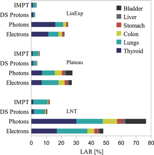
In , the average of the nominal LAR of the patient population is presented in order to compare the overall impact of the investigated treatment techniques. The thyroid and lungs contributed the most to the total risk from all techniques in our patient population. For the electron and photon treatments, the LARs for the residual organs were also influential. The total accumulated risk including all organs was about six times higher compared to the proton techniques by all models. For the linear-exponential model the nominal accumulated risks were 25% and 22% for the photon and electron techniques, whilst the corresponding values for the IMPT and DS protons were 4% and 3%. The photon technique resulted in higher risks compared to electrons assuming a linear dose-response whilst the linear-exponential and plateau response lead to similar risks.
Figure 6. The total life attributable risk of cancer incidence by all models and stratified by technique. The results are averaged over the investigated population group and weighted 2:1 for boys and girls, respectively. Bone is not included as LAR data was not available.
Discussion
The results of the current investigation indicated a significant risk of developing radiation-induced cancer in the lung, thyroid or colon following CSI with conventional electron and photon treatment techniques. Substantially lower cancer risk estimates were obtained with both DS proton technique and IMPT.
The LARs estimated in our study are rather high compared to the indicated risks from follow-up data for this patient group [Citation6]. Packer et al. found a cumulative secondary tumour incidence of 4.2% post- radiation treatment in a population of 379 paediatric medulloblastoma patients [Citation7]. All secondary tumours occurred in the regions exposed to radiation including thyroid and bone cancer. Stavrou et al. also reported a significant increase in subsequent cancer with four of 88 patients diagnosed with secondary cancer within 10 years post-radiation treatment [Citation24]. The existing clinical data can only to some extent provide the basis for precise quantitative description of radiation-induced cancer. However, these statistics indicate that the predictions from the linear models overestimate the risk, in particular with respect to the organs receiving the largest doses. Though further knowledge is warranted in order to understand the progression of secondary cancer, we consider the estimates acquired from the non-linear models as more realistic. These results were also in accordance with similar dosimetric studies [Citation21,Citation25,Citation26]. Some of these latter reports had different endpoints; however, the relative differences between the proton techniques and conventional techniques were reasonably consistent with our results.
The advantage of proton therapy techniques over conventional electron and photon techniques with respect to the risk estimates can be explained by the increased target dose conformity obtained with protons. This is to a great extent attributable to the sharp dose gradients following the Bragg Peaks at the distal end of the target volume. The beneficial proton dose distributions in our treatment plans support the results from other studies with overall distinctive normal tissue sparing [Citation16,Citation27,Citation28].
From the OED analysis, lower secondary cancer risks were predicted from the proton treatments for all organs included in the analysis with one exception. The results for the bones indicate that the incidence of bone cancer is not expected to decrease when moving to a proton therapy regimen. As the target volume must include the bones of the vertebrae, this dose burden is difficult to avoid unless a method to circumvent the sharp dose gradients in the vertebrae is established.
Most of the current clinical experience of using protons for medulloblastoma is from DS protons. However, as IMPT currently is implemented in many new protons centres, this modality is expected to become more available. Newhauser et al. [Citation25] considered the risk of secondary cancer from CSI by comparing passive scattered protons to IMPT in a three-year-old boy and estimated a slightly lower risk from IMPT. The potential superiority of spot-scanned protons versus DS protons was also demonstrated in RT treatments of the whole brain as component of CSI [Citation29]. These differences between the proton techniques were also reflected in our results for most of the organs analysed. However, we have also shown that this is not necessarily the case. In our results the lung dose and estimated OEDs for the IMPT plans were higher compared to the DS protons. In addition to variations in results due to optimisation criteria in calculation algorithms, the outcome of an IMPT plan will depend on the beam configurations and machine constraints. The spot sizes for the delivered beam affects the quality of the dose distribution, with significantly better dose conformity and sparing of organs at risk by using smaller beam sizes [Citation30]. Even when applying a narrow proton beam initially, this is broadened by the multiple Coulomb scattering in the air gap and range shifter plates [Citation31], as applied in our study. An (appropriate) energy selection system with limited requirements for applying range shifters could possibly reduce the beam spot sizes, and hence the lung doses for the IMPT treatment.
In our study we have not included the relatively small, yet non-negligible dose from secondary neutrons that may accompany the therapeutic beams. These neutrons are produced both in the collimator material of the beam delivery unit and in the patient during the radiation treatment, and a complete model for calculating the neutron dose is not included in most planning systems in clinical use today [Citation32]. Photon energies of 6 MV is below the photoneutron production threshold and neutrons are not present at such energies. For the electron technique, however; neutron contamination from the treatment unit imposes a risk in the order of 0.01% for a typical radiation treament [Citation33]. Taddei et al. estimated the lifetime risk of fatal secondary cancer from neutrons after passive scattered proton CSI, with reported risks corresponding to 2.5% from a total dose of 23.4 Gy [Citation34]. For CSI patients the externally received equivalent dose from neutrons can be a factor of 5–6 times higher than the neutron dose generated in the patient, predominantly deposited in the lungs, colon and stomach. For spot-scanned protons the neutron contribution originates within the patient, and is approximately reduced by an order of magnitude over a lateral distance of 10 cm from the target volume [Citation35]. Additional risks from neutrons should not be considered isolated, but rather be added to the nominal dose distribution, causing a slight shift towards higher doses. Assuming that the dose deposited to the lungs is uniformly distributed from the scattered neutrons, this would shift the entire dose distribution towards higher doses. By applying a linear dose-response model the neutron dose would be added to the residual risk simply by adding the isolated risk. However, for the non-linear models the impact of adding doses to the volumes already receiving high doses has little impact on the total risk. In such a paradigm, the neutron dose is the most effective in volumes receiving low doses and plays more of a role for the volumes far away from the primary treatment field. For organs close to the target volume, neutron dose inhomogeneity can potentially have an effect on the risk.
Clinical proton therapy applies a generic RBE weighting factor of 1.1 to the clinical target volume. In the current analysis we have assumed this therapeutic dose factor as valid for our organs at risk both concerning (general) dose constraints as well as the dose-risk response of secondary cancer induction. In the proximity of the radiation field, the RBE of protons shows little variation from the assumed weighting factor, except for small volumes in the distal fall-off of the beam [Citation36]. Considering that most of the organs included in the current analysis is in proximity of the treatment field we found the RBE of 1.1 as the most appropriate to apply. The situation is more complex when it comes to the organs further away from the primary treatment field with increasing uncertainties in the RBE at very low doses [Citation36]. This indicates that the uncertainties can be higher for some of the organs with reduced doses associated with the proton techniques (such as the bladder). A more conservative approach is also attainable with the assumption that the secondary cancer risk should be weighted with a higher RBE compared to therapeutic dose to the target volume. With applying a radiation weighting factor of 2, which is currently recommended by the ICRP for radiation protection purposes [Citation37], the secondary cancer risk estimates for the proton techniques would still be substantially lower compared to the electron and photon techniques.
In agreement with results published by others [Citation13,Citation28], we achieved lower doses to the heart, oesophagus and liver by applying electron spinal fields as an alternative to photons. Our estimations also showed distinctively lower mean doses to the thyroid and bladder from the electrons compared to the conventional photon plans. The difference in total risk between the electron and photon techniques was, however, similar to the variations between organs belonging to different patients. The model dependent variations affected, e.g. the results for the liver and colon with higher risks associated with the linear response compared to the plateau and linear-exponential response. This discrepancy is probably due to the volume fraction receiving doses above 4–5 Gy where the dose-response levels off or start decreasing for the non-linear models. These dose levels amplify the mean dose and thereby the OEDLinear, while given less weight using the OEDPlateau and the OEDLin-Exp (Supplementary Appendix C available online at http://informahealthcare.com/doi/abs/10.3109/0284186X.2014.928420). Our study did not include photon techniques with higher conformity. However, previous studies have shown that the risk is expected to increase with VMAT compared to conventional photons [Citation21,Citation28], likely attributable to the increase in volumes receiving doses in range of 2–5 Gy [Citation38].
Treatment planning systems are designed for creating clinical treatment plans and the main focus is on the organs near the primary beams and in proximity of the target. Most of the organs included in our analysis are close to the target and using dose distributions from the treatment planning system is considered to provide reasonably good estimates of secondary cancer risk. The dose calculation algorithms applied in the treatment planning systems in clinical use today have known inaccuracies, in particular in the low dose range. Studies have shown that the expected uncertainty can be as high as 10–50% in volumes that, according to the dosimetric data given by the treatment planning system, receive less than 5% of the dose delivered to the target volume [Citation32]. This may indicate that some of our organs receiving low doses have larger uncertainties than presented here. This will potentially affect the dose-volume distributions for liver and bladder in the proton treatment plans, indicating that the dose to these organs may be better estimated in combination with other data collection methods like experimental measurements or Monte Carlo simulations [Citation25,Citation26].
Given a linear dose-response, a general reduction of doses (reflected by the mean dose) should be the aim in order to decrease the risk of radiation-induced cancer. Concerning non-linear dose-responses, the approach in order to minimise the risks would be to optimise the dose-volume distributions while avoiding dose levels contributing the most to the secondary cancer risk. With the inherent large uncertainties of the underlying mechanisms of cancer induction and dose-response, such an approach is quite a challenge. In addition, the optimisation procedure is technically difficult without compromising the remaining organs at risk and target volume coverage [Citation21]. Nevertheless, with improved models as well as more flexible treatment techniques, a potential benefit from this approach should not be ruled out.
As an alternative to individually based optimisation as employed in general treatment planning, the secondary cancer risk models could play a part in the evaluation of new treatment modalities, e.g. VMAT and proton therapy. Unlike many side effects that have shorter latent time periods, the long term consequences due to secondary cancer could potentially not be observable until many decades after treatment. However, from considering the general dose-distribution characteristics one may provide reasonable estimates of the expected outcome by comparing to modalities of which we have longer experience with and extended patient follow-up data.
The approach of considering the relative measure of OED does not provide information on the levels of severity for each individual patient or cancer site. Notwithstanding the large confidence intervals of the absolute risk measures in our study, both age- and gender- dependent variations in the nominal LAR values were observed. Considering the effects of increasing risks with decreasing age at exposure, the girls have higher risk factors compared to the boys. For the current patient population, the fluctuations in the risk depend stronger on patient gender compared to the relatively small variations due to age differences. Rather than evaluating the absolute risk, these aspects can be considered in a more qualitative approach. As an example, reducing doses to the thyroid and the lungs for females at a very young age could be targeted from considering the high impact of radiation doses to these organs.
In conclusion, both proton therapy techniques were found to have advantages over photon and electrons with respect to sparing organs at risk during CSI. This resulted in an overall lower risk of radiation- induced cancer estimated from the proton techniques, with the exception of secondary bone cancer with similar risks for all the compared radiation methods. Considering the spectrum of risk responses, the differences between the DS protons and IMPT were small, comparable to the difference between the photon and electron techniques. Uncertainties involving the absolute risk estimates advocate the use of relative measures as the preferred method for comparing risks.
http://informahealthcare.com/doi/abs/10.3109/0284186X.2014.928420
Download PDF (1.4 MB)Declaration of interest: The authors report no conflicts of interest. The authors alone are responsible for the content and writing of the paper.
References
- IRSA – International RadioSurgery Association. [cited 2014 20 Jan]. Available from: http://www.irsa.org/childhood_tumors.html.
- Packer RJ, Gajjar A, Vezina G. Phase III study of craniospinal therapy followed by adjuvant chemotherapy for newly diagnosed average-risk medulloblastoma. Clin Oncol 2006; 24:4202–8.
- Armstrong G. Long-term survivors of childhood central nervous system malignancies: The experience of the Childhood Cancer Survivor Study. Eur J Paediatr Neuro 2010;14:298–303.
- Grau C, Overgaard J. Postirradiation sensorineural hearing loss: A common but ignored late radiation complication. Int J Radiat Oncol Biol Phys 1996;36:515–7.
- Huang E, Teh BS, Strother D, Davis QG, Chiu JK, Lu HH, et al. Intensity-modulated radiation therapy for pediatric medulloblastoma: Early report of the reduction of ototoxicity. Int J Radiat Oncol Biol Phys 2002;52:599–605.
- Strodtbeck K, Sloan A, Rogers L, Fisher PG, Stearns D, Campbell L, et al. Risk of subsequent cancer following a primary CNS tumor. J Neuro-Oncol 2013;112:285–95.
- Packer RJ, Zhou T, Holmes E, Vezina G, Gajjar A. Survival and secondary tumors in children with medulloblastoma receiving radiotherapy and adjuvant chemotherapy: Results of Children’s Oncology Group trial A9961. Neuro Oncol 2013;15:97–103.
- Pierce DA, Preston DL. Radiation-related cancer risks at low doses among atomic bomb survivors. Radiat Res 2000; 154:178–86.
- Preston DL, Mattsson A, Holmberg E, Shore R, Hildreth NG, Boice JD, Jr. Radiation effects on breast cancer risk: A pooled analysis of eight cohorts. Radiat Res 2002;158:220–35.
- Hall EJ, Wuu CS. Review radiation-induced second cancers: The impact of 3D-CRT and IMRT. Int J Radiat Oncol Biol Phys 2003;56:83–8.
- Schneider U, Zwalden D, Ross D, Kaser-Hotz B. Estimation of radiation-induced cancer from three-dimensional dose distributions: Concept of organ equivalent dose. Int J Radiat Oncol Biol Phys 2005;61:1510–5.
- Schneider U, Kaser-Hotz B. Radiation risk estimates after radiotherapy: Application of the organ equivalent dose concept to plateau dose-response relationships. Radiat Environ Bioph 2005;44:235–9.
- Chojnacka M, Skowrońska-Gardas A, Morawska-Kaczyńska M, Zygmuntowicz-Piętka A, Pędziwiatr K, Semaniak A. Craniospinal radiotherapy in children: Electron- or photon-based technique of spinal irradiation. Rep Pract Oncol Radiother 2010;15:21–4.
- Wilkinson JM, Lewis J, Lawrence GP, Lucraft HH, Murphy E. Craniospinal irradiation using a forward planned segmented field technique. Br J Radiol 2007;80:209–15.
- Fogliata A, Bergström S, Cafaro I, Clivio A, Cozzi L, Dipasquale G, et al. Cranio-spinal irradiation with volumetric modulated arc therapy: A multi-institutional treatment experience. Radiother Oncol 2011;99:79–85.
- Yoon M, Shin DH, Kim J, Kim JW, Kim DW, Park SY, et al. Craniospinal irradiation techniques: A dosimetric comparison of proton beams with standard and advanced photon radiotherapy. Int J Radiat Oncol Biol Phys 2011;81:637–46.
- Giebeler A, Newhauser WD, Amos RA, Mahajan A, Homann K, Howell RM. Standardized treatment planning methodology for passively scattered proton craniospinal irradiation. Radiat Oncol 2013;8:32.
- Brown AP1, Barney CL, Grosshans DR, McAleer MF, de Groot JF, Puduvalli VK, et al. Proton beam craniospinal irradiation reduces acute toxicity for adults with medulloblastoma. Int J Radiat Oncol Biol Phys 2013;86:277–84.
- Timmermann B, Lomax AJ, Nobile L, Grotzer MA, Weiss M, Kortmann RD, et al. Novel technique of craniospinal axis proton therapy with the spot-scanning system: Avoidance of patching multiple fields and optimized ventral dose distribution. Strahlenther Onkol 2007;183:685–8.
- International Commission on Radiation Units and Measurements Report 78 Prescribing, recording and reporting proton beam therapy. J ICRU 2007;7:95–122.
- Brodin NP, Rosenschöld P, Aznar M, Kiil-Berthelsen A, Vogelius IR, Nilsson P, et al. Radiobiological risk estimates of adverse events and secondary cancer for proton and photon radiation therapy for pediatric medulloblastoma. Acta Oncol 2011;50:806–16.
- Health risks from exposure to low levels of ionizing radiation: BEIR VII, Phase 2. estimating cancer risk. Washington, DC: National Academy of Science; 2006. p 267–312.
- Clausen N, Garwics S, Glomstein A, Jonmundsson G, Kruus S, Yssing M. Medulloblastoma in Nordic children. A population-based epidemiologic study with long-term follow-up of the survivors.Acta Paediatr Scand Suppl 1990;371:1–27.
- Stavrou T, Bromley CM, Nicholson HS, Byrne J, Packer R, Goldstein AM, et al. Prognostic factors and secondary malignancies in childhood medulloblastoma. J Pediatr Hematol Oncol 2001;23:431–6.
- Newhauser WD. The risk of developing a second cancer after receiving craniospinal proton irradiation. Phys Med Biol 2009;54:2277–91.
- Zhang R, Howell RM, Giebeler A, Taddei PJ, Mahajan A, Newhauser WD. Comparison of risk of radiogenic second cancer following photon and proton craniospinal irradiation for a pediatric medulloblastoma patient.Phys Med Biol 2013;58:807–23.
- Howell RM, Giebeler A, Koontz-Raisig W, Mahajan A, Etzel CJ, D’Amelio Jr AM, et al. Comparison of therapeutic dosimetric data from passively scattered proton and photon craniospinal irradiations for medulloblastoma. Radiat Oncol 2012;7:116.
- Mu X, Björk-Eriksson T, Nill S, Oelfke U, Johansson KA, Gagliardi G, et al. Does electron and proton therapy reduce the risk of radiation induced cancer after spinal irradiation for childhood medulloblastoma?A comparative treatment planning study. Acta Oncol 2005;44:554–62.
- Dinh J, Stoker J, Georges RH, Sahoo N, Zhu RX, Rath S, et al. Comparison of proton therapy techniques for treatment of the whole brain as a component of craniospinal radiation. Radiat Oncol 2013;8:289.
- Moteabbed M, Yock TI, Paganetti H. When does pencil beam scanning improve the outcome of pediatric head and neck /CNS patients over passive beam scattered proton therapy? 2013; astro2013.abstractsnet.com/handouts/pdfs/010091.pdf ASTRO 2013.
- Safai S, Bula C, Meer D, Pedroni E. Improving the precision and performance of proton pencil beam scanning. (Particle Beam Therapy I). Transl Cancer Res 2012;1.
- Howell RM, Scarboro SB, Kry SF. 2010. Accuracy of out-of-field dose calculations by a commercial treatment planning system. Phys Med Biol 2012;55:6999–7008.
- Expósito MR, Romero-Hermida MI, Terrón JA, Esposito D, Planes D, Lagares JI, et al. Neutron contamination in medical linear accelerators operating at electron mode. World Congress on Medical Physics and Biomedical Engineering May 26–31, 2012, Beijing, China IFMBE Proceedings 2013;39:1225–8.
- Taddei PJ, Mirkovic D, Fontenot JD, Giebeler A, Yuynshui Z, Kornguth D, et al. Stray radiation dose and second cancer risk for a pediatric patient receiving craniospinal irradiation with proton beams. Phys Med Biol 2009;54:2259–75.
- Ytre-Hauge KS. Measurements and Monte Carlo simulations of neutron doses from radiation therapy with photons, protons and carbon ions. PhD dissertation.University of Bergen; 2013.
- Paganetti H. Assessment of the risk for developing a second malignancy from scattered and secondary radiation in radiation therapy. Health Phys 2012;103:652–61.
- The 2007 Recommendations of the International Commission on Radiological Protection. ICRP Publication 103. Ann ICRP. 2007;37:1–332.
- Lee KY, Brooks CJ, Bedford LB, Warrington AP, Saran FH. Development and evaluation of multiple isocentric volumetric modulated arc therapy technique for craniospinal axis radiotherapy planning. Int J Radiat Oncol Biol Phys 2012;82:1006–12.