
Abstract
Background. The position and residual motion of the chest wall of breast cancer patients during treatment in deep inspiration breath-hold (DIBH) were investigated.
Material and methods. The study included 58 left-sided breast cancer patients treated with DIBH three-dimensional (3D) conformal radiotherapy in 15 or 25 fractions. The DIBH levels were monitored using an external marker block placed on the chest, either shifted 5 cm to the right at the level of the xiphoid process (Group 1, 27 consecutive patients) or placed medially on the inferior part of the sternum (Group 2, 31 consecutive patients). At every third treatment fraction, continuous portal images were acquired. The time-resolved chest wall position during treatment was compared with the planned position to determine the inter-fraction setup errors and the intra-fraction motion of the chest wall.
Results. The DIBH compliance was 95% during both recruitment periods. A tendency of smaller inter-fraction setup errors and intra-fraction motion was observed for group 2 (medial marker block position). However, apart from a significantly reduced inter-field random shift (σ = 1.7 mm vs. σ = 0.9 mm, p = 0.005), no statistically significant differences between the groups were found. In a combined analysis, the group mean inter-fraction setup error was M = − 0.1 mm, with random and systematic errors of σ = 1.7 mm and Σ = 1.4 mm. The group mean inter-field shift was M = 0.0 (σ = 1.3 mm and Σ = 1.1 mm) and the group mean standard deviation of the intra-field motion was 0.5 mm. The absolute setup error had a maximum of 16.3 mm, exceeding 5 mm in 2.2% of the imaged fields.
Conclusion. Compared to free breathing treatments, the primary benefit of the DIBH technique was the separation of the heart from the target rather than more accurate targeting. Despite a small gating window, occasional large errors in the chest wall position were observed for some patients, illustrating limitations of the external marker block as surrogate in a broad patient population.
During the past decade, studies have shown improved survival rates for early breast cancer patients treated with systemic therapy and radiotherapy (RT) after surgery [Citation1,Citation2]. RT is therefore nowadays included in most breast cancer treatment schemes [Citation3,Citation4]. Meanwhile, a recent study has shown a dose-response relationship between mean heart dose and the risk of long-term cardiac toxicity [Citation5]. This is supported by observations of higher cardiac toxicity for left-sided compared to right-sided breast cancer patients treated with RT. A higher risk of radiation-related cardiac mortality was observed with former treatment strategies (higher fraction dose, larger fields) [Citation3,Citation6]. Even with the current treatment technique in Scandinavia (the tangential field technique) a higher risk of cardiac morbidity may be seen in certain patient subgroups [Citation7–9]. Furthermore, cardiac toxicity after systemic therapy agents used in combination with RT is possible [Citation10]. Caution is therefore warranted for treatment of patients with left-sided breast cancer and a reduction of the cardiac dose is desired [Citation11]. One way of achieving this is the use of respiratory gated RT [Citation12]. Deep inspiration gating techniques are often preferred, because the resulting separation of the breast from the heart allows target coverage with reduced cardiac dose for most left-sided breast cancer patients [Citation13,Citation14]. The gating methods may utilize external surrogates (optical [Citation15] or magnetic markers [Citation16], surface imaging [Citation17]) or breathing control devices [Citation18] to ensure a reproducible gating level. To retain equally high treatment accuracy as for non-gated treatments, image guidance as well as quality assurance should be performed [Citation19,Citation20]. For conformal treatment fields, verification of the in-field anatomy can be accomplished without additional dose to the patient by portal imaging. In this study, continuous portal imaging during treatment delivery was used to determine the treatment accuracy and residual motion for breast cancer treatments in external marker-guided deep inspiration breath-hold (DIBH) with daily image-guided radiotherapy (IGRT) setup. To our knowledge, this is the first study on setup accuracy and residual motion during treatment in a large group of gated left-sided breast cancer patients treated in a clinical routine setting.
Material and methods
Patient characteristics and gating technique
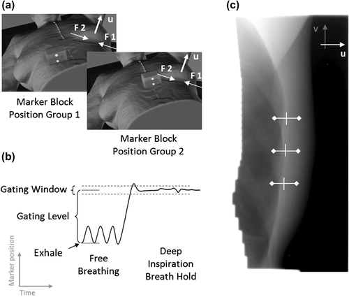
This study includes 58 left-sided breast cancer patients treated with postoperative RT in DIBH using an optical gating method [Real-Time Position Management (RPM) system version 7.5, Varian Medical Systems, Palo Alto, CA, USA]. The RPM system monitored the vertical position of an external marker block placed on the patient and allowed treatment delivery only when this marker was located within a predefined gating window (). The patients were included in two groups. Group 1 consisted of 27 consecutive patients treated between November 2012 and February 2013 with the marker block placed contra-laterally on the chest at the level of the xiphoid process to avoid the treatment area. Group 2 consisted of 31 consecutive patients treated between August and October 2013 with the marker block placed medially on the inferior part of the sternum to investigate if this position would better represent the internal target position (). For both groups, the marker block was stabilized with tape and, if necessary, a foam wedge was used to avoid tilting. Prior to the planning computed tomography (CT) scan each patient received a 30-minute training session, to find the individual maximal comfortable marker block position relative to exhale (gating level, ) for the breath-hold (BH). This gating level was determined by the patient's ability to maintain a stable BH for at least 15 seconds. During training, CT scanning and treatment, the patients were immobilized with the left arm abducted approximately 130° on a standard breast board (Candor ApS, Denmark). Visual feedback (video eyewear) was used to assist the patient in maintaining a stable gating level.
Figure 1. (a) The external marker block position for patient group 1 and 2. Field 1 (F1), field 2 (F2) and the u direction are indicated. (b) A schematic drawing of the marker block motion before and during a DIBH, indicating the gating level and gating window. (c) An example of a portal image of field 2. The relative deviation of the chest wall was found by comparison of the pixel intensities in three regions of interest as shown. The u (and v) directions are indicated by arrows.
Treatment planning and image-guided setup
A gated CT scan with a slice distance of 3 mm (Brilliance CT Big Bore, Philips, Netherlands) was acquired during a single DIBH. The actual gating level and gating window () during the CT scan were recorded for each patient and subsequently applied during the treatments. A single-isocenter technique was planned for all patients, involving two 6 MV conformal fields in a tangential field arrangement (see ), supplemented by a supraclavicular anterior field in case of locoregional irradiation. To improve dose homogeneity, 15 MV segments were added as field-in-field IMRT fields in the directions of the conformal fields. The patients received either 40 Gy in 15 fractions or 50 Gy in 25 fractions (). Daily IGRT setup consisted of an MV image of the lateral field and an orthogonal kV image [Citation21] recorded during two separate short DIBH periods of approximately 5 s duration.
Table I. Patient characteristics and treatment information for patient groups 1 and 2.
Portal imaging and BH monitoring during treatment
During every third fraction, continuous portal images of the two conformal tangential fields were acquired (portal imagers: PortalVision AS500 or AS1000, Varian Medical Systems). The imaged tangential fields, in the following referred to as field 1 (lateral field) and field 2 (medial field), were the first and last field delivered, respectively. The portal images were acquired with frequencies of 2 Hz (AS500) or 1 Hz (AS1000) and pixel lengths of 0.52 mm (AS500) or 0.26 mm (AS1000) when scaled to isocenter distance. The RPM signal was recorded during the entire treatment and saved for offline analysis.
Data analysis and statistics
In order to find the absolute deviation of the chest wall position during treatment, the continuous portal images were first analyzed using an in-house developed Matlab program. This program utilized the distinct pixel intensity variations of the chest wall to determine its position in the u-direction in three manually defined regions of interest relative to the chest wall position in the first portal image (see example in ). The u-direction is the direction in the imager plane that is perpendicular to the gantry rotation axis (). In this study, it is a linear combination of the left-right (LR) direction (approximately one third) and anterior-posterior (AP) direction (approximately two thirds) in patient coordinates. From this relative position, the absolute deviation of the chest wall position was determined manually by comparison with the digitally reconstructed radiograph (DRR) of the treatment fields in the clinical image analysis application (ARIA, Varian Medical Systems). The inter-fraction setup error and intra-fraction motion were quantified as described in the following paragraphs. The intra-fraction motion of the chest wall was subdivided into an inter-field shift component and intra-field motion component. A Student's t-test was used to test for statistical significance.
Inter-fraction setup error
The inter-fraction setup error after IGRT correction was quantified as the group mean (M), systematic (Σ) and random (σ) errors of the mean chest wall position in the u-direction during delivery of field 1 and 2 combined [Citation22]. It represents the overall treatment accuracy, including uncertainties in the IGRT setup procedure, the reproducibility of the internal gating level from IGRT imaging to treatment, and technical inaccuracies.
Inter-field shift
The inter-field shift was quantified as M, Σ and σ of the difference in mean setup error of the chest wall between field 1 and 2. It represents the intra-fractional reproducibility of the internal gating level, but includes also some geometrical inaccuracies of the field aperture (gantry sag, MLC misalignment).
Intra-field motion
The intra-field motion was represented by the motion range (the difference between the maximum and minimum position) of the chest wall during field delivery and by the root- mean-square average of the motion (one standard deviation) during field delivery over all fields, fractions and patients. It describes the residual internal motion during BH, i.e. the stability of the BH, and was compared with the external gating window.
Results
In the recruitment periods, all left-sided breast cancer patients referred to RT completed the DIBH training session, but three of these patients had difficulties with the BH technique and were therefore scanned and treated in free breathing. The remaining 27 (group 1) and 31 (group 2) patients were treated in DIBH and included in this study (). Key characteristics of group 1 and 2 are given in while the inter-fraction setup error, the inter-field shift and the intra-field motion are presented in . Due to missing data, 11 fields and 5 fractions were excluded from the intra- and inter-field analysis, respectively. shows the distributions of the absolute inter-fraction setup errors (a), the absolute inter-field shifts (b), and the intra-field motion range (c) for both patient groups. Although the figure shows a general tendency of smaller setup errors, shifts and chest wall motion for group 2, no statistically significant differences between the two groups were found except from a significantly reduced inter-field random shift (p = 0.005). The majority of patients in this study showed small setup errors during the treatment course (example in ) with mean setup errors below 5 mm in 97.8% of the recorded fields, but some patients occasionally demonstrated large setup errors and motion as illustrated in . The maximum setup error observed in this study was -16.3 mm. Large setup errors were only in some cases associated with an unstable RPM signal that entered and exited the gating window during field delivery as shown in the examples in . Large intra-field motion ranges indicated that the external RPM signal was a non-accurate representation of the internal chest wall stability. The intra-field chest wall motion exceeded the external gating window for 15 of 27 patients (56%) in group 1 and 10 of 31 patients (32%) in group 2. Seven of these 25 patients contributed 89% of all mean setup errors above 5 mm.
Figure 2. Cumulative frequencies of (a) absolute inter-fraction setup error, (b) absolute inter-field shift and (c) intra-field motion range for group 1 and 2.
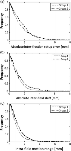
Figure 3. The time resolved setup errors of the chest wall for (a) all imaged fractions and (b) the first treatment fraction for a typical patient. For the first fraction, the figure also shows (c) the external marker block position and (d) the resulting gate signal, which is in “enable state” when the marker block is inside the gating window [- - - -].
![Figure 3. The time resolved setup errors of the chest wall for (a) all imaged fractions and (b) the first treatment fraction for a typical patient. For the first fraction, the figure also shows (c) the external marker block position and (d) the resulting gate signal, which is in “enable state” when the marker block is inside the gating window [- - - -].](/cms/asset/85f56072-76cd-4630-9af5-1ca69dfbbe5c/ionc_a_1045625_f0003_b.gif)
Figure 4. Box plots of the patient individual (a) inter-fraction setup error (b) inter-field shift and (c) intra-field motion range. The box indicates the 25th and 75th percentiles, while the extending narrow bars indicate the most extreme data points that are not considered outliers [o].
![Figure 4. Box plots of the patient individual (a) inter-fraction setup error (b) inter-field shift and (c) intra-field motion range. The box indicates the 25th and 75th percentiles, while the extending narrow bars indicate the most extreme data points that are not considered outliers [o].](/cms/asset/6271f3df-af3d-4afe-97bc-e13a3d278818/ionc_a_1045625_f0004_b.gif)
Figure 5. Three examples of internal chest wall setup error and external marker block position during fractions with large inter-field shifts (a, c) and intra-field motion (b, c). The gating window (---) and resulting binary beam-enable signal (bottom graph) are indicated for each case.
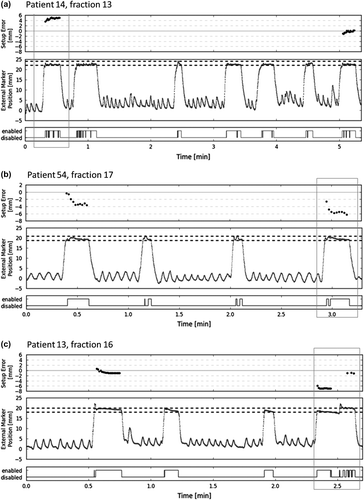
Table II. Gating and imaging characteristics for patient groups 1 and 2.
Table III. The inter-fraction setup error (fields 1 + 2), inter-field shift, and intra-field motion for patient group 1 and 2, individually and combined.
Discussion
This study investigated the internal accuracy of left-sided breast cancer treatments in optically monitored DIBH with daily IGRT for 58 patients in a clinical routine setting. Continuous portal images of the two tangential fields were acquired during every third treatment fraction and analyzed to find the accuracy and stability of the BH during treatment. To investigate the impact of the external marker block position on treatment accuracy, the marker block position was changed between a first group of 27 patients and a second group of 31 patients. To our knowledge, this is the first study using in-treatment portal images to determine the treatment accuracy of RPM-gated DIBH. No prior patient selection was applied and with a compliance rate of 95%, this study therefore includes patients of a wide variety of ages and cooperation ability.
The overall group mean inter-fraction setup error and inter-field shift observed in this study are comparable to those observed in our institution for breast cancer patients treated with the same IGRT technique in free breathing (M = −0.7 mm, Σ = 1.1 mm, σ = 1.5 mm) [Citation21]. The main advantage of DIBH relative to free breathing treatments was therefore the physical separation of the target from the heart during BH. The reproducibility of DIBH with the RPM system has previously been studied in 10 patients by McIntosh et al. [Citation23] using weekly lateral kV images. They found a DIBH reproducibility of 1 mm (range 0–3 mm) in the AP direction. The mean reproducibility of the BH in the present study was smaller, while the observed range of setup errors was larger. Based on the margin recipe in [Citation24], the applied PTV margin of 5 mm was sufficient (2.5Σ+ 0.7σ = 4.7 mm). However, larger deviations were observed for some of the patients in this study () indicating that the chest wall position was not always accurately defined by the external marker block. This was supported by reviewing the RPM signal during treatment of sessions with large inter-field shifts and intra-field motion. The RPM signal did not represent the extent of the chest wall error (as treatment otherwise would not have been possible) and only in some cases did the external marker block motion indicate BH instability (see ). No indications that smoking might affect the ability to cooperate to DIBH gating were found, however, the number of smokers among the patients was low. Further study is required to determine the influence of Astma/COPD. For group 2, a tendency towards smaller inter-fraction setup errors and intra-fraction motion as well as a reduced number of outliers with large internal chest wall motion during BH was observed. Although this indicates a better control of the BH with the medial marker block position, which is closer to the target, only the inter-field random shift was significantly reduced for group 2. Other studies on DIBH reproducibility have utilized different gating methods, such as breathing control devices and other external surrogates [active breathing control (ABC) system, magnetic sensors, 3D surface imaging system (AlignRT), fluoroscopy imaging, visual monitoring [Citation16,Citation17,Citation18,Citation25,Citation26,Citation27]. The overall treatment accuracy in this study for a broad patient population with a compliance of 95% was in accordance with these previously published smaller studies on DIBH. Large patient-specific setup errors as observed in the present study were either not reported or not observed in the previous studies, with the exception of one patient in [Citation16]. As a result of this study, offline review of continuous portal images acquired during three successive treatment fractions was implemented in our clinic as a QA procedure for selected patients who display difficulty with the DIBH method during training or CT scanning. Furthermore, a standard gating window of 2 mm is now used for all patients instead of the previously used individual gating window. The medial marker position has been adopted as standard in our clinic, since no skin reactions due to the modest bolus effect of the marker block in the treatment field were observed. The smaller mean gating level for group 2 () might be explained by the different marker block position.
Although there are many advantages of using continuous portal imaging for position monitoring during treatment delivery, the method is restricted to conformal fields and requires x-ray visible landmarks such as the chest wall. The 2D nature of portal images complicates the analysis of 3D motion. However, this study focussed on the setup error in the u-direction which is most relevant for the cardiac dose from lateral opposing tangential fields.
In conclusion, the overall treatment accuracy of DIBH-gated IGRT in a clinical setting with no patient selection was comparable to the accuracy found in smaller studies using different DIBH techniques and for free breathing treatments in our institution. The occurrence and frequency of large inter-fraction setup errors and intra-fraction motion was patient-specific and indicated that the surrogate motion was not able to reliably represent the chest wall motion for all patients.
Acknowledgments
Supported by CIRRO – The Lundbeck Foundation Center for Interventional Research in Radiation Oncology, The Danish Council for Strategic Research, and The Danish Cancer Society.
Declaration of interest: The authors report no conflicts of interest. The authors alone are responsible for the content and writing of the paper.
References
- Early Breast Cancer Trialists’ Collaborative Group (E. B. C. T. C. G), Davies C, Godwin J, Gray R, Clarke M, Cutter D, et al. Relevance of breast cancer hormone receptors and other factors to the efficacy of adjuvant tamoxifen: Patient-level meta-analysis of randomised trials. Lancet 2011;378: 771–84.
- Early Breast Cancer Trialists’ Collaborative Group (E. B. C. T. C. G), Peto R, Davies C, Godwin J, Gray R, Pan HC, et al Comparisons between different polychemotherapy regimens for early breast cancer: Meta-analyses of long-term outcome among 100,000 women in 123 randomised trials. Lancet 2012;379:432–44.
- Early Breast Cancer Trialists’ Collaborative Group (E.B.C. T.C.G). Effects of radiotherapy and surgery in early breast cancer. An overview of the randomized trials. N Engl J Med 1995;333:1444–55.
- Voogd AC, Nielsen M, Peterse JL, Blichert-Toft M, Bartelink H, Overgaard M, et al. Differences in risk factors for local and distant recurrence after breast-conserving therapy or mastectomy for stage I and II breast cancer: Pooled results of two large European randomized trials. J Clin Oncol 2001;19:1688–97.
- Darby SC, Ewertz M, McGale P, Bennet AM, Blom-Goldman U, Brønnum D, et al. Risk of ischemic heart disease in women after radiotherapy for breast cancer. N Engl J Med 2013;368:987–98.
- Cuzick J, Stewart H, Rutqvist L, Houghton J, Edwards R, Redmond C, et al. Cause-specific mortality in long-term survivors of breast cancer who participated in trials of radiotherapy. J Clin Oncol 1994;12:447–53.
- McGale P, Darby SC, Hall P, Adolfsson J, Bengtsson N, Bennet AM, et al. Incidence of heart disease in 35,000 women treated with radiotherapy for breast cancer in Denmark and Sweden. Radiother Oncol 2011;100:167–75.
- Correa CR, Litt HI, Hwang WT, Ferrari VA, Lawrence JS, Harris EE. Coronary artery findings after left-sided compared with right-sided radiation treatment for early-stage breast cancer. J Clin Oncol 2007;25:3031–7.
- Nilsson G, Holmborg L, Garmo H, Duvernoy O, Sjögren I, Lagerqvist B, et al. Distribution of coronary artery stenosis after radiation for breast cancer. J Clin Oncol 2012;30:380–6.
- Carver JR, Shapiro CL, Ng A, Jacobs L, Schwartz C, Virgo KS, et al. American Society of Clinical Oncology clinical evidence review on the ongoing care of adult cancer survivors: Cardiac and pulmonary late effects. J Clin Oncol 2007;25:3991–4008.
- Demirci S, Nam J, Hubbs JL, Nguyen T, Marks LB. Radiation-induced cardiac toxicity after therapy for breast cancer: Interaction between treatment era and followup-duration Int J Radiat Oncol Biol Phys 2009;73:980–7.
- Ohara K, Okumura T, Akisada M, Inada T, Mori T, Yokota H, et al. Irradiation synchronized with respiration gate. Int J Radiat Oncol Biol Phys 1989;17:853–7.
- Hjelstuen MHB, Mjaaland I, Vikstroem J, Dybvik KI. Radiation during deep inspiration allows loco-regional treatment of left breast and axillary-, supraclavicular- and internal mammary lymph nodes without compromising target coverage or dose restrictions to organs at risk. Acta Oncol 2012;51:333–44.
- Nissen HD, Appelt AL. Improved heart, lung and target dose with deep inspiration breath hold in a large clinical series of breast cancer patients. Radiother Oncol 2013;106:28–32.
- Berson AM, Emery R, Rodriguez L, Richards GM, Ng T, Sanghavi S, et al. Clinical experience using respiratory gated radiation therapy: Comparison of free-breathing and breath-hold techniques. Int J Radiat Oncol Biol Phys 2004;60:419–26.
- Remouchamps VM, Huyskens DP, Mertens I, Destine M, Van Esch A, Salamon E, et al. The use of magnetic sensors to monitor moderate deep inspiration breath hold during breast irradiation with dynamic mlc compensators. Radiother Oncol 2007;82:341–8.
- Alderliesten T, Sonke J, Betgen A, Honnef J, van Vliet-Vroegindeweij C, Remeijer P. Accuracy evaluation of a 3-dimensional surface imaging system for guidance in deep-inspiration breath-hold radiation therapy. Int J Radiat Oncol Biol Phys 2013;85:536–42.
- Remouchamps VM, Letts N, Yan D, Vicini FA, Moreau M, Zielinski JA, et al. Three-dimensional evaluation of intra- and interfraction immobilization of lung and chest wall using active breathing control: A reproducibility study with breast cancer patients. Int J Radiat Oncol Biol Phys 2003;57: 968–78.
- Korreman SS, Juhler-Noettrup T, Persson GF, Navrsted Pedersen A, Enmark M, Nystrom H, et al. The role of image guidance in respiratory gated radiotherapy. Acta Oncol 2008;47:1390–6.
- Jiang SB, Wolfgang J, Mageras GS. Quality assurance challenges for motion-adaptive radiation therapy: Gating, breath holding, and four-dimensional computed tomography. Int J Radiat Oncol Biol Phys 2008;71(1 Suppl):S103–7.
- Thomsen MS, Harrov U, Fledelius W, Poulsen PR. Inter- and intra-fraction geometric errors in daily image-guided radiotherapy of free-breathing breast cancer patients measured with continuous portal imaging. Acta Oncol 2014;53:802–8.
- van Herk M. Errors and margins in radiotherapy. Semin Radiat Oncol 2004;14:52–64.
- McIntosh A, Shoushtari AN, Benedict SH, Read PW, Wijesooriya K. Quantifying the reproducibility of heart position during treatment and corresponding delivered heart dose in voluntary deep inhalation breath hold for left breast cancer patients treated with external beam radiotherapy. Int J Radiat Oncol Biol Phys 2011;81:e569–76.
- van Herk M, Remeijer P, Rasch C, Lebesque JV. The probability of correct target dosage: Dose-population histograms for deriving treatment margins in radiotherapy. Int J Radiat Oncol Biol Phys 2000;47:1121–35.
- Betgen A, Alderliesten T, Sonke J, van Vliet-Vroegindeweij C, Bartelink H, Remeijer P. Assessment of set-up variability during deep inspiration breath hold radiotherapy for breast cancer patients by 3d-surface imaging. Radiother Oncol 2013;106:225–30.
- Bartlett FR, Colgan RM, Carr K, Donovan EM, McNair HA, Locke I, et al. The UK Heartspare Study: Randomised evaluation of voluntary deep-inspiratory breath-hold in women undergoing breast radiotherapy. Radiother Oncol 2013;108:242–7.
- Borst GR, Sonke J, den Hollander S, Betgen A, Remeijer P, van Giersbergen A, et al. Clinical results of image-guided deep inspiration breath hold breast irradiation. Int J Radiat Oncol Biol Phys 2010;78:1345–51.