
Abstract
Background. The management of cardiovascular risk factors is important for prevention of atherosclerotic cardiovascular diseases (ACVD). Visceral fat accumulation plays an important role in the clustering of cardiovascular risk factors, leading to ACVD. The present study investigated the gender- and age-specific relationship between obesity-related cardiovascular risk factor accumulation and computed tomography (CT)-measured fat distribution in a large-scale Japanese general population.
Methods and results. Fat distribution was measured on CT scans in 12,443 subjects (males/females = 10,080/2,363), who underwent medical health check-up at 9 centers in Japan. The investigated obesity-related cardiovascular risk factors were hyperglycemia, dyslipidemia, and elevated blood pressure. Visceral fat area (VFA) for all males and old females showed almost symmetric distribution, while that of young females showed skewed distribution with a marked left shift. Only a small proportion of young females had large visceral fat and cardiovascular risk accumulation. The mean number of risk factors exceeded 1.0 at around 100 cm2 for VFA in all groups, irrespective of gender, age (cut-off age 55), and BMI (cut-off BMI 25 kg/m2).
Conclusions. In this large-scale Japan-wide general population study, an absolute VFA value of about 100 cm2 equated with obesity-related cardiovascular risk factor accumulation, irrespective of gender, age, and BMI.
Clinical trial registration information. https://upload.umin.ac.jp/cgi-open-bin/ctr/ctr.cgi?function=brows&action=brows&type=summary&recptno=R000002780&language=E.
| Abbreviations | ||
| ACVD | = | atherosclerotic cardiovascular diseases |
| AUC | = | area under the curve |
| BMI | = | body mass index |
| CT | = | computed tomography |
| HDL-C | = | high-density lipoprotein cholesterol |
| LDL-C | = | low-density lipoprotein cholesterol |
| ROC | = | receiver operating characteristic |
| SFA | = | subcutaneous fat area |
| TG | = | triglyceride |
| VFA | = | visceral fat area |
| WC | = | waist circumference |
Key messages
Visceral fat area of approximately 100 cm2 equates with the presence of one or more obesity-related cardiovascular risk factors, irrespective of gender, age, and BMI.
In regular health check-ups, the absolute visceral fat area should be set up at 100 cm2 as a criterion for screening multiple obesity-related cardiovascular risk factors, rather than using the relative value calculated from the receiver operating characteristic curve analysis.
Introduction
Management of cardiovascular risk factors is important for primary prevention of cardiovascular diseases. Elevated serum level of low-density lipoprotein cholesterol (LDL-C) is a major target for the prevention of atherosclerotic cardiovascular diseases (ACVD) (Citation1). The second target is non-LDL-C multiple risk factor syndrome accompanying glucose intolerance, hypertriglyceridemia/low high-density lipoproteincholesterolemia, and hypertension, associated with obesity, which has increased worldwide with a life-style of overeating and low physical activity.
Vague (Citation2) was the first to link adipose tissue distribution to complications in obese subjects. Subsequent studies, including our study, demonstrated that abdominal obesity (increased waist-to-hip ratio) rather than body mass index is related to obesity-related disorders and ACVD (Citation3–6). We introduced a method for the evaluation of fat distribution using computed tomography (CT) scanning, which allowed us to analyze the amounts of intra-abdominal visceral fat and subcutaneous fat (Citation7). We also demonstrated that the ratio of abdominal visceral fat area (VFA)/subcutaneous fat area (SFA) (Citation8) correlated significantly with glucose intolerance, hypertriglyceridemia (Citation9), hypertension (Citation10), cardiac dysfunction (Citation11), and sleep apnea syndrome (Citation12) in obese subjects.
Several investigators, including our group, have shown that visceral adiposity measured by CT correlates with ACVD and multiple risk factor syndrome even in overweight and mildly obese individuals (Citation13–15). We also reported that reduction of visceral fat correlated with improvement of hyperglycemia, hypertriglyceridemia and hypertension (Citation16). Therefore, it is necessary to evaluate visceral and subcutaneous fat in relation to the cluster of obesity-related cardiovascular risk factors to select subjects from the general population suitable for programs designed to reduce visceral fat and prevent future ACVD. As a first step towards this goal, the present study was designed to determine the absolute values of VFA, SFA, and VFA/SFA (V/S) ratio in a large population of both genders of different age groups, with or without obesity
Subjects and methods
Study populations
The study subjects comprised all 12,443 consecutive Japanese (males/females: 10,080/2,363; age range 19–88 years), who underwent medical health check-up (or ‘Ningen (Human)-Dock’) and CT scan at one of nine centers across Japan (Ibaraki, Chiba, Tokyo, Toyama, Osaka, Kagawa, Fukuoka) between January 2006 and December 2006. No exclusion or inclusion criteria were set, and the study also included subjects on treatment for diabetes, hypertension, and/or dyslipidemia. The purpose of the medical health check-up program is to promote public health through early detection of diseases and risk factors. Medical service of this kind, known as a ‘Ningen (Human)-Dock’, is common in Japan. The cost of the medical examination was covered by the employers and the employees for the promotion of health and prevention of disease among the employees. summarizes the profiles of male and female participants. The study was approved by the Human Ethics Committee of Osaka University, and informed consent was obtained from all participants to post a notice to the Japan Society of Ningen-Dock and Japan Society of Health Evaluation and Promotion. This research (called the VACATION-J study: Visceral Fat Accumulation and Coronary Artery Disease Investigation in Japanese) is registered with University Hospital Medical Information Network, number UMIN 000002273.
Table I. Clinical characteristics of all subjects.
Anthropometry and laboratory tests
Height (m) and weight (kg) were measured in standing position, and body mass index (BMI) was calculated (= weight (kg)/height (m2)). Waist circumference (WC) was measured at the level of the umbilicus with a non-stretchable tape in late expiration while standing. VFA and SFA were computed or measured manually or automatically using commercial software on CT scans taken at the umbilical level in the supine position, based on the Japanese guideline of obesity treatment (Japan Society for the Study of Obesity, in Japanese). Briefly, 5- to 10-mm thick slices (120 kVp; 400 mAs; 200 mA × 2 s) were acquired. To identify fat-containing pixels, an image display window width of 800–1,000 Housfield units (HU) and a window center of 0 HU were used. VFA and SFA were measured on CT scan obtained by different brands of CT scanners (Aquillion16, 64, Toshiba Medical Systems Corporation, Tokyo, Japan; Xvision/GX, Toshiba Medical Systems Corporation, Tokyo; Xvision/REAL, Toshiba Medical Systems Corporation, Tokyo; Asteion S4, Toshiba Medical Systems Corporation, Tokyo; Light Speed VCT, GE Healthcare UK, Buckinghamshire, England; and Robusto, Hitachi Medical Corporation, Tokyo). The CT scans were acquired either at inspiration or expiration depending on the participating institution. Blood pressure was measured with a standard mercury sphygmomanometer on the right or left arm after the subjects had rested in a sitting position for at least 10 minutes. After an overnight fasting or at least 5-hour fasting, venous blood samples were collected for measurement of blood glucose, high-density lipoprotein cholesterol (HDL-C), and triglyceride (TG), while the subject was in the sitting position. The present study investigated the presence of three obesity-related cardiovascular risk factors: 1) elevated blood pressure (i.e. hypertension, representing systolic blood pressure ≥ 130 mmHg and/or diastolic blood pressure ≥ 85 mmHg), or regular treatment with anti-hypertensive agents; 2) dyslipidemia (fasting TG of ≥ 1.69 mmol/L or HDL-C < 1.04 mmol/L), or regular treatment with hypolipidemic agents; and 3) abnormal glucose levels (fasting plasma glucose concentration of ≥ 6.1 mmol/L), or regular treatment with anti-diabetic agents. Medical history was surveyed by using a self-administered questionnaire.
Statistical analysis
Significance level was set at P < 0.05. Continuous variables are presented as mean ± SD () or mean ± SEM (), as indicated. Differences in the mean number of obesity-related cardiovascular risk factors between males and females were analyzed by the Kruskal-Wallis test (). All statistical analyses were performed with StatView-J 5.0 (Statistical Analysis System Inc., Cary, NC, USA) or the Statistical Package for Social Sciences (version 11.0.1J; SPSS, Chicago, IL, USA).
Figure 1. Association between visceral fat area (VFA) and obesity-related cardiovascular risk factors (top), and the histogram of VFA (bins of 25 cm2) (bottom) for all males (A), young males (age < 55, B), old males (age ≥ 55, C), all females (D), young females (age < 55, E), and old females (age ≥ 55, F). Open bars: subjects with less than 1.0 risk factor; solid bars: subjects with more than 1.0 risk factor. Data are mean ± SEM.
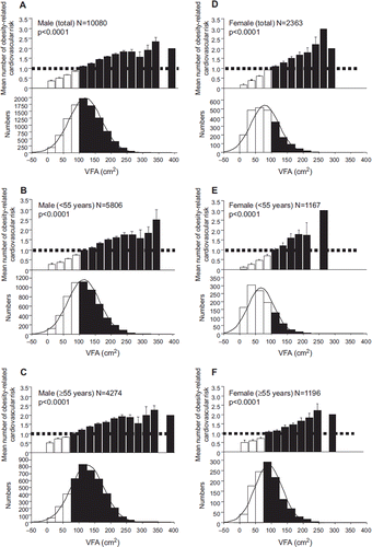
Figure 2. Association between subcutaneous fat area (SFA) and obesity-related cardiovascular risk factors (top), and the histogram of SFA (bins of 25 cm2) (bottom) for all males (A), young males (age < 55, B), old males (age ≥ 55, C), all females (D), young females (age < 55, E), and old females (age ≥ 55, F). Data are mean ± SEM.
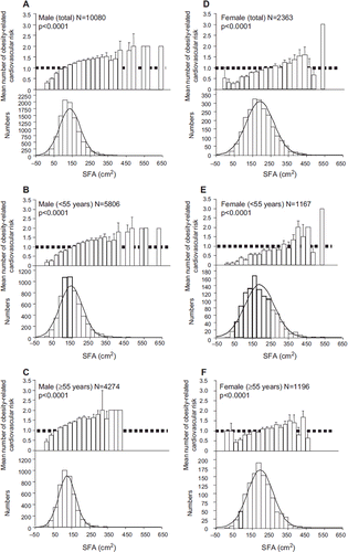
Figure 3. Association between VFA, SFA and obesity-related cardiovascular risk factors in non-obese (BMI < 25 kg/m2) and obese subjects (BMI ≥ 25 kg/m2). The mean number of obesity-related cardiovascular risk factors and VFA in bins of 25 cm2 for non-obese and obese males (A, top and bottom), and for non-obese and obese females (C, top and bottom), SFA in bins of 25 cm2 in non-obese and obese males (B, top and bottom), and in non-obese and obese females (D, top and bottom). Data are mean ± SEM.
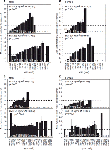
Figure 4. Correlation between waist circumference (WC) and VFA for all males (A), young males (age < 55, B), old males (age ≥ 55, C), all females (D), young females (age < 55, E), and old females (age ≥ 55, F). WC was measured at the level of the umbilicus with a non-stretchable tape in late expiration while standing (in cm) according to the Examination Committee of Criteria for ‘Obesity Disease’ in Japan (Citation19).
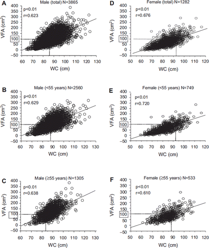
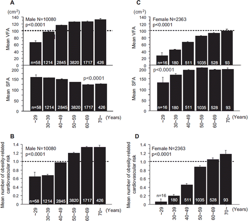
Figure 5. The mean VFA and SFA by age in males (A), and females (C). Prevalence of obesity-related cardiovascular risk factors by age in males (B) and females (D). Data are mean ± SEM.
Results
Number of obesity-related cardiovascular risk factors according to VFA levels and body fat distribution
summarizes the characteristics of the subjects enrolled in this study. First, we evaluated the association between the number of obesity-related cardiovascular risk factors, and VFA () and SFA () for the entire population, the young group (age < 55) and the old group (age ≥ 55) of males and females. Each group was divided into 25 cm2 bins of VFA and SFA.
The average number of obesity-related cardiovascular risk factors was more than 1.0 at 100–125 cm2 for VFA in all age and gender groups: all males (, top), young males (, top), old males (, top), all females (, top), young females (, top), and old females ( top) (P < 0.0001 for trend, Kruskal-Wallis test). The mean number of obesity-related cardiovascular risk factors increased linearly with VFA, without an obvious inflection point, in all groups. VFA was distributed almost symmetrically in all males (n = 10,080, median 115.9 cm2) (, bottom), young males (n = 5,806) (, bottom), and old males (n = 4,274) (, bottom). On the other hand, the median value of VFA was 74.2 cm2 for all females (n = 2,363) (, bottom), and VFA was skewed with a left shift in young females, who were assumed to be approximately pre- or menopausal (n = 1,167, median 59.8 cm2) (, bottom). VFA was distributed almost symmetrically in the old female group (n = 1,196, median 86.8 cm2) (, bottom), similar to males.
, bottom, shows the distribution of SFA. The SFA in females was greater than that of males at any age, with wide variability. The increase in the mean number of obesity-related risk factors based on increase in SFA was milder both for males and females (, top) (P < 0.0001 for trend, Kruskal-Wallis test), in contrast to the linear relationship observed in VFA. The rate of increase in the obesity-related risk factors was milder in females with SFA ≥ 200 cm2 than those with < 200 cm2. Interestingly, the mean number of obesity-related risk factors in relation to increase in SFA reached a plateau in males (, top). The mean number of obesity-related risk factors was more than 1.0 at 100–125 cm2 for SFA in all males (, top), at 225–250 cm2 in all females (, top), and at more than 300 cm2 in young females (, top). In old females, the increase in SFA was less associated with increase in risk factors ().
Association between obesity-related risk factors and VFA/SFA based on BMI
We and others reported previously the importance of fat distribution in obesity (Citation17,Citation18). We divided subjects into non-obese and obese groups according to BMI (cut-off value 25 kg/m2) based on the Japanese criteria (Citation19). For males and females, the mean number of obesity-related cardiovascular risk factors increased linearly with VFA and was more than 1.0 at around 100 cm2 for VFA, irrespective of BMI (, top and bottom, and 3C, top and bottom) (P < 0.0001 for trend, Kruskal-Wallis test). For non-obese males and females, the mean number of obesity-related cardiovascular risk factors increased with SFA (, top, and 3D, top) (P < 0.0001 for trend, Kruskal-Wallis test), but was less than 1.0 irrespective of SFA in non-obese females (, top). On the other hand, there was no increment in obesity-related risk factors with increase in SFA in obese males (, bottom) and obese females (, bottom) (n.s., Kruskal-Wallis test).
Correlation between WC and VFA
WC was measured in 5,147 subjects of the total of 12,443 subjects. VFA of approximately 100 cm2 corresponded to WC of about 85 cm in males and 90 in females. WC correlated significantly with VFA in all males and females (, ), young (, ) and old (, ) males and females.
Body fat distribution, VFA, SFA, and prevalence of obesity-related cardiovascular risk factors by age
The mean VFA increased with age in both males (, top) (P < 0.0001 for trend, Kruskal-Wallis test) and females (, top) (P < 0.0001 for trend, Kruskal-Wallis test), increased gradually with age in males to 100 cm2 but then showed a plateau between 50 and > 70 years of age (, top). In contrast, in females, VFA continued to increase with age, though the average VFA was less than 100 cm2 (, top). SFA decreased gradually with age in males (, bottom) (P < 0.0001 for trend, Kruskal-Wallis test), but increased in females (, bottom) (P < 0.0001 for trend, Kruskal-Wallis test). The mean number of obesity-related cardiovascular risk factors was more than 1.0 at over-40s for males (), while at over-60s for females ().
Discussion
Several single-center and relatively small-scale Japanese population studies (average age ∼50 years) reported that the WC and VFA correlated with the number of obesity-related cardiovascular risk factors (Citation20–22). The present study is the first report of VFA and SFA measured accurately by CT scan and analyzed in large-scale population in multiple centers across Japan. The results demonstrated a symmetrical distribution of VFA in both males and older females, with a median value of 85–120 cm2. However, younger females aged less than 55 years, assumed to be pre- and menopausal, had markedly smaller VFA, and the VFA in this population showed a skewed distribution with a median value of 59.8 cm2. The latter group also had considerably fewer obesity-related cardiovascular risk factors. Nevertheless, the average number of obesity-related cardiovascular risk factors exceeded 1.0 at VFA around 100 cm2 for all groups of males and females, irrespective of age. The numbers of obesity-related cardiovascular risk factors also increased proportionally with the increase in VFA, irrespective of gender, age, and BMI.
Although the average number of obesity-related cardiovascular risk factors was more than 2.0 for all males with VFA of 300 cm2 (), and total females with VFA of 250 cm2 (), based on Japanese criteria of the metabolic syndrome, the concept of the Japanese remedy for the metabolic syndrome, i.e. visceral fat reduction, is made available for subjects with atherogenic metabolic profile at the early stage of disease in a community health care center. Therefore, the VFA value that corresponds to one or more obesity-related cardiovascular risk factors () (100 cm2) should be important for primary prevention of ACVD events.
The results demonstrated that intra-abdominal visceral fat accumulation has an important role in the development of metabolic disorders, such as diabetes, lipid disorders, and hypertension. In order to clarify how visceral fat accumulation causes metabolic and cardiovascular diseases, we analyzed previously the gene expression profile in subcutaneous and visceral adipose tissues (Citation23). The results showed that adipose tissue, especially visceral adipose tissue, over-expresses various genes that encode bioactive substances such as cytokines (collectively termed adipocytokines), and especially adiponectin, which has anti-diabetic, -hypertensive and -atherogenic, -inflammatory, and -oncogenic functions (Citation24). Thus, dysregulated production of adipocytokines seems to provide the link between visceral fat accumulation and metabolic disorders and CVD. Although the exact amount of visceral fat can be evaluated on a CT scan measurement, it is important of course to consider the risks (i.e. radiation exposure), benefits, and costs (Citation25).
The present study showed that the mean number of obesity-related risk factors was more than 1.0 at VFA/SFA ratio of 0.4 in obese males and females (Supplementary Figure 1B and 1D) (P < 0.0001 for trend, Kruskal-Wallis test), which is the cut-off value used for discriminating visceral and subcutaneous type obesity, as reported by our group in obese subjects (Citation8). On the other hand, in non-obese males and females, the mean number of obesity-related risk factors was more than 1.0 at a V/S ratio of about 1.0 (Supplementary Figure 1A and 1C) (P < 0.0001 for trend, Kruskal-Wallis test). The recent Framingham Heart Study of 3,001 participants reported that abdominal subcutaneous fat was not associated with a linear increase in risk factors, especially in obese people, suggesting a possible protective effect for subcutaneous fat (Citation26,Citation27). Consistently, in the present study, the mean number of obesity-related risk factors increased linearly with VFA, but the increment of the risk factors reached almost a plateau in spite of the increase in SFA (), especially in obese subjects (). These findings suggest a somewhat protective or reservoir effect for accumulated subcutaneous fat in obese subjects.
In this study, females were older and had a larger SFA than males. Therefore, for a similar VFA, the WC is larger in females than males. Since VFA measured by the CT scan cannot be measured in a routine health check-up, WC is widely used as a surrogate marker of VFA. Of note, 100 cm2 VFA is nearly equivalent to ∼85 cm WC in males and ∼90 cm in females (). It has been reported that SFA is lower and VFA is larger in Japanese males than in Caucasian-Americans at similar WC levels (Citation28). Therefore, the cut-off value for WC should be lower in Japanese males compared to Caucasians in relation to visceral fat accumulation. Despite the ethnic difference in the cut-off values, a significant increase in the frequency of ACVD with WC for both genders was a common finding in 168,000 patients from 63 countries worldwide (Citation29). WC, which correlates with the frequency of ACVD in females, was always larger than in males, across countries (Citation29).
Sakurai et al. (Citation22) reported a low prevalence of visceral fat accumulation and obesity-related metabolic abnormalities in middle-aged Japanese women (age 35–59 years). The current study also demonstrated that young Japanese females have relatively less visceral fat and fewer obesity-related cardiovascular risk factors ( and ). Therefore, the cut-off value of VFA based on the receiver operating characteristic (ROC) curve analysis of Japanese female subjects should be read with caution. Indeed, in young females of the present study, the cut-off level of VFA yielding the maximal sensitivity plus specificity for predicting the prevalence of two or more obesity-related abnormalities using ROC curve analysis was 75.8 cm2 (sensitivity 0.716; specificity 0.704; area under the curve (AUC) 0.776; 95% CI 0.741–0.811). At that relative value of VFA (75.8 cm2), the mean number of obesity-related cardiovascular risks was low (∼0.5) (). This was due to the low proportion of subjects with VFA ≥ 100 cm2 and those with two or more risk factors (20.3% and 16.3%, respectively). Therefore, based on the present results and previous work using bioelectrical impedance for estimation of VFA (Citation30), in young females with a significantly low prevalence of obesity-related abnormalities, the low cut-off value of VFA estimated by the ROC curve analysis yields a significant proportion of false positives, with women at no risk being incorrectly labeled as abnormal. The VFA cut-off level from the ROC curve analysis might be influenced by the characteristics of the study participants. Hence, in regular health check-up, especially in Japanese females, the absolute cut-off value of VFA obtained from the ROC curve analysis may not be practical for screening multiple obesity-related cardiovascular risk factors. These results suggest that a common cut-off value of visceral fat reduction should be set in absolute rather than relative terms, such as the ROC curve analysis. Using a common absolute cut-off value for VFA, various life-style interventions, such as caloric restriction and exercise, could be implemented to prevent ACVD, as we discussed previously (‘Adipo-Do-It’) (Citation31).
In conclusion, in this large-scale analysis of Japanese population, the absolute cut-off value for VFA that can be used to predict more than one obesity-related cardiovascular risk factor is ∼100 cm2, irrespective of gender, age, and BMI.
Limitations of the study
There are several limitations to this study. First, all subjects in this study were Japanese, thus any differences from other ethnicities are unknown. Next, the present study separated subjects into younger and older groups (cut-off age value of 55 years), based on the presumption that the menopause occurs at around 50–55 years in Japanese women, though little information was available regarding the exact time of menopause, or history of previous hysterectomy, ovariectomy, birth experience, or use of pills in this population. Finally, because the study was retrospective in nature, the CT protocol was not harmonized among the participating centers. In this regard, the CT scans were acquired at either inspiration or expiration depending on the participating institution, as described in the Subjects and methods section.
Supplementary Figure 1
Download PDF (218.3 KB)Acknowledgements
We thank the radiation technologists and the staff of each center for analysis of CT scans. We also thank Mrs Naho Imaeda from our laboratory for the excellent clerical assistance, and all members of the Third Laboratory (Funahashi Adiposcience Laboratory) for the helpful discussions on this project.
Declaration of interest: This research was supported in part by a Grant-in-Aid for Scientific Research, Health and Labor Sciences Research Grants from the Ministry of Health, Labor and Welfare (to T. Funahashi). The authors declare no conflict of interest.
References
- Baigent C, Keech A, Kearney PM, Blackwell L, Buck G, Pollicino C, . Cholesterol Treatment Trialists (CTT) Collaborators. Efficacy and safety of cholesterol-lowering treatment: prospective meta-analysis of data from 90,056 participants in 14 randomised trials of statins. Lancet. 2005; 366:1267–78.
- Vague J. La differenciation sexuelle, feateur determinant des formes de l'obesite. Presse Med. 1947;55:339–40.
- Björntorp P. Classification of obese patients and complications related to the distribution of surplus fat. Am J Clin Nutr. 1987;45:112–25.
- Kissebah AH, Vydelingum N, Murray R, Evans DJ, Hartz AJ, Kalkhoff RK, . Relation of body fat distribution to metabolic complications of obesity. J Clin Endocrinol Metab. 1982;54:254–60.
- Kaplan NM. The deadly quartet. Upper-body obesity, glucose intolerance, hypertriglyceridemia, and hypertension. Arch Intern Med. 1989;149:1514–20.
- Kotani K, Tokunaga K, Fujioka S, Kobatake T, Keno Y, Yoshida S, . Sexual dimorphism of age-related changes in whole-body fat distribution in the obese. Int J Obes Relat Metab Disord. 1994;18:207–12.
- Tokunaga K, Matsuzawa Y, Ishikawa K, Tarui S. A novel technique for the determination of body fat by computed tomography. Int J Obes. 1983;7:437–45.
- Matsuzawa Y, Fujioka S, Tokunaga K, Tarui S. Classification of obesity with respect to morbidity. Proc Soc Exp Biol Med. 1992;200:197–201.
- Fujioka S, Matsuzawa Y, Tokunaga K, Tarui S. Contribution of intra-abdominal fat accumulation to the impairment of glucose and lipid metabolism in human obesity. Metabolism. 1987;36:54–9.
- Kanai H, Matsuzawa Y, Kotani K, Keno Y, Kobatake T, Nagai Y, . Close correlation of intra-abdominal fat accumulation to hypertension in obese women. Hypertension. 1990;16:484–90.
- Nakajima T, Fujioka S, Tokunaga K, Matsuzawa Y, Tarui S. Correlation of intraabdominal fat accumulation and left ventricular performance in obesity. Am J Cardiol. 1989;64:369–73.
- Shinohara E, Kihara S, Yamashita S, Yamane M, Nishida M, Arai T, . Visceral fat accumulation as an important risk factor for obstructive sleep apnoea syndrome in obese subjects. J Intern Med. 1997;241:11–18.
- Nakamura T, Tokunaga K, Shimomura I, Nishida M, Yoshida S, Kotani K, . Contribution of visceral fat accumulation to the development of coronary artery disease in non-obese men. Atherosclerosis. 1994;107:239–46.
- Zamboni M, Armellini F, Sheiban I, De Marchi M, Todesco T, Bergamo-Andreis IA, . Relation of body fat distribution in men and degree of coronary narrowings in coronary artery disease. Am J Cardiol. 1992;70:1135–8.
- Marques MD, Santos RD, Parga JR, Rocha-Filho JA, Quaglia LA, Miname MH, . Relation between visceral fat and coronary artery disease evaluated by multidetector computed tomography. Atherosclerosis. 2010;209:481–6.
- Kanai H, Tokunaga K, Fujioka S, Yamashita S, Kameda-Takemura KK, Matsuzawa Y. Decrease in intra-abdominal visceral fat may reduce blood pressure in obese hypertensive women. Hypertension. 1996;27:125–9.
- Peiris AN, Sothmann MS, Hoffmann RG, Hennes MI, Wilson CR, Gustafson AB, . Adiposity, fat distribution, and cardiovascular risk. Ann Intern Med. 1989;110: 867–72.
- Larsson B, Svärdsudd K, Welin L, Wilhelmsen L, Björntorp P, Tibblin G. Abdominal adipose tissue distribution, obesity, and risk of cardiovascular disease and death: 13 year follow up of participants in the study of men born in 1913. Br Med J (Clin Res Ed). 1984;288:1401–4.
- Examination Committee of Criteria for ‘Obesity Disease’ in Japan; Japan Society for the Study of Obesity. New criteria for ‘obesity disease’ in Japan. Circ J. 2002;66:987–92.
- Miyawaki T, Hirata M, Moriwaki K, Sasaki Y, Aono H. Saito N, . Metabolic syndrome in Japanese diagnosed with visceral fat measurement by computed tomography. Proc Japan Acad. 2005;81:471–9.
- Oka R, Kobayashi J, Yagi K, Tanii H, Miyamoto S, Asano A, . Reassessment of the cutoff values of waist circumference and visceral fat area for identifying Japanese subjects at risk for the metabolic syndrome. Diabetes Res Clin Pract. 2008;79:474–81.
- Sakurai M, Takamura T, Miura K, Kaneko S, Nakagawa H. Middle-aged Japanese women are resistant to obesity-related metabolic abnormalities. Metabolism. 2009;58:456–9.
- Maeda K, Okubo K, Shimomura I, Mizuno K, Matsuzawa Y, Matsubara K. Analysis of an expression profile of genes in the human adipose tissue. Gene. 1997;190:227–35.
- Matsuzawa Y. Establishment of a concept of visceral fat syndrome and discovery of adiponectin. Proc Jpn Acad Ser B Phys Biol Sci. 2010;86:131–41.
- Hunink MG, Gazelle GS. CT screening: a trade-off of risks, benefits, and costs. J Clin Invest. 2003;111:1612–9.
- Fox CS, Massaro JM, Hoffmann U, Pou KM, Maurovich-Horvat P, Liu CY, . Abdominal visceral and subcutaneous adipose tissue compartments: association with metabolic risk factors in the Framingham Heart Study. Circulation. 2007;116:39–48.
- Porter SA, Massaro JM, Hoffmann U, Vasan RS, O'Donnel CJ, Fox CS. Abdominal subcutaneous adipose tissue: a protective fat depot? Diabetes Care. 2009;32:1068–75.
- Kadowaki T, Sekikawa A, Murata K, Maegawa H, Takamiya T, Okamura T, . Japanese men have larger areas of visceral adipose tissue than Caucasian men in the same levels of waist circumference in a population-based study. Int J Obes. 2006;30:1163–5.
- Balkau B, Deanfield JE, Després JP, Bassand JP, Fox KA, Smith SC Jr, . International Day for the Evaluation of Abdominal Obesity (IDEA): a study of waist circumference, cardiovascular disease, and diabetes mellitus in 168,000 primary care patients in 63 countries. Circulation. 2007; 116:1942–51.
- Okauchi Y, Kishida K, Funahashi T, Noguchi M, Ogawa T, Ryo M, . Absolute value of bioelectrical impedance analysis-measured visceral fat area with obesity-related cardiovascular risk factors in Japanese workers. J Atheroscler Thromb. 2010: Sep 7 (Epub ahead of print). Available from: http://www.jstage.jst.go.jp/article/jat/advpub/0/1009060282/_pdf
- Funahashi T, Matsuzawa Y. Metabolic syndrome: clinical concept and molecular basis. Ann Med. 2007;39:482–94.

