Abstract
In all mammals including humans, most white and brown adipocytes are found together in visceral and subcutaneous depots (adipose organ) despite the well known difference in their function, respectively of storing energy and producing heat. A growing body of evidence suggests that the reason for such anatomical arrangement is their plasticity, which under appropriate stimulation allows direct conversion of one cell type into the other. In conditions of chronic cold exposure white-to-brown conversion meets the need for thermogenesis, whereas an obesogenic diet induces brown-to-white conversion to meet the need for storing energy. White-to-brown transdifferentiation is of medical interest, because the brown phenotype of the adipose organ is associated to obesity resistance, and drugs inducing this phenotype curb murine obesity and related disorders. Type 2 diabetes is the most common disorder associated to visceral obesity. Macrophages infiltrating the adipose organ are responsible for the low-grade chronic inflammation related to the removal of dead adipocytes, which leads to insulin resistance and T2 diabetes. Adipocyte death is closely related to their growth up to the critical death size. The critical death size of visceral adipocytes is smaller than that of subcutaneous adipocytes, likely accounting for the greater morbidity related to visceral fat.
Key messages
White and brown adipocytes have different functional roles but can directly convert each other.
This transdifferentiation could play a key role in the future therapy of obesity and type 2 diabetes.
Evolutionary considerations on white adipose tissue
The availability of cheap food is a relatively recent phenomenon. This means that for millions of years until 50–100 years ago those individuals who were able to efficiently accumulate energy molecule stores had better chances of surviving the fasting periods between meals. The smallest-sized molecules having the greatest energy content are fatty acids, and the cells specializing in their accumulation are white adipocytes. These cells are endowed with special properties that allow accumulation of energy reserves capable of sustaining intervals of weeks between meals. The genes that provide for the rapid development of white adipose tissue (WAT) have enabled the survival of the human species, but in the current obesogenic environment of the Western world they are participating in the spread of epidemic diabesity.
White adipose tissue: the energy-storing tissue
The properties of white adipocytes that make them especially well suited to this purpose are substantially an ability to accumulate and rapidly release fatty acids and to store them in the cytoplasm as triglycerides. A crucial feature subserving these abilities is a capacity to expand; indeed, in situations requiring triglyceride accumulation, such as genetic obesity and obesity induced by a fat-rich diet, adipocytes can grow in volume by 6–7 times. Hypertrophy has considerable consequences on adipocyte endocrine activity, entailing a reduction in adiponectin secretion and an increase in leptin secretion. While the latter change involves a reduction in instinctive food-seeking behaviour (Citation1), the physiological significance of the diminished adiponectin secretion is less clear (Citation2). The effect of leptin is of short duration, due to the early development of leptin resistance (Citation3). From the morphological standpoint, more than 90% of the white fat cell volume is made up of a single, spherical, lipid vacuole ( and ) separated from the rest of the cytoplasm by a non-membranous electron-dense barrier containing functionally important proteins such as perilipin (Citation4). The thin cytoplasm contains the nucleus, characteristically squeezed by the large lipid vacuole, a usually under-developed Golgi apparatus, rough endoplasmic reticulum made up of short, isolated cisternae, rare lysosomes, and thin, elongated mitochondria with short, randomly arranged cristae. Numerous pinocytotic vesicles are found at the level of the outer cytoplasmic membrane, where a distinct basal membrane is also present. Several of these features can also be seen during the development of adipocyte precursors, or preadipocytes (Citation5,Citation6). The least differentiated preadipocyte stage seems to be the one where the cell is integrated into the wall of WAT capillaries. Tang et al. (Citation7) recently demonstrated that the majority of adipocytes descend from a pool of proliferating progenitors that are already committed, either prenatally or early in postnatal life. In agreement with earlier data by our group (Citation5) these progenitors reside in the mural cell compartment of the adipose vasculature, but are not found in the vasculature of other tissues, and show specific molecular markers (Citation8). Thus, the adipose vasculature appears to function as a progenitor niche. During adipose tissue development the walls of blood vessels are often studded with poorly differentiated cells, which lie in a doubling of the basal membrane of the capillary wall (pericytic position). These cells are often considerably larger than endothelial cells, they have an electron-lucent cytoplasm with scant organelles, of which the most characteristic are mitochondria (with features very similar to those described above), and predominantly scattered glycogen granules. Some of these cells are found partly facing the surrounding interstitium, suggesting a stage where they become detached from the capillary. All intermediate stages, where the cell cytoplasm progressively fills with lipids, can be seen in cells lying in perivascular position. Such phases are marked by a rapid disappearance of glycogen and a transient multilocularity of cytoplasmic lipids. The cytoplasm rapidly becomes thinner, and the nucleus is soon indented. Often, small adipocytes—still measuring 5–10 microns in diameter (roughly one-tenth of the adult diameter)—already display all the morphological features of mature adipocytes. Even in their very earliest stage of development preadipocytes are easily distinguished from any other poorly differentiated cell element having a high nucleus–cytoplasm ratio in the adipose tissue, since they are characteristically surrounded by a distinct basal membrane. Both preadipocytes and mature adipocytes are immunoreactive for S-100 protein (Citation9). A white adipocyte undergoing intense lipolysis acquires a distinctive morphology, with cell projections characterized by the presence of several microvilli-like structures that can be seen since the early phase of lipolysis, when the lipid vacuole is still quite large (Citation10). Cells in a more advanced stage of delipidization may exhibit numerous projections, and the lipid vacuole progressively shrinks. Smooth endoplasmic reticulum becomes abundant and is characteristically arranged around the lipid vacuoles (Citation10). Although the ability to contain triglycerides is uncommon in mammalian cells, adipocytes share it with hepatocytes and striated muscle cells. It is well established that variable amounts of triglycerides are found in the liver of healthy subjects and that the muscles of athletes contain a larger amount of triglycerides than those of controls (Citation11). Consequently these organs can also be considered as physiological sites of lipid accumulation.
Figure 1. Mouse adipose organ: anterior subcutaneous depot, interscapular area. Light microscopy of a section where the transition between brown (BAT) and white adipose tissue (WAT) is visible. Note the different cytoplasmic lipid accumulation: mostly unilocular in WAT and multilocular in BAT. Only BAT is intensely stained by UCP1 antibodies (immunohistochemistry ABC method, primary antibody: anti-sheep UCP1 dilution 1:4000, kind gift of Dr D. Ricquier, Paris). Bar = 25 μm.
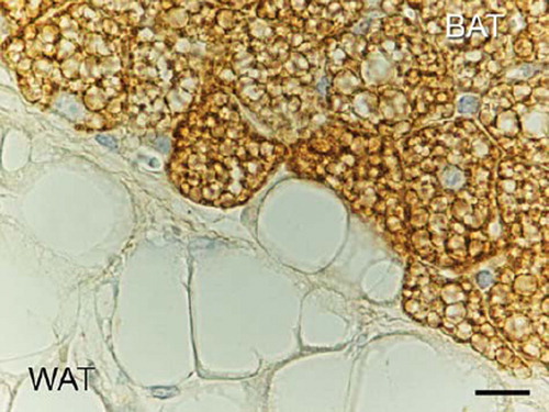
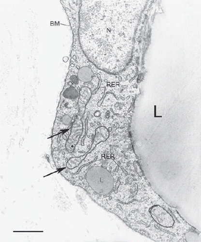
Figure 2. Rat adipose organ: epididymal depot. Electron microscopy of the peripheral part of a white adipocyte. Elongated mitochondria with randomly oriented cristae are visible (arrows). N = nucleus; RER = rough endoplasmic reticulum; large L = main lipid droplet; small L = peripheral lipid droplet; BM = basal membrane. Bar = 0.7 μm.
Evolutionary considerations on brown adipose tissue
Humans live in widely different environments at temperatures that range from 50°C above to 50°C below zero, but the human body requires a constant internal temperature of ∼37°C. This is achieved by internal mechanisms capable of dissipating heat when body temperature tends to rise, and of thermogenesis when it falls. In addition, environmental changes in temperature are perceived by skin thermoreceptors as disagreeable or adverse stimuli, inducing thermoregulatory behaviours aimed to adapt to the environment. The pilosebaceous system is an outstanding example of the evolution of human thermogenic apparatuses, which from great profusion in early man has progressively diminished to near disappearance in modern man. The change can be explained by the growing ability to find protection from cold that has characterized the evolution of the human species (Citation12). The reduction of the pilosebaceous apparatus has probably been accompanied by a progressive decline of other thermogenic mechanisms including brown adipose tissue (BAT), a major thermogenic mechanism in mammals (see below), (Citation13). However, it should be noted that as late as two–three centuries ago winter temperatures in Western households were considerably lower than at the present time; for instance, in the Viennese Imperial Palace of the Hapsburgs the average winter temperature in the mid-eighteenth century was 14–15°C, suggesting that the dramatic reduction in the need for thermogenesis in human evolution is more likely to coincide with the availability of reasonably priced food and the reduced need for physical exertion. Considering the energy balance that regulates body-weight, such reduced energy dispersion, due to diminished energy expenditure for heat generation, could thus be considered as one of the mechanisms favouring the current diabesity pandemic.
Brown adipose tissue: the thermogenic tissue
Triglycerides usually serve as an essential substrate for the fundamental function of brown adipocytes. The name adipocyte has been given to this cell type because it contains large amounts of lipids; however, this happened long before the discovery of its role, which is quite different from that of white adipocytes. In fact the purpose of these cells is to produce heat (Citation14,Citation15). Morphologically, brown adipocytes differ from white fat cells essentially in their multilocular cytoplasmic lipids and the numerous special mitochondria. These have a spherical or ovoid shape and are rich in laminar cristae which contain a protein, UCP1, subserving their thermogenic activity (Citation16,Citation17) (). The numerous mitochondria and rich vascularity of the tissue are the main reasons for the brown colour of BAT, setting it apart from WAT even on gross inspection. When BAT is activated (e.g. by cold), UCP1 reduces the electrochemical gradient between the external compartment and the mitochondrial matrix due to fatty acid beta oxidation, uncoupling oxidative phosphorylation. Therefore the sole final product of fatty acid combustion is heat dissipation. Given the large amount of the substrate, the numerous, cristae-rich mitochondria ( and ) burn considerable amounts of fat, resulting in intense thermogenesis. The other organelles of brown adipocytes have similar characteristics as those of white adipocytes, including a distinct basal membrane on the outer side of the plasma membrane. The spherical central nucleus of brown adipocytes is not usually squeezed by the lipid vacuoles in the cytoplasm. Mature brown adipocytes are approximately half the size of white adipocytes. Their nucleus and size therefore denote a lower degree of elasticity and expansibility compared with white fat cells. However, both cell types are characterized by a fairly variable morphology depending on functional status (Citation10,Citation18). Exposure to temperatures below thermoneutrality activates BAT directly, through adrenergic fibres that stimulate thermogenesis via neuro-adipose junctions (). Noradrenaline acts via beta3 receptors (Citation19). Beta3 agonists have similar pharmacological effects as cold exposure (Citation20). Thermogenically active cells are characterized by numerous, small lipid vacuoles and plentiful spherical mitochondria rich in laminar cristae containing abundant UCP1. The closer the temperature to thermoneutrality, the larger and less numerous the lipid vacuoles; at the same time mitochondria shrink and cristae numbers decrease in parallel with the decreased synthesis of UCP1 and its immunoreactivity (Citation10,Citation15). The morphology of the under-stimulated brown adipocyte is therefore more akin to that of the white adipocyte, and only in this morphological condition is the brown adipocyte immunoreactive for protein S-100 (Citation21). Leptin cannot be detected with immunohistochemical methods in the brown adipocytes of cold-exposed animals, exactly as UCP1 is undetectable in white adipocytes, but leptin is expressed in white-like brown adipocytes just as UCP1 is expressed in brown-like white adipocytes (see below) (Citation22–24). The development of brown adipocytes is quite similar to that of their white counterparts, the least differentiated brown precursors also being found in the same niches as white precursors: the capillary walls of the developing adipose tissue. It has recently been shown that interscapular BAT brown adipocytes arise from progenitors expressing myoblast markers and are not involved in white adipogenesis (Citation25,Citation26). Brown preadipocytes of classic interscapular BAT are characterized by a structural marker that is not found in white preadipocytes, i.e. large mitochondria whose morphology foreshadows that of mature cell mitochondria (pretypical mitochondria) (Citation27). Other characteristics are the presence of glycogen granules and the basal membrane. Therefore, these cells are not only clearly distinguishable morphologically from the other cell types in the adipose tissue, but they are also easily discerned from white preadipocytes based on their characteristic pretypical mitochondria.
Figure 3. Rat adipose organ: anterior subcutaneous depot, interscapular area. Electron microscopy of cytoplasm of a brown adipocyte. Several big mitochondria packed with cristae are visible. L = lipid droplet (some indicated). Bar = 1 μm.
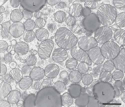
Figure 4. Mouse adipose organ: anterior subcutaneous depot, interscapular area. High-resolution scanning electron microscopy of a cytoplasm of brown adipocyte. 3D aspect of the classic mitochondria. Note the laminar cristae. Bar = 0.4 μm.
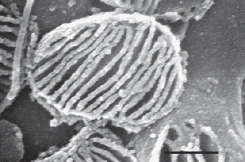
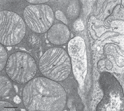
Figure 5. Rat adipose organ: anterior subcutaneous depot, interscapular area. Electron microscopy of a classic neuro-adipose junction. A synapse-like enlargement of a parenchymal nerve fibre is in tight contact with the plasma membrane of a brown adipocyte. V = synaptic vesicles; m = mitochondria (some indicated); N = nucleus of endothelial cell; CAP = capillary lumen. Bar = 0.4 μm.
The concept of adipose organ
In small mammals WAT and BAT are found in subcutaneous and trunk depots delimited by a connective capsule and characterized by discrete anatomical shapes (Citation28). Two main subcutaneous depots (anterior and posterior), a minor subcutaneous limb depot, and several trunk (visceral) depots can be dissected in mice and rats (murine adipose organ) (). The anterior depot encompasses the interscapular area, which is considered as the classic BAT depot. The visceral depots include the mediastinal depot in the chest, the omental, mesenteric, retroperitoneal and abdomino-pelvic depot (perirenal + periovarian + parametrial + perivesical) in females, and the epididymal depot in males. It should be noted that the trunk depots are also characterized by two distinct anatomical positions: peri-coelomic (pericardial, pleural, and peritoneal) and perivascular around the aorta and its main branches (carotid, subclavian, intercostal, and renal). The subcutaneous and trunk depots are both mixed, i.e. part WAT and part BAT. All depots are provided with specific vessels and nerves, although the white portions are less densely vascularized and innervated (Citation29,Citation30). The nerves are predominantly adrenergic (Citation31–34). In the murine adipose organ most of the anterior subcutaneous depot is made up of BAT, whereas the posterior subcutaneous depot is mostly white. Of the trunk depots the mediastinal is predominantly brown, especially in mice, whereas the omental and mesenteric are mostly white. The abdomino-pelvic depot is equally composed of BAT and WAT, whereas the epididymal depot is nearly completely white (Citation35,Citation36). The proportions of the two tissues in each depot depend largely on genetic background, age, gender, and environmental conditions (temperature, diet, exercise). Skin, lymph nodes, bone-marrow, parotid gland, parathyroid glands, thymus, and pancreas contain variable amounts of white adipocytes.
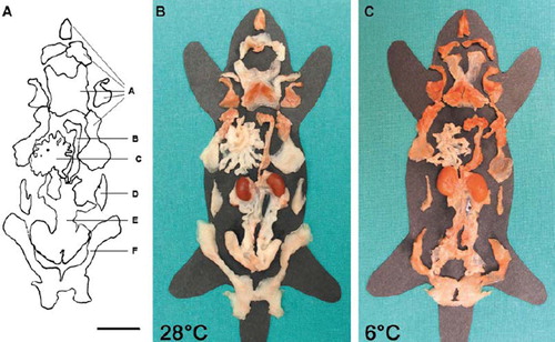
Figure 6. Gross anatomy of mouse adipose organ (adult female Sv129 mice). Middle: mouse kept at warm temperature (28°C for 10 days). Right: mouse kept at cold temperature (6°C for 10 days). Note the different colour of the organ, due to the increase of brown adipose tissue and decrease of white adipose tissue contained in the organ. The organ is made up of two subcutaneous depots (A and F): anterior (A) (composed of: deep cervical, superficial cervical, interscapular, subscapular, axillo-thoracic) and posterior (F) (composed of: dorso-lumbar, inguinal, gluteal); and of several visceral depots: mediastinal (B), mesenteric (C), retroperitoneal (D), and abdomino-pelvic (composed of: perirenal, periovarian, parametrial, perivesical) (E); reproduced from Murano et al., Adipocytes. 2005;1(2):121–30, with permission (Citation36). Bar = 1 cm.
The human adipose organ
A similar compartmentalization of adipose tissue into subcutaneous and trunk depots appears to be shared by humans, although their size and composition are considerably different. The subcutaneous depot is substantially continuous, with accumulations in the mammary and gluteofemoral areas in women. In adults BAT seems to be confined to the trunk (Citation37–40), in line with the different thermogenic requirements of the human organism due to its different surface/volume ratio compared with small mammals. Humans and rodents share an identical cell composition of the adipose organ, including different vessel and nerve density in WAT and BAT (Citation41). White and brown preadipocytes are also identical in structure and niche (Citation42,Citation43). In addition, genetic background, age, gender, and environmental conditions play a role in the relative proportions of the two tissues found in the different depots. An inverse correlation has been found between BAT and body mass index (Citation38,Citation39,Citation41), and human BAT activity—and, possibly, amount—is greater in individuals with pheochromocytoma (Citation44,Citation45) and in those with acute and chronic cold exposure (Citation46).
Brown adipose tissue negatively correlates with the development of obesity and diabetes
In 1979 Mike Stock demonstrated that food consumption is a functional stimulus for BAT, suggesting that it may have an anti-obesity action (Citation47). In 1993 Brad Lowell showed that mice lacking BAT become obese (Citation48) and, subsequently, that mice lacking beta-adrenergic receptors become rapidly and massively obese—albeit eating the same amount of food as control mice—and are more active (Citation24). In addition, the lack of UCP1 is sufficient by itself to induce a proneness to obesity in animals kept in thermoneutral conditions (Citation49). It has also been shown that knock-out of the insulin receptor from BAT leads to diabetic mice (Citation50) and that mice displaying greater resistance to obesity and diabetes have greater amounts of brown adipocytes (Citation51,Citation52) or UCP1 (Citation53) in the adipose organ.
The adipose organ is plastic: physiological reversible transdifferentiation
The presence of both BAT and WAT in the various depots of the adipose organ suggested to us that the two tissues might be capable of turning into one another, i.e. that in conditions of chronic cold exposure the amount of BAT in the organ could increase via WAT-to-BAT transdifferentiation and, vice versa, that BAT could turn into WAT in case of exposure to an obesogenic diet, to enable greater energy accumulation. In our opinion the term transdifferentiation should be used to describe direct transformation of a differentiated cell into another, mature cell with different morphological and functional characteristics in physiological conditions. Other authors include an additional step, i.e. de-differentiation (Citation54). Indeed, it is well documented that the relative amount of BAT increases in cold-exposed animals () and in those treated with beta3 agonists and that WAT increases in obese animals. Such changes involve simultaneous reductions in WAT and BAT, respectively (Citation55,Citation56). Quantitative studies have shown that the BAT increase seen in cold-exposed animals corresponds to a reduction in WAT that is unrelated to apoptosis (Citation36). In addition, in animals treated with beta3 agonists 80%–95% of newly formed brown adipocytes do not display proliferation markers and present all the morphological stages of white-to-brown transition (Citation57,Citation58).
Such direct WAT-to-BAT conversion is especially interesting for two main reasons:
It could address the problem of the pharmacological therapy of obesity and type 2 diabetes (Citation59,Citation60);
It would be a new basic physiological cell property with a broad scope for biomedical application.
The adipose organ also offers another example of physiological and reversible transdifferentiation. The female mammary gland is made up of subcutaneous adipose tissue containing white and brown adipocytes, as described above, and is coursed by branched ducts originating in the nipple. About 90% of the gland is constituted of adipose cells. During pregnancy the number of adipocytes diminishes, mainly due to the development of the lobuloalveolar component, i.e. the part of the gland that provides for milk synthesis and secretion. During lactation the majority of adipocytes thus disappear and the vast majority of the organ is occupied by a well-developed milk-producing lobuloalveolar component. After lactation the organ gradually recovers its initial anatomy, with reappearance of adipocytes and disappearance of the lobuloalveolar component. Data from our laboratory, based mainly on cell lineage investigations and on the study of explants of labelled adipose tissue, support the notion that during pregnancy and lactation adipocytes turn into milk-secreting lobuloalveolar epithelial cells and that in the post-lactation period the epithelial cells of the milk-secreting component turn into adipocytes. These data suggest a new instance of physiological and reversible transdifferentiation in the adipose organ (Citation61,Citation62).
The mechanism underlying the cold-induced transformation of WAT into BAT seems to be closely related to the activity of the sympathetic nervous system. In fact, as described above, brown adipocytes are closely regulated by the activity of parenchymal noradrenergic fibres that are in direct contact with them (). The number of such fibres in the adipose organ is proportional to the overall amount of brown adipocytes found in its various depots. In cold-exposed animals the density of parenchymal fibres increases with the number of brown adipocytes in the different depots (Citation35). This suggests that the sympathetic nervous system could play a role in the phenotypic differentiation of adipocyte precursors; adipocytes developing in a highly innervated area (such as the interscapular area of the adipose organ) could thus acquire the brown adipocyte phenotype, whereas those developing in poorly innervated areas of the adipose organ, e.g. epididymal fat, could assume the phenotype of white adipocytes. It has recently been shown that brown adipocyte precursors arising in ‘classic’ interscapular BAT are marked by the expression of myf5, which is also expressed by skeletal muscle precursors. Brown adipocytes arising in WAT following an adrenergic stimulus did not express myf5 in lineage studies, suggesting a different lineage origin. This agrees with the possibility that brown adipocytes arising in WAT as a result of adrenergic stimulation derive directly from transdifferentiation of white adipocytes lacking myf5 expression during development. However, some researchers support the hypothesis that they derive from myf-negative brown precursors present in WAT (Citation63,Citation64). In the opinion of some authors the multilocular cells expressing UCP1—the BAT marker gene and protein—arising in the depots traditionally viewed as ‘white’ cannot be considered as ‘classic’ brown adipocytes; accordingly, terms such as brite (brown to white) (Citation64) and beige (the intermediate colour between white and brown) (Citation65) have been coined for them, essentially to stress their different molecular and embryological lineage features compared with the cells found in the interscapular depot, which is traditionally considered as typical BAT. We believe that such differences could reflect the presence in the tissue of cells in intermediate transdifferentiation stages and that these very elements can be responsible for the molecular differences described (Citation63). It should also be stressed that, like all the other depots, the anterior subcutaneous depot containing interscapular BAT (a ‘classic’ brown adipocyte depot) is mixed, i.e. part WAT and part BAT. In the white part of this depot cold exposure induces the emergence of new UCP1-positive multilocular adipocytes having the morphological characteristics of those arising in other subcutaneous or visceral depots, including all the intermediate stages of white-to-brown transformation. These new brown adipocytes belong anatomically to the anterior subcutaneous fat depot that includes the interscapular area viewed as the classic site of brown adipocytes. We wonder if they should therefore be considered as classic brown adipocytes, even if they display all the morphological and immunohistochemical features of the new brown (or brite, or beige) adipocytes that emerge in WAT of other depots (). We agree that brown adipocytes developing as such should be distinguished from those deriving from transdifferentiation, but believe that all UCP1-positive multilocular adipocytes, which therefore are potentially endowed with thermogenic activity, should be considered as brown adipocytes. Our data suggest that in most of the organ the development of new brown adipocytes from preadipocytes appears to be a minor phenomenon (Citation63); however, a more important role in some depots (such as the interscapular area) cannot be excluded. Furthermore, even though the origin of interscapular brown adipocytes is different from that of brite adipocytes, it should be emphasized that the transdifferentiation properties of adipocytes do not seem to be confined to those found in predominantly white depots. Indeed the interscapular brown adipocytes of obese mice are transformed into unilocular white-like adipocytes immunoreactive for leptin (one of the major genes expressed in white adipocytes) (Citation22). Of note, classic multilocular brown adipocytes are not immunoreactive for leptin (Citation22). Furthermore the interscapular fat of mice lacking beta adrenoceptors is formed by leptin-immunoreactive unilocular adipocytes (Citation24). These data suggest that interscapular classic brown adipocytes can turn into white-like adipocytes capable of producing leptin, which is not produced by classic brown adipocytes. Therefore adipocyte transdifferentiation is not limited to the transformation of WAT into BAT in white depots but can also be extended to the reverse phenomenon, even in interscapular BAT. On the other hand it should not be forgotten that interscapular BAT is only a part of the anterior subcutaneous depot, a mixed depot like all adipose organ depots, at least in some murine strains (Citation35,Citation36). However, some researchers tend to separate white from brown adipose cells due essentially to different embryonic origins of the two cell lines (Citation25,Citation26,Citation66).
Figure 7. Mouse adipose organ: anterior subcutaneous depot, interscapular area. Immunofluorescence multiple labelling showing parenchymal noradrenergic nerve fibres (tyrosine hydroxylase immunoreactive: red) in tight association with UCP1 immunoreactive brown adipocytes (green) with nuclei in blue (TOTO nuclear counterstaining). Bar = 6 μm.
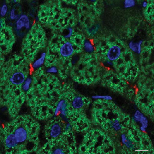
Figure 8. Mouse adipose organ (adult Sv129 female): anterior subcutaneous depot at the periphery of the interscapular area (i.e. white adipose tissue surrounding the classic interscapular brown adipose tissue). UCP1 immunohistochemistry as in . Three days of cold exposure (6°C). Newly formed brown adipocytes appear among white adipocytes. In this depot some newly formed brown adipocytes appear to differentiate from preadipocytes forming small multilocular UCP1 immunoreactive cells (arrows). Some appear to derive from direct transformation of white adipocytes with intermediate stages: UCP1-negative paucilocular cells (asterisk) and UCP1-positive paucilocular cells (double asterisk). Bar = 16 μm.
The obese adipose organ and type 2 diabetes
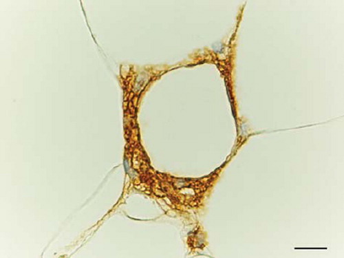
In 2003 two American teams independently documented macrophage infiltration of the human and the murine adipose organ. The phenomenon induces a low-grade chronic inflammation that is associated with insulin resistance, which generally leads to type 2 diabetes. Such reaction appears to be related mainly to macrophage-produced cytokines (e.g. TNF-alpha and IL-6) capable of interfering with the normal activity of insulin receptor (Citation67,Citation68). The cause of macrophage infiltration has remained unknown until we discovered that it was part of a process triggered by adipocyte death. We found that more than 90% of macrophages immunoreactive for MAC2 (activated macrophages) surround adipocyte debris mainly represented by a large lipid droplet (). We designated this characteristic picture as a crown-like structure (CLS) (Citation69). Cell death induces the recruitment of macrophages, whose physiological function is to remove cell debris. Since adipocytes are fairly larger than macrophages, these cells, as in foreign body reaction, give rise to syncytial multinucleated giant cells that are more appropriate for the removal of giant debris (the lipid droplet). Although CLSs are also detected in the WAT of lean animals, they are 30 times more frequent in the WAT of obese animals and directly correlate to adipocyte size but not to the obese condition per se. Accordingly, the WAT of lean animals having hypertrophic adipocytes due to the absence of hormone-sensitive lipase (HSL) has the same CLS density as the WAT of obese animals (Citation69), and mice or humans with hyperplastic obesity do not show an increased number of CLSs (Citation69) (and our unpublished observations). It has recently been shown that human adipocytes are also subject to turn-over (Citation70,Citation71), and CLS frequency is greater in individuals with hypertrophic adipocytes (Citation69). Obese humans with a hypertrophic adipose tissue (i.e. few, large adipocytes) suffer from a greater amount of metabolic complications than obese humans with a hyperplastic adipose tissue (numerous small adipocytes) (Citation72).
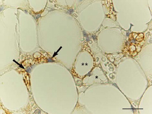
Figure 9. Obese mouse (db/db) adipose organ: omentum. MAC2/Galectin 3 immunoreactive macrophages surround a dead adipocyte forming a classic crown-like structure (CLS). Bar = 15 μm.
The earliest clinical studies of obesity already found that accumulation of visceral fat was associated with greater morbidity (i.e. type 2 diabetes, hypertension, and cardiovascular disease) than accumulation of subcutaneous fat (Citation73). However, the reason for this difference is still unknown. The direct correlation between adipocyte size and CLS density applies both to the subcutaneous and to the visceral fat of obese animals, and the hypertrophic adipocytes of subcutaneous fat are larger than those of visceral fat in genetically obese mice (Citation74). CLSs should therefore be more numerous in the former depots. On the contrary, they are much more frequent in visceral depots (Citation74). A high-fat diet is associated with greater CLS infiltration in visceral than subcutaneous fat in mice and coincides with the development of insulin resistance (Citation75). Altogether, these data suggest that CLSs form as a result of adipocyte death, possibly due to achievement of the maximum cell size, or critical death size (CDS), and that a different CDS applies to visceral and subcutaneous adipocytes. In addition, the greater amount of macrophages and macrophage-secreted cytokines found in visceral fat is in line with the greater morbidity associated with these depots (Citation75,Citation76). It is also well established that despite the strong phenotypic similarities of the two best known and most extensively explored murine models of genetic obesity, ob/ob and db/db, the latter model is associated with a greater tendency to develop type 2 diabetes. This is in line with the fact that the visceral fat of db/db animals is 3.4 times richer in CLSs than ob/ob fat (Citation74) and, notably, that db/db is the genetic obesity model displaying the more marked adipocyte hypertrophy (Citation77).
The reason for the lower degree of expansibility of visceral adipocytes remains unknown, but some data seem to connect this aspect to the plasticity of the adipose organ described above. Visceral WAT differs anatomically from subcutaneous WAT: it has smaller adipocytes and a larger supply of nerves and vessels; visceral fat therefore has many of the characteristics of BAT-turned-WAT (Citation22). We have hypothesized that the white adipocytes derived from brown adipocytes may be less expansible, and have a smaller CDS, than originally white adipocytes. In relation to this it is interesting to note that:
Epicardial (visceral) fat belongs to the mediastinal depot, which in young lean mice consists entirely of brown adipocytes (Citation35).
In obese mice the mediastinal depot is rich in CLSs (our unpublished observations made in collaboration with B. Walker et al.)
UCP1 protein and mRNA have been demonstrated in the human epicardium (Citation78).
However, this construct fails to explain both why ‘pure’ white visceral depots, like the epididymal, are as prone to adipocyte death as other visceral depots (Citation75) and the role of brown adipocytes in subcutaneous fat. In addition, macrophage infiltration of the obese adipose organ may not be the sole mechanism involved in the arising of insulin resistance and type 2 diabetes, since in surgically treated obese individuals the diabetes often resolves prior to the weight loss (Citation79,Citation80), and other mechanisms, like those related to the gut bacterial population (Citation81,Citation82), may also be involved. It should also be noted that the major human depots (omental and mesenteric) contain large amounts of lymphatic tissue having different characteristics from other areas of lymphatic tissue (Citation83). According to some researchers these lymphatic tissues have specific immunological properties (Citation84). It cannot therefore be ruled out that the adipocytes found in such depots, which are particularly prone to hypertrophy-related death, may have different properties compared with the adipocytes of other depots due to a collaborative role with lymphatic tissue (Citation85). Therefore, if the function of omental and mesenteric fat were also immunological, it might be a sort of ectopic depot for excess lipids, possibly predating the ectopic depots in the liver and muscle fibres, respectively, in line with the expandability theory of Vidal-Puig and co-workers (Citation86). Liver steatosis correlates closely and directly with visceral fat accumulation (Citation87); the CDSs of hepatocytes might also play a role here and even constitute the trigger for cell death and the activation of inflammatory mechanisms.
The mechanisms leading to adipocyte death are still unclear but appear to be connected with hypertrophy, which might induce cell distress, possibly due to a relative hypoxic condition given that WAT perfusion does not increase in obese individuals (Citation88). Notably, cell death with CLS formation also takes place in normal WAT, and normal tissues are unlikely to possess death mechanisms other than apoptosis. However, the hypertrophy characterizing the obese WAT may be pathological, involving an acceleration of apoptosis.
In conclusion, the mammalian adipose organ is a complex structure endowed with special properties, like physiological reversible transdifferentiation. In obese individuals it is affected by low-grade chronic inflammation, which results in a distinctive histopathological appearance (CLSs). The inflammation seems to be related to the clinical complications of obesity, such as type 2 diabetes. Such conditions could benefit from interventions targeting the plastic properties of the adipose organ. The recent discovery of molecules regulating differentiation such as PRDM16 and BMP7 (Citation89,Citation90) and transdifferentiation such as Rb and COX2 (Citation91–93), plus the fact that the human adipose organ shares several similarities with the murine organ, encourages the hope that further advances will be achieved in the near future.
Declaration of interest: The research leading to some of these findings has received funding from the European Community's 7th Framework Programme (FP7/2007-2013) under grant agreement no. 201608. The author declares no other conflicts of interest.
References
- Friedman JM. Leptin at 14 y of age: an ongoing story. Am J Clin Nutr. 2009;89:973S–9S.
- Matsuzawa Y. Adiponectin: a key player in obesity related disorders. Curr Pharm Des. 2010;16:1896–901.
- Maffei M, Halaas J, Ravussin E, Pratley RE, Lee GH, Zhang Y, . Leptin levels in human and rodent: measurement of plasma leptin and ob RNA in obese and weight-reduced subjects. Nat Med. 1995;1:1155–61.
- Greenberg AS, Egan JJ, Wek SA, Garty NB, Blanchette-Mackie EJ, Londos C. Perilipin, a major hormonally regulated adipocyte-specific phosphoprotein associated with the periphery of lipid storage droplets. J Biol Chem. 1991; 266:11341–6.
- Cinti S, Cigolini M, Bosello O, Bjorntorp P. A morphological study of the adipocyte precursor. J Submicrosc Cytol. 1984;16:243–51.
- Cinti S, Cigolini M, Gazzanelli G, Bosello O. An ultrastructural study of adipocyte precursors from epididymal fat pads of adult rats in culture. J Submicrosc Cytol. 1985;17:631–6.
- Tang W, Zeve D, Suh JM, Bosnakovski D, Kyba M, Hammer RE, . White fat progenitor cells reside in the adipose vasculature. Science. 2008;322:583–6.
- Rodeheffer MS, Birsoy K, Friedman JM. Identification of white adipocyte progenitor cells in vivo. Cell. 2008;135:240–9.
- Cinti S, Cigolini M, Morroni M, Zingaretti MC. S-100 protein in white preadipocytes: an immunoelectronmicroscopic study. Anat Rec. 1989;224:466–72.
- Cinti S. The adipose organ. Milan: Kurtis; 1999.
- Delmonico MJ, Harris TB, Visser M, Park SW, Conroy MB, Velasquez-Mieyer P, . Longitudinal study of muscle strength, quality, and adipose tissue infiltration. Am J Clin Nutr. 2009;90:1579–85.
- Kushlan JA. The evolution of hairlessness in man. The American Naturalist. 1980;116:727–9.
- Lean J, James P. Brown adipose tissue in man. Trayhurn P, Nicholls DG, Brown adipose tissue. Edward Arnold; London. 1986. 339–65.
- Ricquier D. Molecular biology of brown adipose tissue. Proc Nutr Soc. 1989;48:183–7.
- Cannon B, Nedergaard J. Brown adipose tissue: function and physiological significance. Physiol Rev. 2004;84:277–359.
- Cinti S, Zancanaro C, Sbarbati A, Cicolini M, Vogel P, Ricquier D, . Immunoelectron microscopical identification of the uncoupling protein in brown adipose tissue mitochondria. Biol Cell. 1989;67:359–62.
- Rial E, Gonzalez-Barroso MM. Physiological regulation of the transport activity in the uncoupling proteins UCP1 and UCP2. Biochim Biophys Acta. 2001;1504:70–81.
- Casteilla L, Muzzin P, Revelli JP, Ricquier D, Giacobino JP. Expression of beta 1- and beta 3-adrenergic-receptor messages and adenylate cyclase beta-adrenergic response in bovine perirenal adipose tissue during its transformation from brown into white fat. Biochem J. 1994;297 (Pt 1):93–7.
- Collins S, Surwit RS. The beta-adrenergic receptors and the control of adipose tissue metabolism and thermogenesis. Recent Prog Horm Res. 2001;56:309–28.
- Cinti S, Cancello R, Zingaretti MC, Ceresi E, De Matteis R, Giordano A, . CL316,243 and cold stress induce heterogeneous expression of UCP1 mRNA and protein in rodent brown adipocytes. J Histochem Cytochem. 2002; 50:21–31.
- Barbatelli G, Morroni M, Vinesi P, Cinti S, Michetti F. S-100 protein in rat brown adipose tissue under different functional conditions: a morphological, immunocytochemical, and immunochemical study. Exp Cell Res. 1993;208:226–31.
- Cinti S, Frederich RC, Zingaretti MC, De Matteis R, Flier JS, Lowell BB. Immunohistochemical localization of leptin and uncoupling protein in white and brown adipose tissue. Endocrinology. 1997;138:797–804.
- Cancello R, Zingaretti MC, Sarzani R, Ricquier D, Cinti S. Leptin and UCP1 genes are reciprocally regulated in brown adipose tissue. Endocrinology. 1998;139:4747–50.
- Bachman ES, Dhillon H, Zhang CY, Cinti S, Bianco AC, Kobilka BK, . betaAR signaling required for diet-induced thermogenesis and obesity resistance. Science. 2002;297: 843–5.
- Timmons JA, Wennmalm K, Larsson O, Walden TB, Lassmann T, Petrovic N, . Myogenic gene expression signature establishes that brown and white adipocytes originate from distinct cell lineages. Proc Natl Acad Sci U S A. 2007; 104:4401–6.
- Seale P, Bjork B, Yang W, Kajimura S, Chin S, Kuang S, . PRDM16 controls a brown fat/skeletal muscle switch. Nature. 2008;454:961–7.
- Cinti S, Morroni M. Brown adipocyte precursor cells: a morphological study. Ital J Anat Embryol. 1995;100 Suppl 1:75–81.
- Cinti S. The adipose organ. Prostaglandins Leukot Essent Fatty Acids. 2005;73:9–15.
- Giordano A, Frontini A, Cinti S. Adipose organ nerves revealed by immunohistochemistry. Methods Mol Biol. 2008;456:83–95.
- Giordano A, Frontini A, Murano I, Tonello C, Marino MA, Carruba MO, . Regional-dependent increase of sympathetic innervation in rat white adipose tissue during prolonged fasting. J Histochem Cytochem. 2005;53:679–87.
- Bartness TJ. Dual innervation of white adipose tissue: some evidence for parasympathetic nervous system involvement. J Clin Invest. 2002;110:1235–7.
- Bartness TJ, Bamshad M. Innervation of mammalian white adipose tissue: implications for the regulation of total body fat. Am J Physiol. 1998;275(5 Pt 2):R1399–411.
- Bartness TJ, Song CK. Thematic review series: adipocyte biology. Sympathetic and sensory innervation of white adipose tissue. J Lipid Res. 2007;48:1655–72.
- Demas GE, Bartness TJ. Direct innervation of white fat and adrenal medullary catecholamines mediate photoperiodic changes in body fat. Am J Physiol Regul Integr Comp Physiol. 2001;281:R1499–505.
- Murano I, Barbatelli G, Giordano A, Cinti S. Noradrenergic parenchymal nerve fiber branching after cold acclimatisation correlates with brown adipocyte density in mouse adipose organ. J Anat. 2009;214:171–8.
- Murano I, Zingaretti CM, Cinti S. The adipose organ of Sv129 mice contains a prevalence of brown adipocytes and shows plasticity after cold exposure. Adipocytes. 2005;1: 121–30.
- Nedergaard J, Bengtsson T, Cannon B. Unexpected evidence for active brown adipose tissue in adult humans. Am J Physiol Endocrinol Metab. 2007;293:E444–52.
- Cypess AM, Lehman S, Williams G, Tal I, Rodman D, Goldfine AB, . Identification and importance of brown adipose tissue in adult humans. N Engl J Med. 2009;360: 1509–17.
- van Marken Lichtenbelt WD, Vanhommerig JW, Smulders NM, Drossaerts JM, Kemerink GJ, Bouvy ND, . Cold-activated brown adipose tissue in healthy men. N Engl J Med. 2009;360:1500–8.
- Virtanen KA, Lidell ME, Orava J, Heglind M, Westergren R, Niemi T, . Functional brown adipose tissue in healthy adults. N Engl J Med. 2009;360:1518–25.
- Zingaretti MC, Crosta F, Vitali A, Guerrieri M, Frontini A, Cannon B, . The presence of UCP1 demonstrates that metabolically active adipose tissue in the neck of adult humans truly represents brown adipose tissue. FASEB J. 2009;23:3113–20.
- Manieri M, Murano I, Fianchini A, Brunelli A, Cinti S. Morphological and immunohistochemical features of brown adipocytes and preadipocytes in a case of human hibernoma. Nutr Metab Cardiovasc Dis. 2010;20:567–74.
- Boiani R, Cinti S, Savage DB, Vidal-Puig A, O'Rahilly S. Abdominal subcutaneous adipose tissue morphology in a patient with a dominant-negative mutation (P467L) in the nuclear receptor peroxisome proliferator-activated receptor-gamma (PPARG) gene. Nutr Metab Cardiovasc Dis. 2010;20:e11–2.
- Kuji I, Imabayashi E, Minagawa A, Matsuda H, Miyauchi T. Brown adipose tissue demonstrating intense FDG uptake in a patient with mediastinal pheochromocytoma. Ann Nucl Med. 2008;22:231–5.
- Ricquier D, Mory G, Nechad M, Combes-George M, Thibault J. Development and activation of brown fat in rats with pheochromocytoma PC 12 tumors. Am J Physiol. 1983;245:C172–7.
- Saito M, Okamatsu-Ogura Y, Matsushita M, Watanabe K, Yoneshiro T, Nio-Kobayashi J, . High incidence of metabolically active brown adipose tissue in healthy adult humans: effects of cold exposure and adiposity. Diabetes. 2009;58:1526–31.
- Rothwell NJ, Stock MJ. A role for brown adipose tissue in diet-induced thermogenesis. Nature. 1979;281:31–5.
- Lowell BB, S-Susulic V, Hamann A, Lawitts JA, Himms-Hagen J, Boyer BB, . Development of obesity in transgenic mice after genetic ablation of brown adipose tissue. Nature. 1993;366:740–2.
- Feldmann HM, Golozoubova V, Cannon B, Nedergaard J. UCP1 ablation induces obesity and abolishes diet-induced thermogenesis in mice exempt from thermal stress by living at thermoneutrality. Cell Metab. 2009;9:203–9.
- Guerra C, Navarro P, Valverde AM, Arribas M, Bruning J, Kozak LP, . Brown adipose tissue-specific insulin receptor knockout shows diabetic phenotype without insulin resistance. J Clin Invest. 2001;108:1205–13.
- Guerra C, Koza RA, Yamashita H, Walsh K, Kozak LP. Emergence of brown adipocytes in white fat in mice is under genetic control. Effects on body weight and adiposity. J Clin Invest. 1998;102:412–20.
- Almind K, Manieri M, Sivitz WI, Cinti S, Kahn CR. Ectopic brown adipose tissue in muscle provides a mechanism for differences in risk of metabolic syndrome in mice. Proc Natl Acad Sci U S A. 2007;104:2366–71.
- Kopecky J, Clarke G, Enerback S, Spiegelman B, Kozak LP. Expression of the mitochondrial uncoupling protein gene from the aP2 gene promoter prevents genetic obesity. J Clin Invest. 1995;96:2914–23.
- Tosh D, Slack JM. How cells change their phenotype. Nat Rev Mol Cell Biol. 2002;3:187–94.
- Cinti S. The adipose organ: morphological perspectives of adipose tissues. Proc Nutr Soc. 2001;60:319–28.
- Cinti S. Adipocyte differentiation and transdifferentiation: plasticity of the adipose organ. J Endocrinol Invest. 2002;25:823–35.
- Granneman JG, Li P, Zhu Z, Lu Y. Metabolic and cellular plasticity in white adipose tissue I: effects of beta3-adrenergic receptor activation. Am J Physiol Endocrinol Metab. 2005;289:E608–16.
- Himms-Hagen J, Melnyk A, Zingaretti MC, Ceresi E, Barbatelli G, Cinti S. Multilocular fat cells in WAT of CL-316243-treated rats derive directly from white adipocytes. Am J Physiol Cell Physiol. 2000;279:C670–81.
- Cypess AM, Kahn CR. Brown fat as a therapy for obesity and diabetes. Curr Opin Endocrinol Diabetes Obes. 2010;17:143–9.
- Enerback S. Human brown adipose tissue. Cell Metab. 2010;11:248–52.
- Morroni M, Giordano A, Zingaretti MC, Boiani R, De Matteis R, Kahn BB, . Reversible transdifferentiation of secretory epithelial cells into adipocytes in the mammary gland. Proc Natl Acad Sci U S A. 2004;101:16801–6.
- De Matteis R, Zingaretti MC, Murano I, Vitali A, Frontini A, Giannulis I, . In vivo physiological transdifferentiation of adult adipose cells. Stem Cells. 2009;27:2761–8.
- Barbatelli G, Murano I, Madsen L, Hao Q, Jimenez M, Kristiansen K, . The emergence of cold-induced brown adipocytes in mouse white fat depots is determined predominantly by white to brown adipocyte transdifferentiation. Am J Physiol Endocrinol Metab. 2010;298: E1244–53.
- Petrovic N, Walden TB, Shabalina IG, Timmons JA, Cannon B, Nedergaard J. Chronic peroxisome proliferator-activated receptor gamma (PPARgamma) activation of epididymally derived white adipocyte cultures reveals a population of thermogenically competent, UCP1-containing adipocytes molecularly distinct from classic brown adipocytes. J Biol Chem. 2009;285:7153–64.
- Seale P, Kajimura S, Spiegelman BM. Transcriptional control of brown adipocyte development and physiological function—of mice and men. Genes Dev. 2009;23:788–97.
- Atit R, Sgaier SK, Mohamed OA, Taketo MM, Dufort D, Joyner AL, . Beta-catenin activation is necessary and sufficient to specify the dorsal dermal fate in the mouse. Dev Biol. 2006;296:164–76.
- Weisberg SP, McCann D, Desai M, Rosenbaum M, Leibel RL, Ferrante AW Jr. Obesity is associated with macrophage accumulation in adipose tissue. J Clin Invest. 2003;112: 1796–808.
- Xu H, Barnes GT, Yang Q, Tan G, Yang D, Chou CJ, . Chronic inflammation in fat plays a crucial role in the development of obesity-related insulin resistance. J Clin Invest. 2003;112:1821–30.
- Cinti S, Mitchell G, Barbatelli G, Murano I, Ceresi E, Faloia E, . Adipocyte death defines macrophage localization and function in adipose tissue of obese mice and humans. J Lipid Res. 2005;46:2347–55.
- Spalding KL, Arner E, Westermark PO, Bernard S, Buchholz BA, Bergmann O, . Dynamics of fat cell turnover in humans. Nature. 2008;453:783–7.
- Arner P, Spalding KL. Fat cell turnover in humans. Biochem Biophys Res Commun. 2010;396:101–4.
- Hoffstedt J, Arner E, Wahrenberg H, Andersson DP, Qvisth V, Lofgren P, . Regional impact of adipose tissue morphology on the metabolic profile in morbid obesity. Diabetologia. 2010;53(12):2496–503.
- Bjorntorp P. Metabolic abnormalities in visceral obesity. Ann Med. 1992;24:3–5.
- Murano I, Barbatelli G, Parisani V, Latini C, Muzzonigro G, Castellucci M, . Dead adipocytes, detected as crown-like structures, are prevalent in visceral fat depots of genetically obese mice. J Lipid Res. 2008;49:1562–8.
- Strissel KJ, Stancheva Z, Miyoshi H, Perfield JW 2nd, Defuria J, Jick Z, . Adipocyte death, adipose tissue remodeling and obesity complications. Diabetes. 2007;56: 2910–8.
- Cancello R, Tordjman J, Poitou C, Guilhem G, Bouillot JL, Hugol D, . Increased infiltration of macrophages in omental adipose tissue is associated with marked hepatic lesions in morbid human obesity. Diabetes. 2006;55: 1554–61.
- Johnson PR, Hirsch J. Cellularity of adipose depots in six strains of genetically obese mice. J Lipid Res. 1972;13: 2–11.
- Sacks HS, Fain JN, Holman B, Cheema P, Chary A, Parks F, . Uncoupling protein-1 and related messenger ribonucleic acids in human epicardial and other adipose tissues: epicardial fat functioning as brown fat. J Clin Endocrinol Metab. 2009;94:3611–5.
- Rubino F, Marescaux J. Effect of duodenal-jejunal exclusion in a non-obese animal model of type 2 diabetes: a new perspective for an old disease. Ann Surg. 2004;239:1–11.
- Rubino F, R'Bibo SL, del Genio F, Mazumdar M, McGraw TE. Metabolic surgery: the role of the gastrointestinal tract in diabetes mellitus. Nat Rev Endocrinol. 2010;6:102–9.
- Turnbaugh PJ, Ley RE, Mahowald MA, Magrini V, Mardis ER, Gordon JI. An obesity-associated gut microbiome with increased capacity for energy harvest. Nature. 2006;444: 1027–31.
- Cani PD, Possemiers S, Van de Wiele T, Guiot Y, Everard A, Rottier O, . Changes in gut microbiota control inflammation in obese mice through a mechanism involving GLP-2-driven improvement of gut permeability. Gut. 2009; 58:1091–103.
- Moro K, Yamada T, Tanabe M, Takeuchi T, Ikawa T, Kawamoto H, . Innate production of T(H)2 cytokines by adipose tissue-associated c-Kit(+)Sca-1(+) lymphoid cells. Nature. 2010;463:540–4.
- Rangel-Moreno J, Moyron-Quiroz JE, Carragher DM, Kusser K, Hartson L, Moquin A, . Omental milky spots develop in the absence of lymphoid tissue-inducer cells and support B and T cell responses to peritoneal antigens. Immunity. 2009;30:731–43.
- Shimotsuma M, Shields JW, Simpson-Morgan MW, Sakuyama A, Shirasu M, Hagiwara A, . Morpho-physiological function and role of omental milky spots as omentum-associated lymphoid tissue (OALT) in the peritoneal cavity. Lymphology. 1993;26:90–101.
- Virtue S, Vidal-Puig A. Adipose tissue expandability, lipotoxicity and the metabolic syndrome—an allostatic perspective. Biochim Biophys Acta. 2010;1801:338–49.
- Marchesini G, Marzocchi R, Agostini F, Bugianesi E. Nonalcoholic fatty liver disease and the metabolic syndrome. Curr Opin Lipidol. 2005;16:421–7.
- Trayhurn P, Wang B, Wood IS. Hypoxia in adipose tissue: a basis for the dysregulation of tissue function in obesity? Br J Nutr. 2008;100:227–35.
- Seale P, Kajimura S, Yang W, Chin S, Rohas LM, Uldry M, . Transcriptional control of brown fat determination by PRDM16. Cell Metab. 2007;6:38–54.
- Tseng YH, Kokkotou E, Schulz TJ, Huang TL, Winnay JN, Taniguchi CM, . New role of bone morphogenetic protein 7 in brown adipogenesis and energy expenditure. Nature. 2008;454:1000–4.
- Mercader J, Ribot J, Murano I, Feddersen S, Cinti S, Madsen L, . Haploinsufficiency of the retinoblastoma protein gene reduces diet-induced obesity, insulin resistance, and hepatosteatosis in mice. Am J Physiol Endocrinol Metab. 2009;297:E184–93.
- Madsen L, Pedersen LM, Lillefosse HH, Fjaere E, Bronstad I, Hao Q, . UCP1 induction during recruitment of brown adipocytes in white adipose tissue is dependent on cyclooxygenase activity. PLoS One. 2010;5:e11391.
- Vegiopoulos A, Muller-Decker K, Strzoda D, Schmitt I, Chichelnitskiy E, Ostertag A, . Cyclooxygenase-2 controls energy homeostasis in mice by de novo recruitment of brown adipocytes. Science. 2010;328:1158–61.