Abstract
Both supraventricular and ventricular rhythm disorders are frequently observed in patients with isolated left ventricular noncompaction (IVNC). Most importantly, these patients are prone to develop life-threatening ventricular arrhythmias, which are amongst their most frequent causes of death. Data regarding risk stratification of ventricular arrhythmias, however, are scarce due to the rareness of the disease. Indeed, even invasive electrophysiological studies may be of limited value in this regard in the majority of patients. Implantable cardioverter defibrillators (ICDs) have been demonstrated to be highly effective for the prevention of sudden arrhythmic death in IVNC and should be considered in patients who are clinically judged to be at high risk for ventricular tachyarrhythmias. These include patients with a severely reduced ejection fraction as well as those with a prior history of sustained ventricular tachycardia or fibrillation, recurrent syncope of unknown etiology, or a family history of ventricular tachyarrhythmias or sudden cardiac death. This review summarizes the electrocardiographic and electrophysiological findings in patients with IVNC and discusses possibilities for risk stratification as well as the rationale for ICD implantation for the prevention of sudden cardiac death.
Key messages
Patients with noncompaction cardiomyopathy have a high propensity to develop malignant ventricular arrhythmias.
Implantable cardioverter defibrillators have been demonstrated to be highly effective for the prevention of sudden arrhythmic death in noncompaction cardiomyopathy.
Arrhythmic risk stratification in patients with noncompaction cardiomyopathy remains a challenge.
Introduction
Isolated left ventricular noncompaction cardiomyopathy (IVNC) is a primary congenital cardiomyopathy (Citation1), with a characteristic morphology consisting of a two-layered myocardial structure with a thin epicardial layer and a non-compacted, severely thickened endocardial layer () (Citation2,Citation3). IVNC is a rare disorder with a prevalence of 0.014% in a large series of patients referred to a tertiary care echocardiography laboratory (Citation4). Recent guidelines from the Heart Failure Society of America have recommended clinical screening, family history, and genetic testing in the management of patients with IVNC (Citation5). As a result, an increasing number of individuals identified during family screening as well as a growing awareness of the disease has led to an increase in the number of patients diagnosed with IVNC. For symptomatic patients, end-stage heart failure and ventricular tachyarrhythmias are the most common causes of death (Citation4,Citation6).
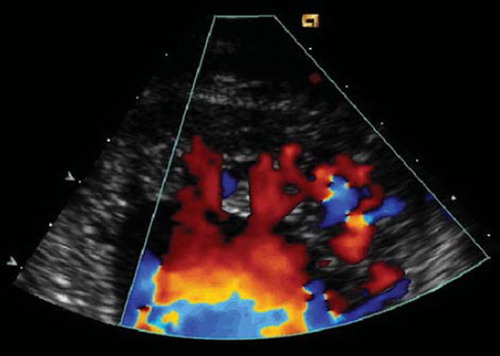
Figure 1. Echocardiographic appearance of IVNC. End-diastolic still frame (close-up view of LV apex) of a 37-year-old woman showing the typical echocardiographic features of IVNC with a thin epicardial and a thickened endocardial layer with prominent deep recesses. Blood flow from the ventricular cavity into the recesses is visualized with color Doppler. Reprinted from Steffel et al. (Citation12), Electrocardiographic characteristics at initial diagnosis in patients with isolated left ventricular noncompaction, American Journal of Cardiology 2009;104:984–89, Reprinted from American Journal of Cardiology, volume 104, Steffel J, Kobza R, Oechslin E, Jenni R, Duru F, Electrocardiographic characteristics at initial diagnosis in patients with isolated left ventricular noncompaction, pages 984–89, 2009 (Citation12), Copyright (2009), with permission from Elsevier.
This review first provides a brief overview on the principles of pathophysiology, clinical presentation, and management of patients with IVNC. We then summarize the typical ECG and electrophysiological (EP) findings as well as the incidence of ventricular tachyarrhythmias in these patients and discuss the available evidence on the management of patients at high risk for arrhythmic death.
Principles of pathophysiology, clinical presentation, and management
During physiological intrauterine development, deep intertrabecular recesses communicating with the ventricular endocardium evolve, which are subsequently converted to capillaries during the process of myocardial condensation (Citation3,Citation7). The premature arrest of this crucial developmental compaction process is believed to result in the typical phenotype encountered in IVNC (Citation8,Citation9).
The clinical presentation at the time of diagnosis as well as the subsequent course of these patients varies considerably, ranging from asymptomatic, coincidental discovery of the disease at one end of the spectrum to severe heart failure and sudden cardiac death at the other end (Citation3,Citation10). Rarely, patients initially present with palpitations or syncope. While a prolonged asymptomatic clinical course is not infrequent in asymptomatic patients, the prognosis in patients with severe heart failure is usually unfavorable (Citation4,Citation6).
The management of heart failure in patients with IVNC is usually not different from that of patients with other etiologies. It includes careful history taking and examination, exercise stress testing, spirometry, and echocardiographic follow-up if necessary. Similarly, treatment of heart failure in patients with IVNC primarily consists of standard medical therapy including diuretics, beta-blockers, ACE inhibitors, etc. In severe cases, mechanical assist device implantation and heart transplantation may be considered (Citation11). The incidence as well as the optimal management and prevention of potentially life-threatening ventricular arrhythmias have so far not been extensively studied.
Electrocardiographic findings
Abnormal base-line ECGs are common in patients with IVNC () (Citation4,Citation6,Citation12–17). Comprehensive electrocardiographic analyses of larger patient numbers newly diagnosed with IVNC, however, have only recently been reported (Citation12,Citation14). When comparing the studies outlined in , marked differences are observed regarding the occurrence of ECG changes in general as well as the frequency of serious electrocardiographic abnormalities. The reason for this discrepancy most likely relates to the difference in study populations included in these analyses. In older studies, IVNC was frequently diagnosed at an advanced symptomatic stage (Citation4,Citation6); in contrast, during the more recent years, individuals affected with IVNC are frequently being identified at a less advanced disease stage due to an increased awareness of the disease or during family screening, leading to a greater representation of asymptomatic subjects in these cohorts (Citation12,Citation14). Hence, both cohorts are clearly subject to a certain degree of selection bias; in view of the more recent time-frame of patient inclusion, however, the latter ones are likely to be more representative of the current spectrum of patients diagnosed with IVNC and as such likely of higher clinical value for present-day physicians involved in the care of these patients.
Table I. Electrocardiographic findings in patients with IVNC. Number of patients and (%) are shown.
ECG changes are observed in almost every level of atrial and/or ventricular depolarization and repolarization. Atrial fibrillation was frequently observed in earlier studies (up to 26%) and was associated with a worse prognosis (Citation4). In more recent years, however, these numbers have decreased substantially, most likely for the above-mentioned reasons. The underlying pathophysiological mechanism for atrial fibrillation in these patients currently remains elusive. Indeed, a development due to direct affection of the atria or as a result of the diseased heart with enlarged atria and increased filling pressures may be conceivable. Similarly, ventricular depolarization and repolarization are frequently altered in patients with IVNC. We previously observed a substantial overlap between electrocardiographic findings of intraventricular conduction delay (LBBB, in particular), atrial conduction delay (i.e. PR interval prolongation or AV block), and prolonged QTc duration on one hand, and the presence of a reduced left ventricular ejection fraction (LVEF), left ventricular (LV) and/or left atrial dilation on the other (Citation12). While IVNC is most likely responsible for the development of heart failure in these subjects, the associated ECG alterations are typically also observed at the advanced stage of other cardiomyopathies (Citation18) and are hence more likely to reflect the severity of the diseased heart in itself, independently of IVNC (Citation12).
Since repolarization abnormalities belong to the most frequently observed ECG changes in patients with IVNC (), it was tempting to believe that such abnormalities correspond to the morphologic wall changes related to IVNC. However, only a moderate degree of such correlation has been reported in this respect (Citation12). In the same way, no specific segmental involvement has been observed in patients with intraventricular conduction abnormalities or LBBB. Although similarly conceivable, electrocardiographic voltage signs of LV hypertrophy do not appear to correlate with ventricular wall thickening on echocardiography but rather with increased ventricular sizes in patients with IVNC (Citation12). Conversely, patients with echocardiographic signs of LV hypertrophy did not show any specific ECG abnormalities, including voltage signs of LV hypertrophy. These findings may be explained by the inability of ECG voltage criteria for LV hypertrophy to differentiate between genuine LV hypertrophy and LV dilatation (Citation18) or an effect of the noncompaction itself. Hence, in summary, no ECG abnormalities have yet been identified to specifically derive the localization of the non-compacted LV segments.
Recently, a Brugada-like ECG pattern has been reported in six patients with IVNC (with largely normal LVEF), one of whom experienced ventricular fibrillation and underwent implantable cardioverter defibrillator (ICD) implantation (Citation14). Although a transmural voltage gradient across the non-compacted versus the compacted endocardium, ischemia, and patchy fibrosis were discussed as a potential mechanism, the last-mentioned currently remains elusive and requires further study.
ECG patterns in children with noncompaction differ from those in the adult population. In particular, marked biventricular hypertrophy with extreme QRS voltage and Wolf–Parkinson–White syndrome appear to be more common in the pediatric population (Citation19). Rhythm disorders in children with noncompaction are presented and reviewed elsewhere in detail (Citation19,Citation20).
Electrophysiology of IVNC
While ventricular arrhythmias are common in IVNC, previous publications have reported wide variations in their occurrence incidence (). This can be due to several factors: It is possible that inclusion of less sick patients in cohorts stemming from more recent years may have a significant impact on the occurrence of ventricular arrhythmias. Moreover, the reported absolute numbers of ventricular arrhythmias in clinical studies may vary due to the type of arrhythmia (non-sustained versus sustained). Finally, the occurrence of ventricular tachycardia (VT) depends on the methodology used for its assessment as well as on the studied patient population. Indeed, a higher incidence of VT in patients with an implantable cardioverter defibrillator (ICD) in place as compared to a population of ‘all-comers’ with a one-time 24-hour ECG is not necessarily unexpected. Hence, in summary, the true occurrence of ventricular arrhythmias in IVNC is difficult to assess in view of different inclusion periods, patient characteristics, and methodology used for VT detection.
Table II. Frequency of ventricular arrhythmias in IVNC.
Invasive EP study may be a valuable tool both for unraveling the mechanisms and for risk stratification of these patients. However, only few reports have investigated the value of EP testing in patients with IVNC (Citation10,Citation21,Citation22). In one case of an apparently healthy 43-year-old man, in whom IVNC was diagnosed during a routine evaluation for sport activity, an EP study and an MRI were performed during work-up (Citation21). Interestingly, electrical abnormalities, such as low voltage and scar areas, were less related to non-compacted myocardium but predominantly to the presence and extent of myocardial fibrosis, indicating that the non-compacted segments themselves are less likely the culprit for malignant arrhythmias. These findings confirm and extend previous studies, in which a decrease in coronary flow reserve was observed in patients with IVNC, which was not confined to non-compacted segments but extended to most segments with wall motion abnormalities (Citation23). Hence, both the non-compacted myocardium and the impaired flow reserve in structurally compacted myocardial segments with resultant ischemia may serve as the arrhythmic substrate in IVNC.
We recently reported on 24 patients with IVNC undergoing EP testing, which represents the largest comprehensive analysis of EP findings in this patient population so far () (Citation10). This study showed that ventricular as well as supraventricular arrhythmias were readily inducible during EP studies. However, the inducibility of a sustained monomorphic VT (two patients, 8%) or a sustained polymorphic VT or fibrillation (two patients, 8%) was relatively low. Non-sustained polymorphic VT, which is generally regarded as a non-specific finding, was more commonly observed (five patients, 21%), mostly under stimulation with several extra-stimuli and/or isoproterenol infusion. Indeed, two of these last-mentioned patients demonstrated ventricular arrhythmias during follow-up, which were adequately treated by their ICDs (Citation10). In contrast, no ventricular arrhythmias or sudden cardiac deaths were observed in the 12 patients without inducible VT on EP testing during 30 (±19) months of clinical follow-up. Although these data do imply that a negative EP study may identify patients with IVNC at low risk of developing malignant tachyarrhythmias, the duration of follow-up in this subset was substantially shorter as compared to that of patients with inducible VT (i.e. 61 months). The fact that a VT was observed in one patient from the latter group 8 years after the EP study demonstrates that malignant arrhythmic events may well occur beyond the 30 months follow-up period for patients without inducible VT. This may be due to stochastic reasons, but may also be due to progression of the disease, which subsequently alters the arrhythmogenic substrate increasing the propensity of developing malignant VT. Therefore, further studies are warranted to determine the role of EP testing in IVNC, especially with respect to the prospective value of a negative EP study in these patients (Citation10).
Table III. EP findings in 24 patients with IVNC. Data from Steffel et al., Europace 2009 (Citation10).
Single case reports have indicated that VT ablation may be performed in patients with IVNC and inducible VT during EP study (Citation24,Citation25). However, experience is too scarce at present to recommend this as a standard procedure. More importantly, VT ablation as a sole means of antiarrhythmic treatment in a patient with IVNC and inducible VT during EP testing does not appear warranted in view of the high propensity of these patients for potentially life-threatening ventricular arrhythmias, and ICD implantation should strongly be considered in these individuals.
EP studies may also be helpful in the diagnosis and treatment of supraventricular arrhythmias () (Citation10). While ventricular pre-excitation with the Wolf–Parkinson–White (WPW) syndrome is frequently observed in children with noncompaction (Citation26), it is rather infrequent in the adult population (Citation4,Citation12,Citation14,Citation16,Citation27,Citation28). However, at least one case of a patient with the WPW syndrome has been described, in which rapidly conducted atrial fibrillation via an accessory pathway led to ventricular fibrillation and cardiac arrest (Citation27). Hence, if pre-excitation is observed, either during careful analysis of the surface ECG or during an EP study, ablation of the accessory pathway should be performed. Furthermore, radiofrequency ablation of the cavo-tricuspid isthmus for typical atrial flutter and slow-pathway modification for AV-nodal re-entry tachycardia can safely and effectively be performed in this patient population (Citation10,Citation29). Hence, there are no indications at present that invasive treatment of such uncomplicated supraventricular arrhythmias should differ in patients with IVNC as compared to the general population.
Antiarrhythmic drug therapy
Few studies have described the efficacy and/or safety of antiarrhythmic drug therapy in patients with IVNC. Beta-blockers appear to be safe and should be considered in particular in the presence of heart failure. For the suppression of ventricular or supraventricular arrhythmias, class III antiarrhythmics, in particular amiodarone, appears to be effective in our own experience and in isolated case reports (Citation25,Citation30). In contrast, there are no data on the safety of class I antiarrhythmic drugs. Even though coronary artery disease or left ventricular hypertrophy, i.e. contraindications for the use of class I antiarrhythmics, are comparatively rare in patients with IVNC, these ventricles may not be considered structurally normal. Therefore, these agents should be avoided, if possible. Whether novel antiarrhythmic drugs such as dronedarone may be an option in the management of these patients remains to be seen.
Rationale for ICD therapy
Together with end-stage heart failure, malignant ventricular arrhythmias are the most common cause of death in patients with IVNC (Citation3,Citation4,Citation11,Citation31). In the largest series so far, we recently reported on the follow-up of 30 IVNC patients who had undergone ICD implantation (Citation32). ICDs were implanted for primary prevention if the left ventricular ejection fraction was less than 35% (n = 18) or for secondary prevention if the patient had a history of survived sudden cardiac death, sustained VT, or documented non-sustained VT causing syncope or near-syncope (n = 12). During the follow-up period of 40 ± 34 months, 11 patients (37%) received appropriate ICD therapies, i.e. anti-tachycardia pacing (ATP) (n = 3), shock therapy (n = 4), and ATP plus shock (n = 4). Appropriate ICD therapy occurred 21 ± 16 months after ICD implantation and was observed both after primary (6/18 patients) and secondary (5/12 patients) ICD implantation. In bivariate regression analysis a reduced ejection fraction was associated with appropriate ICD therapy; however, no predictors were observed on multivariate analysis. Inappropriate ICD therapy occurred in four patients (13%) due to atrial fibrillation (n = 2), and due to atrial tachycardia and T wave over-sensing, each in one patient. Hence, ICD implantation appears safe and effective in the largest cohort of patients with IVNC to date. Interestingly, in a previous smaller study of 11 patients with IVNC and ICD, no appropriate therapies were applied during a mean follow-up of 2.5 years, while two patients experienced inappropriate ICD therapies (Citation13). The mean ejection fraction in the latter study was 41% ± 15%, indicating a sicker patient population in the former study, which may have contributed to the higher risk for arrhythmias and consecutive appropriate ICD therapies.
Based on the results of several large prospective, randomized clinical trials (Citation33,Citation34), current heart failure guidelines recommend empirical ICD implantation in patients with a severely reduced ejection fraction (Citation35). Although it is presently unclear whether these guidelines are also applicable to patients with IVNC, it is hard to argue against prophylactic ICD implantation in patients fulfilling these criteria. Furthermore, ICD implantation appears warranted in patients clinically judged to be at high risk for malignant arrhythmias or sudden cardiac death, including those with a prior history of sustained VT or VF, recurrent syncope of unknown etiology, or a family history of ventricular arrhythmias or sudden cardiac death. This is acknowledged by the current ACC/AHA/HRS guidelines for device-based therapy of cardiac arrhythmias, in which ICD implantation is considered a reasonable strategy to reduce the risk of sudden cardiac death in patients with IVNC (Grade IIb, level of evidence C) (Citation36).
Cardiac resynchronization therapy
Cardiac resynchronization therapy (CRT) has been shown to decrease morbidity and mortality in symptomatic heart failure patients (NYHA III–IV) with a severely reduced left ventricular ejection fraction (≤ 35%) and a wide QRS complex (QRS ≥ 120 ms) (Citation37,Citation38). Whether these benefits also extend to patients with IVNC, however, has not yet been prospectively assessed. In an early study, four IVNC patients treated with CRT showed an improvement in ejection fraction (20.8 ± 7.8 to 36.0 ± 13.6%; P < 0.05) and heart failure symptoms (Citation39). In our recent study of IVNC patients receiving an ICD, implantation of a biventricular device was considered in patients presenting with the above-mentioned Cardiac Resynchronization im Heart Failure (CARE-HF) criteria or in the presence of echocardiographic signs for dyssynchrony. Using these selection criteria, 6 of 30 patients were implanted with an ICD with CRT (CRT-D) (Citation32). In these patients, ejection fraction improved from 22% ± 5% to 37% ± 15% (P = 0.094) and functional NYHA class from 2.5 ± 0.5 to 1.6 ± 0.8 (P = 0.011) during follow-up of 18 ± 11 months after implantation (Citation32). These pilot experiences indicate that patients with IVNC and heart failure may profit from CRT-D to a similar degree as those with congestive heart failure of other etiologies. Whether selection criteria and responder characteristics are different in the former and the latter group of patients, however, currently remains elusive and will require further studies. With the available data to date, implantation of a CRT-D should be considered in patients fulfilling current eligibility criteria for these devices as indicated above (Citation35).
Summary and clinical implications
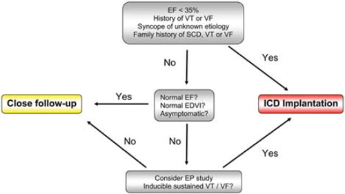
Both supraventricular and ventricular rhythm disorders are frequent in patients with IVNC. With respect to the former, therapeutic strategies (including ablation, if deemed indicated) generally follow those for patients without IVNC. In contrast, data regarding risk stratification and treatment of ventricular arrhythmias, which are amongst the most frequent causes of death in these patients, are scarce due to the rareness of the disease. For the prevention of sudden arrhythmic death, ICDs have been demonstrated to be highly effective. Unfortunately, to date, no reliable predictors of future arrhythmic events have been identified, and identifying patients most likely to profit from an ICD remains a challenge. Implantation of an ICD appears warranted in patients with a severely reduced EF, as well as in those at high risk of ventricular arrhythmias. In patients who do not fall into either of these categories, an individualized approach is necessary. Close clinical follow-up may be considered, especially in asymptomatic patients with normal left ventricular function and diameters (). In addition, an EP study for further risk stratification may be considered; indeed, patients in whom ventricular arrhythmia cannot be provoked using an aggressive stimulation protocol may be at low risk for future life-threatening arrhythmic events. However, close follow-up by a cardiologist is warranted, since long-term experience regarding the natural history of these patients is still missing. To improve methods of arrhythmic risk stratification in patients with IVNC, data from prospective studies and registries are required.
Figure 2. Proposed algorithm for arrhythmic risk stratification in IVNC.
EF = ejection fraction; VT = ventricular tachycardia; VF = ventricular fibrillation; SCD = sudden cardiac death; EDVI = end-diastolic volume index; ICD = implantable cardioverter defibrillator; EP = electrophysiological.
Declaration of interest: The authors state no conflict of interest and have received no payment in preparation of this manuscript.
References
- Maron BJ, Towbin JA, Thiene G, Antzelevitch C, Corrado D, Arnett D, . Contemporary definitions and classification of the cardiomyopathies: an American Heart Association Scientific Statement from the Council on Clinical Cardiology, Heart Failure and Transplantation Committee; Quality of Care and Outcomes Research and Functional Genomics and Translational Biology Interdisciplinary Working Groups; and Council on Epidemiology and Prevention. Circulation. 2006;113:1807–16.
- Jenni R, Oechslin E, Schneider J, Attenhofer Jost C, Kaufmann PA. Echocardiographic and pathoanatomical characteristics of isolated left ventricular noncompaction: a step towards classification as a distinct cardiomyopathy. Heart. 2001;86:666–71.
- Jenni R, Oechslin EN, van der Loo B. Isolated ventricular noncompaction of the myocardium in adults. Heart. 2007;93:11–15.
- Oechslin EN, Attenhofer Jost CH, Rojas JR, Kaufmann PA, Jenni R. Long-term follow-up of 34 adults with isolated left ventricular noncompaction: a distinct cardiomyopathy with poor prognosis. J Am Coll Cardiol. 2000; 36:493–500.
- Hershberger RE, Lindenfeld J, Mestroni L, Seidman CE, Taylor MR, Towbin JA. Genetic evaluation of cardiomyopathy—a Heart Failure Society of America practice guideline. J Card Fail. 2009;15:83–97.
- Murphy RT, Thaman R, Blanes JG, Ward D, Sevdalis E, Papra E, . Natural history and familial characteristics of isolated left ventricular noncompaction. Eur Heart J. 2005;26:187–92.
- Sedmera D, Pexieder T, Vuillemin M, Thompson RP, Anderson RH. Developmental patterning of the myocardium. Anat Rec. 2000;258:319–37.
- Freedom RM, Yoo SJ, Perrin D, Taylor G, Petersen S, Anderson RH. The morphological spectrum of ventricular noncompaction. Cardiol Young. 2005;15:345–64.
- Ichida F. Left ventricular noncompaction. Circ J. 2009;73: 19–26.
- Steffel J, Kobza R, Namdar M, Wolber T, Brunckhorst C, Luscher TF, . Electrophysiological findings in patients with isolated left ventricular noncompaction. Europace. 2009;11:1193–200.
- Kovacevic-Preradovic T, Jenni R, Oechslin EN, Noll G, Seifert B, Attenhofer Jost CH. Isolated left ventricular noncompaction as a cause for heart failure and heart transplantation: a single center experience. Cardiology. 2009;112:158–64.
- Steffel J, Kobza R, Oechslin E, Jenni R, Duru F. Electrocardiographic characteristics at initial diagnosis in patients with isolated left ventricular noncompaction. Am J Cardiol. 2009;104:984–9.
- Stanton C, Bruce C, Connolly H, Brady P, Syed I, Hodge D, . Isolated left ventricular noncompaction syndrome. Am J Cardiol. 2009;104:1135–8.
- Shoji M, Yamashita T, Uejima T, Asada K, Semba H, Otsuka T, . Electrocardiography characteristics of isolated noncompaction of ventricular myocardium in Japanese adult patients. Circ J. 2010;74:1431–5.
- Lofiego C, Biagini E, Pasquale F, Ferlito M, Rocchi G, Perugini E, . Wide spectrum of presentation and variable outcomes of isolated left ventricular noncompaction. Heart. 2007;93:65–71.
- Aras D, Tufekcioglu O, Ergun K, Ozeke O, Yildiz A, Topaloglu S, . Clinical features of isolated ventricular noncompaction in adults long-term clinical course, echocardiographic properties, and predictors of left ventricular failure. J Card Fail. 2006;12:726–33.
- Duru F, Candinas R. Noncompaction of ventricular myocardium and arrhythmias. J Cardiovasc Electrophysiol. 2000;11:493.
- Madias JE. The resting electrocardiogram in the management of patients with congestive heart failure: established applications and new insights. Pacing Clin Electrophysiol. 2007; 30:123–8.
- Pignatelli RH, McMahon CJ, Dreyer WJ, Denfield SW, Price J, Belmont JW, . Clinical characterization of left ventricular noncompaction in children: a relatively common form of cardiomyopathy. Circulation. 2003;108:2672–8.
- Celiker A, Ozkutlu S, Dilber E, Karagoz T. Rhythm abnormalities in children with isolated ventricular noncompaction. Pacing Clin Electrophysiol. 2005;28:1198–202.
- Casella M, Pieroni M, Dello Russo A, Pennestri F, Meduri A, Natale L, . Characterization of the electroanatomic substrate in a case of noncompaction left ventricle. J Cardiovasc Med (Hagerstown). 2008;9:636–8.
- Khan IA, Biddle WP, Najeed SA, Abdul-Aziz S, Mehta NJ, Salaria V, . Isolated noncompaction cardiomyopathy presenting with paroxysmal supraventricular tachycardia—case report and literature review. Angiology. 2003;54:243–50.
- Jenni R, Wyss CA, Oechslin EN, Kaufmann PA. Isolated ventricular noncompaction is associated with coronary microcirculatory dysfunction. J Am Coll Cardiol. 2002;39: 450–4.
- Lim HE, Pak HN, Shim WJ, Ro YM, Kim YH. Epicardial ablation of ventricular tachycardia associated with isolated ventricular noncompaction. Pacing Clin Electrophysiol. 2006;29:797–9.
- Fiala M, Januska J, Bulkova V, Pleva M. Septal ventricular tachycardia with alternating LBBB-RBBB morphology in isolated ventricular noncompaction. J Cardiovasc Electrophysiol. 2010;21:704–7.
- Ichida F, Hamamichi Y, Miyawaki T, Ono Y, Kamiya T, Akagi T, . Clinical features of isolated noncompaction of the ventricular myocardium: long-term clinical course, hemodynamic properties, and genetic background. J Am Coll Cardiol. 1999;34:233–40.
- Fichet J, Legras A, Bernard A, Babuty D. Aborted sudden cardiac death revealing isolated noncompaction of the left ventricle in a patient with wolff-Parkinson-white syndrome. Pacing Clin Electrophysiol. 2007;30:444–7.
- Brembilla-Perrot B, Codreanu A, Marie PY, Beurrier D, Husson JL, Hutin O, . [Association of Wolff-Parkinson-White syndrome with isolated noncompaction of the left ventricle: a case report]. Arch Mal Coeur Vaiss. 2006;99: 626–8.
- Enriquez SG, Entem FR, Cobo M, Olalla JJ. Uncommon etiology of syncope in a patient with isolated ventricular noncompaction. Pacing Clin Electrophysiol. 2007;30:577–9.
- Sato Y, Matsumoto N, Matsuo S, Imai S, Yoda S, Tani S, . Subendomyocardial perfusion abnormality and necrosis detected by magnetic resonance imaging in a patient with isolated noncompaction of the ventricular myocardium associated with ventricular tachycardia. Cardiovasc Revasc Med. 2009;10:66–8.
- Kobza R, Jenni R, Erne P, Oechslin E, Duru F. Implantable cardioverter-defibrillators in patients with left ventricular noncompaction. Pacing Clin Electrophysiol. 2008;31: 461–7.
- Kobza R, Steffel J, Erne P, Schoenenberger AW, Hurlimann D, Luscher TF, . Implantable cardioverter-defibrillator and cardiac resynchronization therapy in patients with left ventricular noncompaction. Heart Rhythm. 2010;7:1545–9.
- Kadish A, Dyer A, Daubert JP, Quigg R, Estes NA, Anderson KP, . Prophylactic defibrillator implantation in patients with nonischemic dilated cardiomyopathy. N Engl J Med. 2004; 350:2151–8.
- Bardy GH, Lee KL, Mark DB, Poole JE, Packer DL, Boineau R, . Amiodarone or an implantable cardioverter-defibrillator for congestive heart failure. N Engl J Med. 2005;352:225–37.
- Epstein AE, Dimarco JP, Ellenbogen KA, Estes NA 3rd, Freedman RA, Gettes LS, . ACC/AHA/HRS 2008 guidelines for device-based therapy of cardiac rhythm abnormalities. Heart Rhythm. 2008;5:e1–62.
- Epstein AE, DiMarco JP, Ellenbogen KA, Estes NA 3rd, Freedman RA, Gettes LS, . ACC/AHA/HRS 2008 guidelines for device-based therapy of cardiac rhythm abnormalities: a report of the American College of Cardiology/American Heart Association Task Force on Practice Guidelines (Writing Committee to Revise the ACC/AHA/NASPE 2002 Guideline Update for Implantation of Cardiac Pacemakers and Antiarrhythmia Devices) developed in collaboration with the American Association for Thoracic Surgery and Society of Thoracic Surgeons. J Am Coll Cardiol. 2008;51:e1–62.
- Cleland JG, Daubert JC, Erdmann E, Freemantle N, Gras D, Kappenberger L, . The effect of cardiac resynchronization on morbidity and mortality in heart failure. N Engl J Med. 2005;352:1539–49.
- Abraham WT, Fisher WG, Smith AL, Delurgio DB, Leon AR, Loh E, . Cardiac resynchronization in chronic heart failure. N Engl J Med. 2002;346:1845–53.
- Oginosawa Y, Nogami A, Soejima K, Aonuma K, Kubota S, Sato T, . Effect of cardiac resynchronization therapy in isolated ventricular noncompaction in adults: follow-up of four cases. J Cardiovasc Electrophysiol. 2008;19:935–8.